4GSX
 
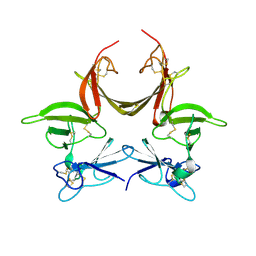
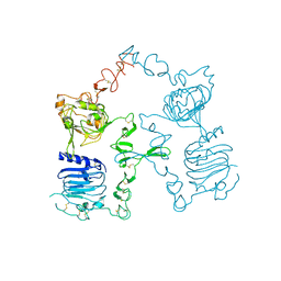
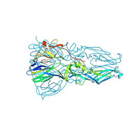 | | High resolution structure of dengue virus serotype 1 sE containing stem | | 分子名称: | 2-acetamido-2-deoxy-beta-D-glucopyranose, CADMIUM ION, CHLORIDE ION, ... | | 著者 | Klein, D.E, Choi, J.L, Harrison, S.C. | | 登録日 | 2012-08-28 | | 公開日 | 2012-12-19 | | 最終更新日 | 2023-09-13 | | 実験手法 | X-RAY DIFFRACTION (1.903 Å) | | 主引用文献 | Structure of a dengue virus envelope protein late-stage fusion intermediate.
J.Virol., 87, 2013
|
|
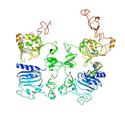
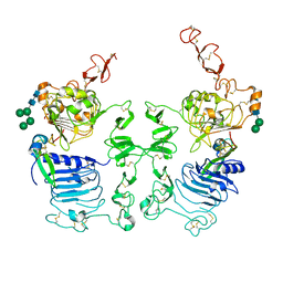
4GT0
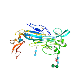
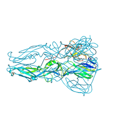 | | Structure of dengue virus serotype 1 sE containing stem to residue 421 | | 分子名称: | 2-acetamido-2-deoxy-beta-D-glucopyranose, CADMIUM ION, CHLORIDE ION, ... | | 著者 | Klein, D.E, Choi, J.L, Harrison, S.C. | | 登録日 | 2012-08-28 | | 公開日 | 2012-12-19 | | 最終更新日 | 2020-07-29 | | 実験手法 | X-RAY DIFFRACTION (2.57 Å) | | 主引用文献 | Structure of a dengue virus envelope protein late-stage fusion intermediate.
J.Virol., 87, 2013
|
|
3CA7
 | |
3C9A
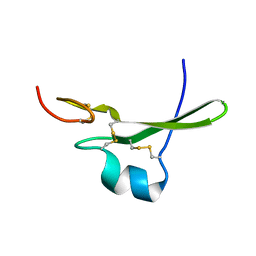
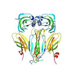
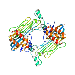
 | | High Resolution Crystal Structure of Argos bound to the EGF domain of Spitz | | 分子名称: | BROMIDE ION, Protein giant-lens, Protein spitz | | 著者 | Klein, D.E, Stayrook, S.E, Shi, F, Narayan, K, Lemmon, M.A. | | 登録日 | 2008-02-15 | | 公開日 | 2008-05-20 | | 最終更新日 | 2017-10-25 | | 実験手法 | X-RAY DIFFRACTION (1.6 Å) | | 主引用文献 | Structural basis for EGFR ligand sequestration by Argos.
Nature, 453, 2008
|
|
3CGU
 | |
3LTG
 | |
3LTF
 | | Crystal Structure of the Drosophila Epidermal Growth Factor Receptor ectodomain in complex with Spitz | | 分子名称: | 2-acetamido-2-deoxy-beta-D-glucopyranose, 2-acetamido-2-deoxy-beta-D-glucopyranose-(1-4)-2-acetamido-2-deoxy-beta-D-glucopyranose, Epidermal growth factor receptor, ... | | 著者 | Alvarado, D, Klein, D.E, Lemmon, M.A. | | 登録日 | 2010-02-15 | | 公開日 | 2010-08-25 | | 最終更新日 | 2023-09-06 | | 実験手法 | X-RAY DIFFRACTION (3.2 Å) | | 主引用文献 | Structural basis for negative cooperativity in growth factor binding to an EGF receptor.
Cell(Cambridge,Mass.), 142, 2010
|
|
5AOQ
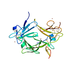
 | | Structural basis of neurohormone perception by the receptor tyrosine kinase Torso | | 分子名称: | 2-acetamido-2-deoxy-beta-D-glucopyranose-(1-4)-2-acetamido-2-deoxy-beta-D-glucopyranose, PREPROPTTH, TORSO | | 著者 | Jenni, S, Goyal, Y, von Grotthuss, M, Shvartsman, S.Y, Klein, D.E. | | 登録日 | 2015-09-11 | | 公開日 | 2015-11-25 | | 最終更新日 | 2020-07-29 | | 実験手法 | X-RAY DIFFRACTION (2.7 Å) | | 主引用文献 | Structural Basis of Neurohormone Perception by the Receptor Tyrosine Kinase Torso.
Mol.Cell, 60, 2015
|
|
3I2T
 | | Crystal structure of the unliganded Drosophila Epidermal Growth Factor Receptor ectodomain | | 分子名称: | 2-acetamido-2-deoxy-beta-D-glucopyranose-(1-4)-2-acetamido-2-deoxy-alpha-D-glucopyranose, 2-acetamido-2-deoxy-beta-D-glucopyranose-(1-4)-2-acetamido-2-deoxy-beta-D-glucopyranose, Epidermal growth factor receptor, ... | | 著者 | Alvarado, D, Klein, D.E, Lemmon, M.A. | | 登録日 | 2009-06-29 | | 公開日 | 2009-09-08 | | 最終更新日 | 2023-09-06 | | 実験手法 | X-RAY DIFFRACTION (2.7 Å) | | 主引用文献 | ErbB2 resembles an autoinhibited invertebrate epidermal growth factor receptor.
Nature, 461, 2009
|
|
7LIR
 | | Structure of the invertebrate ALK GRD | | 分子名称: | 2-acetamido-2-deoxy-beta-D-glucopyranose, 2-acetamido-2-deoxy-beta-D-glucopyranose-(1-4)-2-acetamido-2-deoxy-beta-D-glucopyranose, ALK tyrosine kinase receptor homolog scd-2, ... | | 著者 | Stayrook, S, Li, T, Klein, D.E. | | 登録日 | 2021-01-27 | | 公開日 | 2021-11-24 | | 最終更新日 | 2021-12-15 | | 実験手法 | X-RAY DIFFRACTION (2.6 Å) | | 主引用文献 | Structural basis for ligand reception by anaplastic lymphoma kinase.
Nature, 600, 2021
|
|
7LS0
 | | Structure of the Human ALK GRD bound to AUG | | 分子名称: | 2-acetamido-2-deoxy-beta-D-glucopyranose, ALK tyrosine kinase receptor fused with ALK and LTK ligand 2, CITRIC ACID | | 著者 | Stayrook, S, Li, T, Klein, D.E. | | 登録日 | 2021-02-17 | | 公開日 | 2021-11-24 | | 最終更新日 | 2023-10-18 | | 実験手法 | X-RAY DIFFRACTION (3.05 Å) | | 主引用文献 | Structural basis for ligand reception by anaplastic lymphoma kinase.
Nature, 600, 2021
|
|
7LRZ
 | |
7MK7
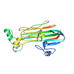
 | | Augmentor domain of augmentor-beta | | 分子名称: | ALK and LTK ligand 1,Maltodextrin-binding protein, alpha-D-glucopyranose-(1-4)-alpha-D-glucopyranose | | 著者 | Krimmer, S.G, Reshetnyak, A.V, Puleo, D.E, Schlessinger, J. | | 登録日 | 2021-04-21 | | 公開日 | 2021-11-24 | | 最終更新日 | 2023-10-18 | | 実験手法 | X-RAY DIFFRACTION (2.42815185 Å) | | 主引用文献 | Structural basis for ligand reception by anaplastic lymphoma kinase.
Nature, 600, 2021
|
|


