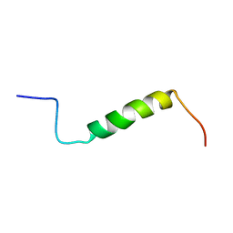
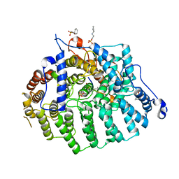
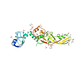
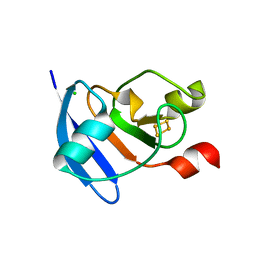
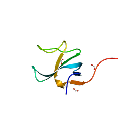
4B6W
 
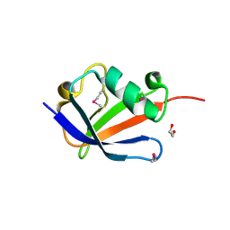 | | Architecture of Trypanosoma brucei Tubulin-Binding cofactor B | | 分子名称: | 1,2-ETHANEDIOL, TUBULIN-SPECIFIC CHAPERONE | | 著者 | Fleming, J.R, Morgan, R.E, Fyfe, P.K, Kelly, S.M, Hunter, W.N. | | 登録日 | 2012-08-15 | | 公開日 | 2012-08-22 | | 最終更新日 | 2013-07-17 | | 実験手法 | X-RAY DIFFRACTION (2.35 Å) | | 主引用文献 | The Architecture of Trypanosoma Brucei Tubulin-Binding Cofactor B and Implications for Function.
FEBS J., 280, 2013
|
|
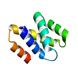
1UNQ
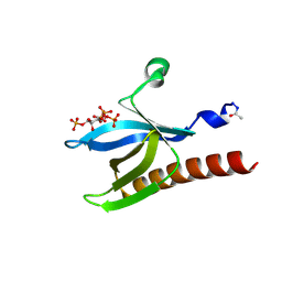 | | High resolution crystal structure of the Pleckstrin Homology Domain Of Protein Kinase B/Akt Bound To Ins(1,3,4,5)-Tetrakisphophate | | 分子名称: | INOSITOL-(1,3,4,5)-TETRAKISPHOSPHATE, RAC-ALPHA SERINE/THREONINE KINASE | | 著者 | Milburn, C.C, Deak, M, Kelly, S.M, Price, N.C, Alessi, D.R, van Aalten, D.M.F. | | 登録日 | 2003-09-12 | | 公開日 | 2004-09-16 | | 最終更新日 | 2023-12-13 | | 実験手法 | X-RAY DIFFRACTION (0.98 Å) | | 主引用文献 | Binding of phosphatidylinositol 3,4,5-trisphosphate to the pleckstrin homology domain of protein kinase B induces a conformational change.
Biochem. J., 375, 2003
|
|
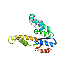
1UNP
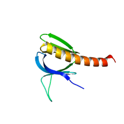 | | Crystal structure of the pleckstrin homology domain of PKB alpha | | 分子名称: | RAC-ALPHA SERINE/THREONINE KINASE | | 著者 | Milburn, C.C, Deak, M, Kelly, S.M, Price, N.C, Alessi, D.R, van Aalten, D.M.F. | | 登録日 | 2003-09-12 | | 公開日 | 2004-09-16 | | 最終更新日 | 2023-12-13 | | 実験手法 | X-RAY DIFFRACTION (1.65 Å) | | 主引用文献 | Binding of phosphatidylinositol 3,4,5-trisphosphate to the pleckstrin homology domain of protein kinase B induces a conformational change.
Biochem. J., 375, 2003
|
|
1UNR
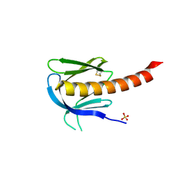 | | Crystal structure of the PH domain of PKB alpha in complex with a sulfate molecule | | 分子名称: | RAC-ALPHA SERINE/THREONINE KINASE, SULFATE ION | | 著者 | Milburn, C.C, Deak, M, Kelly, S.M, Price, N.C, Alessi, D.R, van Aalten, D.M.F. | | 登録日 | 2003-09-15 | | 公開日 | 2004-09-16 | | 最終更新日 | 2023-12-13 | | 実験手法 | X-RAY DIFFRACTION (1.25 Å) | | 主引用文献 | Binding of phosphatidylinositol 3,4,5-trisphosphate to the pleckstrin homology domain of protein kinase B induces a conformational change.
Biochem. J., 375, 2003
|
|
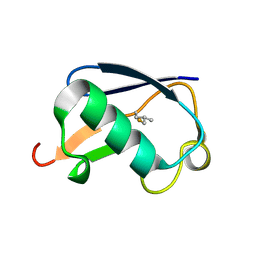
2KYA
 | | Solution structure of the leader sequence of the patellamide precursor peptide, PatE1-34 | | 分子名称: | Patellamide protein | | 著者 | Houssen, W.E, Wright, S.H, Kalverda, A.P, Thompson, G.S, Kelly, S.M, Jaspars, M. | | 登録日 | 2010-05-21 | | 公開日 | 2010-09-01 | | 最終更新日 | 2024-05-01 | | 実験手法 | SOLUTION NMR | | 主引用文献 | Solution Structure of the Leader Sequence of the Patellamide Precursor Peptide, PatE(1-34).
Chembiochem, 11, 2010
|
|
1OGW
 | | Synthetic Ubiquitin with fluoro-Leu at 50 and 67 | | 分子名称: | UBIQUITIN | | 著者 | Alexeev, D, Ramage, R, Young, D.W, Sawyer, L. | | 登録日 | 2003-05-13 | | 公開日 | 2003-05-30 | | 最終更新日 | 2023-12-13 | | 実験手法 | X-RAY DIFFRACTION (1.32 Å) | | 主引用文献 | Synthesis, Structural and Biological Studies of Ubiquitin Mutants Containing (2S, 4S)-5-Fluoroleucine Residues Strategically Placed in the Hydrophobic Core
Chembiochem, 4, 2003
|
|
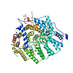
7PVC
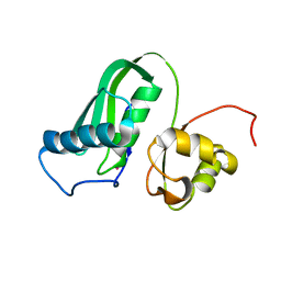 | |
2JPS
 | | NAB2 N-terminal domain | | 分子名称: | Nuclear polyadenylated RNA-binding protein NAB2 | | 著者 | Grant, R, Marshall, N.J, Yang, J, Fasken, M, Kelly, S, Harreman, M.T, Neuhaus, D, Corbett, A.H, Stewart, M. | | 登録日 | 2007-05-23 | | 公開日 | 2008-03-18 | | 最終更新日 | 2023-12-20 | | 実験手法 | SOLUTION NMR | | 主引用文献 | Structure of the N-terminal Mlp1-binding domain of the Saccharomyces cerevisiae mRNA-binding protein, Nab2
J.Mol.Biol., 376, 2008
|
|
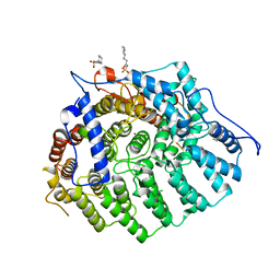
3Q79
 | |
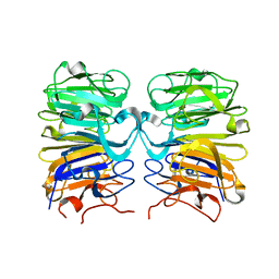
3Q7A
 | | Cryptococcus neoformans protein farnesyltransferase in complex with FPP and L-778,123 | | 分子名称: | (2R)-3-(cyclohexylamino)-2-hydroxypropane-1-sulfonic acid, 4-[(5-{[4-(3-CHLOROPHENYL)-3-OXOPIPERAZIN-1-YL]METHYL}-1H-IMIDAZOL-1-YL)METHYL]BENZONITRILE, FARNESYL DIPHOSPHATE, ... | | 著者 | Hast, M.A, Beese, L.S. | | 登録日 | 2011-01-04 | | 公開日 | 2011-08-03 | | 最終更新日 | 2024-02-21 | | 実験手法 | X-RAY DIFFRACTION (2 Å) | | 主引用文献 | Structures of Cryptococcus neoformans Protein Farnesyltransferase Reveal Strategies for Developing Inhibitors That Target Fungal Pathogens.
J.Biol.Chem., 286, 2011
|
|
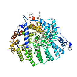
3Q78
 | |
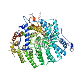
3Q75
 | |
3Q7F
 | |
3Q73
 | | Cryptococcus neoformans protein farnesyltransferase, apo enzyme | | 分子名称: | (2S)-3-(cyclohexylamino)-2-hydroxypropane-1-sulfonic acid, Farnesyltransferase, alpha subunit, ... | | 著者 | Hast, M.A, Beese, L.S. | | 登録日 | 2011-01-04 | | 公開日 | 2011-08-03 | | 最終更新日 | 2024-02-21 | | 実験手法 | X-RAY DIFFRACTION (2.3 Å) | | 主引用文献 | Structures of Cryptococcus neoformans Protein Farnesyltransferase Reveal Strategies for Developing Inhibitors That Target Fungal Pathogens.
J.Biol.Chem., 286, 2011
|
|
3SFX
 | | Cryptococcus neoformans protein farnesyltransferase in complex with FPT-II and tipifarnib | | 分子名称: | (2R)-3-(cyclohexylamino)-2-hydroxypropane-1-sulfonic acid, 6-[(S)-AMINO(4-CHLOROPHENYL)(1-METHYL-1H-IMIDAZOL-5-YL)METHYL]-4-(3-CHLOROPHENYL)-1-METHYLQUINOLIN-2(1H)-ONE, Cryptococcus neoformans protein farnesyltransferase alpha subunit, ... | | 著者 | Hast, M.A, Beese, L.S. | | 登録日 | 2011-06-14 | | 公開日 | 2011-08-03 | | 最終更新日 | 2024-02-28 | | 実験手法 | X-RAY DIFFRACTION (2 Å) | | 主引用文献 | Structures of Cryptococcus neoformans Protein Farnesyltransferase Reveal Strategies for Developing Inhibitors That Target Fungal Pathogens.
J.Biol.Chem., 286, 2011
|
|
3SFY
 | |
4N58
 | | Crystal Structure of Pectocin M2 at 1.86 Angstroms | | 分子名称: | (4S)-2-METHYL-2,4-PENTANEDIOL, CHLORIDE ION, FE2/S2 (INORGANIC) CLUSTER, ... | | 著者 | Grinter, R, Roszak, A.W, Zeth, K, Cogdell, C.J, Walker, D. | | 登録日 | 2013-10-09 | | 公開日 | 2014-06-04 | | 最終更新日 | 2024-02-28 | | 実験手法 | X-RAY DIFFRACTION (1.86 Å) | | 主引用文献 | Structure of the atypical bacteriocin pectocin M2 implies a novel mechanism of protein uptake.
Mol.Microbiol., 93, 2014
|
|
4N59
 | | The Crystal Structure of Pectocin M2 at 2.3 Angstroms | | 分子名称: | CHLORIDE ION, FE2/S2 (INORGANIC) CLUSTER, Pectocin M2, ... | | 著者 | Zeth, K, Grinter, R, Roszak, A.W, Cogdell, R.J, Walker, D. | | 登録日 | 2013-10-09 | | 公開日 | 2014-06-04 | | 最終更新日 | 2023-09-20 | | 実験手法 | X-RAY DIFFRACTION (2.3 Å) | | 主引用文献 | Structure of the atypical bacteriocin pectocin M2 implies a novel mechanism of protein uptake.
Mol.Microbiol., 93, 2014
|
|
4ZHP
 | | The crystal structure of Potato ferredoxin I with 2Fe-2S cluster | | 分子名称: | FE2/S2 (INORGANIC) CLUSTER, Potato Ferredoxin I | | 著者 | Grinter, R, Josts, I, Roszak, A.W, Cogdell, R.J, Walker, D. | | 登録日 | 2015-04-26 | | 公開日 | 2016-08-31 | | 最終更新日 | 2024-01-10 | | 実験手法 | X-RAY DIFFRACTION (2.46 Å) | | 主引用文献 | Structure of the bacterial plant-ferredoxin receptor FusA.
Nat Commun, 7, 2016
|
|
4ZGV
 | | The Crystal Structure of the Ferredoxin Receptor FusA from Pectobacterium atrosepticum SCRI1043 | | 分子名称: | Ferredoxin receptor, LAURYL DIMETHYLAMINE-N-OXIDE, octyl beta-D-glucopyranoside | | 著者 | Grinter, R, Josts, I, Roszak, A.W, Cogdell, R.J, Walker, D. | | 登録日 | 2015-04-24 | | 公開日 | 2016-08-31 | | 最終更新日 | 2024-10-09 | | 実験手法 | X-RAY DIFFRACTION (3.2 Å) | | 主引用文献 | Structure of the bacterial plant-ferredoxin receptor FusA.
Nat Commun, 7, 2016
|
|
4ZHO
 | | The crystal structure of Arabidopsis ferredoxin 2 with 2Fe-2S cluster | | 分子名称: | CHLORIDE ION, FE2/S2 (INORGANIC) CLUSTER, Ferredoxin-2, ... | | 著者 | Grinter, R, Josts, I, Roszak, A.W, Cogdell, R.J, Walker, D. | | 登録日 | 2015-04-26 | | 公開日 | 2016-08-31 | | 最終更新日 | 2024-05-08 | | 実験手法 | X-RAY DIFFRACTION (2.34 Å) | | 主引用文献 | Structure of the bacterial plant-ferredoxin receptor FusA.
Nat Commun, 7, 2016
|
|
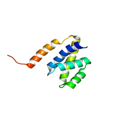
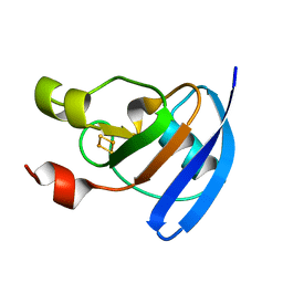
2V75
 | | N-terminal domain of Nab2 | | 分子名称: | NUCLEAR POLYADENYLATED RNA-BINDING PROTEIN NAB2 | | 著者 | Grant, R.P, Marshall, N.J, Stewart, M. | | 登録日 | 2007-07-26 | | 公開日 | 2008-01-29 | | 最終更新日 | 2024-05-01 | | 実験手法 | X-RAY DIFFRACTION (1.8 Å) | | 主引用文献 | Structure of the N-Terminal Mlp1-Binding Domain of the Saccharomyces Cerevisiae Mrna-Binding Protein, Nab2.
J.Mol.Biol., 376, 2008
|
|
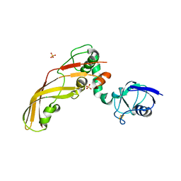
1E6C
 | | K15M MUTANT OF SHIKIMATE KINASE FROM ERWINIA CHRYSANTHEMI | | 分子名称: | (4R)-2-METHYLPENTANE-2,4-DIOL, (4S)-2-METHYL-2,4-PENTANEDIOL, CHLORIDE ION, ... | | 著者 | Maclean, J, Krell, T, Coggins, J.R, Lapthorn, A.J. | | 登録日 | 2000-08-10 | | 公開日 | 2001-06-20 | | 最終更新日 | 2023-12-13 | | 実験手法 | X-RAY DIFFRACTION (1.8 Å) | | 主引用文献 | Biochemical and X-Ray Crystallographic Studies on Shikimate Kinase: The Important Structural Role of the P-Loop Lysine
Protein Sci., 10, 2001
|
|
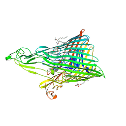
4D9S
 | | Crystal structure of Arabidopsis thaliana UVR8 (UV Resistance locus 8) | | 分子名称: | UVB-resistance protein UVR8 | | 著者 | Arvai, A.S, Christie, J.M, Pratt, A.J, Hitomi, K, Getzoff, E.D. | | 登録日 | 2012-01-11 | | 公開日 | 2012-04-04 | | 最終更新日 | 2023-09-13 | | 実験手法 | X-RAY DIFFRACTION (1.701 Å) | | 主引用文献 | Plant UVR8 Photoreceptor Senses UV-B by Tryptophan-Mediated Disruption of Cross-Dimer Salt Bridges.
Science, 335, 2012
|
|
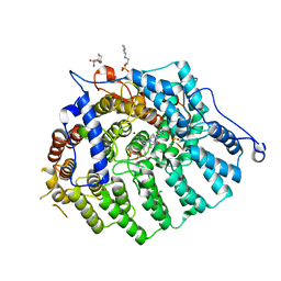
4B6M
 | |


