7T92
 
 | |
7SK4
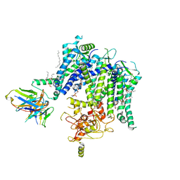
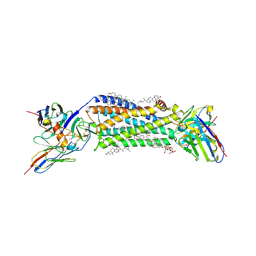
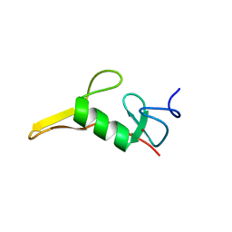
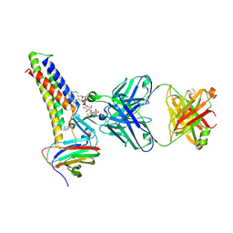
 | | Cryo-EM structure of ACKR3 in complex with chemokine N-terminal mutant CXCL12_LRHQ, an intracellular Fab, and an extracellular Fab | | 分子名称: | Atypical chemokine receptor 3, CHOLESTEROL, CID24 Fab heavy chain, ... | | 著者 | Yen, Y.C, Schafer, C.T, Gustavsson, M, Handel, T.M, Tesmer, J.J.G. | | 登録日 | 2021-10-19 | | 公開日 | 2022-07-27 | | 最終更新日 | 2025-06-04 | | 実験手法 | ELECTRON MICROSCOPY (3.3 Å) | | 主引用文献 | Structures of atypical chemokine receptor 3 reveal the basis for its promiscuity and signaling bias.
Sci Adv, 8, 2022
|
|
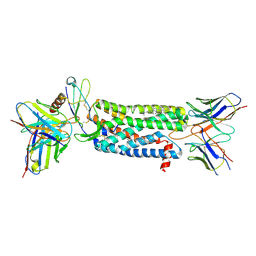
7SK6
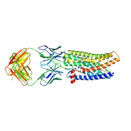
 | | Cryo-EM structure of human ACKR3 in complex with chemokine N-terminal mutant CXCL12_LRHQ and an intracellular Fab | | 分子名称: | Atypical chemokine receptor 3, CID24 Fab heavy chain, CID24 Fab light chain, ... | | 著者 | Yen, Y.C, Schafer, C.T, Gustavsson, M, Handel, T.M, Tesmer, J.J.G. | | 登録日 | 2021-10-19 | | 公開日 | 2022-07-27 | | 最終更新日 | 2024-11-20 | | 実験手法 | ELECTRON MICROSCOPY (4 Å) | | 主引用文献 | Structures of atypical chemokine receptor 3 reveal the basis for its promiscuity and signaling bias.
Sci Adv, 8, 2022
|
|
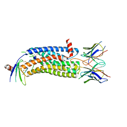
7SK3
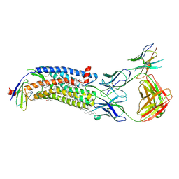
 | | Cryo-EM structure of ACKR3 in complex with CXCL12, an intracellular Fab, and an extracellular Fab | | 分子名称: | Atypical chemokine receptor 3, CHOLESTEROL, CID24 Fab heavy chain, ... | | 著者 | Yen, Y.C, Schafer, C.T, Gustavsson, M, Handel, T.M, Tesmer, J.J.G. | | 登録日 | 2021-10-19 | | 公開日 | 2022-07-27 | | 最終更新日 | 2025-05-28 | | 実験手法 | ELECTRON MICROSCOPY (3.8 Å) | | 主引用文献 | Structures of atypical chemokine receptor 3 reveal the basis for its promiscuity and signaling bias.
Sci Adv, 8, 2022
|
|
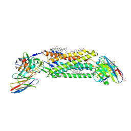
7SK8
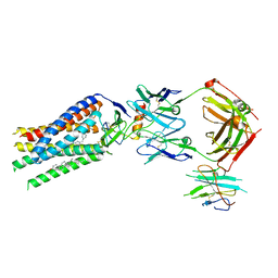
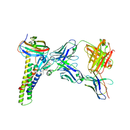
 | | Cryo-EM structure of human ACKR3 in complex with CXCL12, a small molecule partial agonist CCX662, an extracellular Fab, and an intracellular Fab | | 分子名称: | (1R)-4-[7-(3-carboxypropoxy)-6-methylquinolin-8-yl]-1-{[2-(4-hydroxypiperidin-1-yl)-1,3-thiazol-4-yl]methyl}-1,4-diazepan-1-ium, Atypical chemokine receptor 3, CHOLESTEROL, ... | | 著者 | Yen, Y.C, Schafer, C.T, Gustavsson, M, Handel, T.M, Tesmer, J.J.G. | | 登録日 | 2021-10-19 | | 公開日 | 2022-07-27 | | 最終更新日 | 2025-05-28 | | 実験手法 | ELECTRON MICROSCOPY (3.3 Å) | | 主引用文献 | Structures of atypical chemokine receptor 3 reveal the basis for its promiscuity and signaling bias.
Sci Adv, 8, 2022
|
|
7SK9
 | | Cryo-EM structure of human ACKR3 in complex with a small molecule partial agonist CCX662, and an intracellular Fab | | 分子名称: | (1R)-4-[7-(3-carboxypropoxy)-6-methylquinolin-8-yl]-1-{[2-(4-hydroxypiperidin-1-yl)-1,3-thiazol-4-yl]methyl}-1,4-diazepan-1-ium, Atypical chemokine receptor 3, CHOLESTEROL, ... | | 著者 | Yen, Y.C, Schafer, C.T, Gustavsson, M, Handel, T.M, Tesmer, J.J.G. | | 登録日 | 2021-10-19 | | 公開日 | 2022-07-27 | | 最終更新日 | 2025-06-04 | | 実験手法 | ELECTRON MICROSCOPY (3.7 Å) | | 主引用文献 | Structures of atypical chemokine receptor 3 reveal the basis for its promiscuity and signaling bias.
Sci Adv, 8, 2022
|
|
7SK5
 | | Cryo-EM structure of ACKR3 in complex with CXCL12 and an intracellular Fab | | 分子名称: | Anti-Fab nanobody, Atypical chemokine receptor 3, CHOLESTEROL, ... | | 著者 | Yen, Y.C, Schafer, C.T, Gustavsson, M, Handel, T.M, Tesmer, J.J.G. | | 登録日 | 2021-10-19 | | 公開日 | 2022-07-27 | | 最終更新日 | 2025-06-04 | | 実験手法 | ELECTRON MICROSCOPY (4 Å) | | 主引用文献 | Structures of atypical chemokine receptor 3 reveal the basis for its promiscuity and signaling bias.
Sci Adv, 8, 2022
|
|
7SK7
 | | Cryo-EM structure of human ACKR3 in complex with CXCL12, a small molecule partial agonist CCX662, and an extracellular Fab | | 分子名称: | (1R)-4-[7-(3-carboxypropoxy)-6-methylquinolin-8-yl]-1-{[2-(4-hydroxypiperidin-1-yl)-1,3-thiazol-4-yl]methyl}-1,4-diazepan-1-ium, Anti-Fab nanobody, Atypical chemokine receptor 3, ... | | 著者 | Yen, Y.C, Schafer, C.T, Gustavsson, M, Handel, T.M, Tesmer, J.J.G. | | 登録日 | 2021-10-19 | | 公開日 | 2022-07-27 | | 最終更新日 | 2024-11-20 | | 実験手法 | ELECTRON MICROSCOPY (3.3 Å) | | 主引用文献 | Structures of atypical chemokine receptor 3 reveal the basis for its promiscuity and signaling bias.
Sci Adv, 8, 2022
|
|
7T9X
 | |
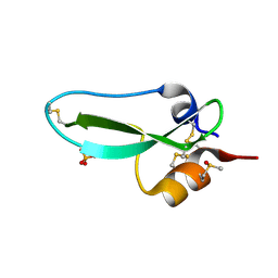
1FAN
 | | CREVICE-FORMING MUTANTS IN THE RIGID CORE OF BOVINE PANCREATIC TRYPSIN INHIBITOR: CRYSTAL STRUCTURES OF F22A, Y23A, N43G, AND F45A | | 分子名称: | BOVINE PANCREATIC TRYPSIN INHIBITOR | | 著者 | Danishefsky, A.T, Wlodawer, A, Kim, K.-S, Tao, F, Woodward, C. | | 登録日 | 1992-08-21 | | 公開日 | 1993-10-31 | | 最終更新日 | 2024-11-20 | | 実験手法 | X-RAY DIFFRACTION (2 Å) | | 主引用文献 | Crevice-forming mutants in the rigid core of bovine pancreatic trypsin inhibitor: crystal structures of F22A, Y23A, N43G, and F45A.
Protein Sci., 2, 1993
|
|
7KEO
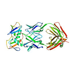 | | Crystal structure of K29-linked di-ubiquitin in complex with synthetic antigen binding fragment | | 分子名称: | PHOSPHATE ION, Synthetic antigen binding fragment, heavy chain, ... | | 著者 | Yu, Y, Zheng, Q, Erramilli, S, Pan, M, Kossiakoff, A, Liu, L, Zhao, M. | | 登録日 | 2020-10-11 | | 公開日 | 2021-07-28 | | 最終更新日 | 2024-11-13 | | 実験手法 | X-RAY DIFFRACTION (2.9 Å) | | 主引用文献 | K29-linked ubiquitin signaling regulates proteotoxic stress response and cell cycle.
Nat.Chem.Biol., 17, 2021
|
|
9PTI
 | |
5EU7
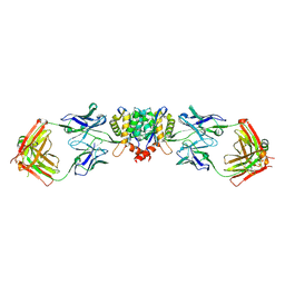 | | Crystal structure of HIV-1 integrase catalytic core in complex with Fab | | 分子名称: | FAB Heavy Chain, FAB light chain, Integrase | | 著者 | Galilee, M, Griner, S.L, Stroud, R.M, Alian, A. | | 登録日 | 2015-11-18 | | 公開日 | 2016-09-28 | | 最終更新日 | 2024-11-20 | | 実験手法 | X-RAY DIFFRACTION (2.64 Å) | | 主引用文献 | The Preserved HTH-Docking Cleft of HIV-1 Integrase Is Functionally Critical.
Structure, 24, 2016
|
|
8T7I
 | | Structure of the S1CE variant of Fab F1 (FabS1CE-F1) | | 分子名称: | 1,2-ETHANEDIOL, CHLORIDE ION, S1CE variant of Fab F1 heavy chain, ... | | 著者 | Singer, A.U, Bruce, H.A, Enderle, L, Blazer, L, Adams, J.J, Sicheri, F, Sidhu, S.S. | | 登録日 | 2023-06-20 | | 公開日 | 2023-11-22 | | 最終更新日 | 2024-10-16 | | 実験手法 | X-RAY DIFFRACTION (2.6 Å) | | 主引用文献 | Engineered antigen-binding fragments for enhanced crystallization of antibody:antigen complexes.
Protein Sci., 33, 2024
|
|
8T7G
 | | Structure of the CK variant of Fab F1 (FabC-F1) | | 分子名称: | 1,2-ETHANEDIOL, CHLORIDE ION, CK variant of Fab F1 heavy chain, ... | | 著者 | Singer, A.U, Bruce, H.A, Blazer, L, Adams, J.J, Sicheri, F, Sidhu, S.S. | | 登録日 | 2023-06-20 | | 公開日 | 2023-11-22 | | 最終更新日 | 2024-11-06 | | 実験手法 | X-RAY DIFFRACTION (2 Å) | | 主引用文献 | Engineered antigen-binding fragments for enhanced crystallization of antibody:antigen complexes.
Protein Sci., 33, 2024
|
|
8T7F
 | | Structure of the S1 variant of Fab F1 | | 分子名称: | 4-(2-HYDROXYETHYL)-1-PIPERAZINE ETHANESULFONIC ACID, S1 variant of Fab F1 heavy chain, S1 variant of Fab F1 light chain, ... | | 著者 | Singer, A.U, Bruce, H.A, Enderle, L, Blazer, L, Adams, J.J, Sicheri, F, Sidhu, S.S. | | 登録日 | 2023-06-20 | | 公開日 | 2023-11-22 | | 最終更新日 | 2024-10-23 | | 実験手法 | X-RAY DIFFRACTION (3.5 Å) | | 主引用文献 | Engineered antigen-binding fragments for enhanced crystallization of antibody:antigen complexes.
Protein Sci., 33, 2024
|
|
8U5B
 | |
8U4V
 | |
7MDJ
 | | The structure of KcsA in complex with a synthetic Fab | | 分子名称: | Fab heavy chain, Fab light chain, POTASSIUM ION, ... | | 著者 | Rohaim, A, Slezak, T, Blackowicz, L, Kossiakoff, A, Roux, B. | | 登録日 | 2021-04-05 | | 公開日 | 2022-02-16 | | 最終更新日 | 2024-11-20 | | 実験手法 | X-RAY DIFFRACTION (2.75 Å) | | 主引用文献 | Engineering of a synthetic antibody fragment for structural and functional studies of K+ channels.
J.Gen.Physiol., 154, 2022
|
|
6E1N
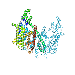
 | | Structure of AtTPC1(DDE) in state 1 | | 分子名称: | 1,2-DIACYL-GLYCEROL-3-SN-PHOSPHATE, CALCIUM ION, PALMITIC ACID, ... | | 著者 | Kintzer, A.F, Green, E.M, Cheng, Y, Stroud, R.M. | | 登録日 | 2018-07-10 | | 公開日 | 2018-09-19 | | 最終更新日 | 2024-12-25 | | 実験手法 | ELECTRON MICROSCOPY (3.7 Å) | | 主引用文献 | Structural basis for activation of voltage sensor domains in an ion channel TPC1.
Proc. Natl. Acad. Sci. U.S.A., 115, 2018
|
|
6E1K
 | | Structure of AtTPC1(DDE) reconstituted in saposin A with cat06 Fab | | 分子名称: | CALCIUM ION, PALMITIC ACID, Two pore calcium channel protein 1, ... | | 著者 | Kintzer, A.F, Green, E.M, Cheng, Y, Stroud, R.M. | | 登録日 | 2018-07-10 | | 公開日 | 2018-09-19 | | 最終更新日 | 2024-12-25 | | 実験手法 | ELECTRON MICROSCOPY (3.3 Å) | | 主引用文献 | Structural basis for activation of voltage sensor domains in an ion channel TPC1.
Proc. Natl. Acad. Sci. U.S.A., 115, 2018
|
|
6CX0
 | | Structure of AtTPC1 D376A | | 分子名称: | (1S,3R)-1-(3-{[4-(2-fluorophenyl)piperazin-1-yl]methyl}-4-methoxyphenyl)-2,3,4,9-tetrahydro-1H-beta-carboline-3-carboxylic acid, CALCIUM ION, Two pore calcium channel protein 1 | | 著者 | Kintzer, A.F, Stroud, R.M. | | 登録日 | 2018-04-02 | | 公開日 | 2018-09-19 | | 最終更新日 | 2023-10-04 | | 実験手法 | X-RAY DIFFRACTION (3.501 Å) | | 主引用文献 | Structural basis for activation of voltage sensor domains in an ion channel TPC1.
Proc. Natl. Acad. Sci. U.S.A., 115, 2018
|
|
6E1M
 | | Structure of AtTPC1(DDE) reconstituted in saposin A | | 分子名称: | 1,2-DIACYL-GLYCEROL-3-SN-PHOSPHATE, CALCIUM ION, PALMITIC ACID, ... | | 著者 | Kintzer, A.F, Green, E.M, Cheng, Y, Stroud, R.M. | | 登録日 | 2018-07-10 | | 公開日 | 2018-09-19 | | 最終更新日 | 2024-12-25 | | 実験手法 | ELECTRON MICROSCOPY (3.3 Å) | | 主引用文献 | Structural basis for activation of voltage sensor domains in an ion channel TPC1.
Proc. Natl. Acad. Sci. U.S.A., 115, 2018
|
|
6E1P
 | | Structure of AtTPC1(DDE) in state 2 | | 分子名称: | CALCIUM ION, PALMITIC ACID, Two pore calcium channel protein 1 | | 著者 | Kintzer, A.F, Green, E.M, Cheng, Y, Stroud, R.M. | | 登録日 | 2018-07-10 | | 公開日 | 2018-09-19 | | 最終更新日 | 2024-12-25 | | 実験手法 | ELECTRON MICROSCOPY (3.7 Å) | | 主引用文献 | Structural basis for activation of voltage sensor domains in an ion channel TPC1.
Proc. Natl. Acad. Sci. U.S.A., 115, 2018
|
|
1BTI
 | | CREVICE-FORMING MUTANTS IN THE RIGID CORE OF BOVINE PANCREATIC TRYPSIN INHIBITOR: CRYSTAL STRUCTURES OF F22A, Y23A, N43G, AND F45A | | 分子名称: | BOVINE PANCREATIC TRYPSIN INHIBITOR | | 著者 | Housset, D, Tao, F, Kim, K.-S, Fuchs, J, Woodward, C, Wlodawer, A. | | 登録日 | 1991-07-11 | | 公開日 | 1993-10-31 | | 最終更新日 | 2024-11-06 | | 実験手法 | X-RAY DIFFRACTION (2.2 Å) | | 主引用文献 | Crevice-forming mutants in the rigid core of bovine pancreatic trypsin inhibitor: crystal structures of F22A, Y23A, N43G, and F45A.
Protein Sci., 2, 1993
|
|


