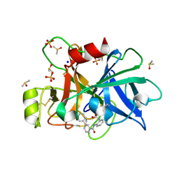
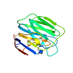
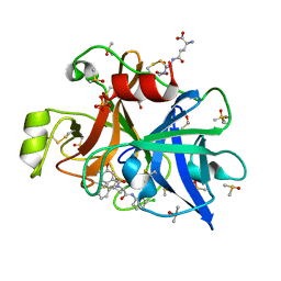
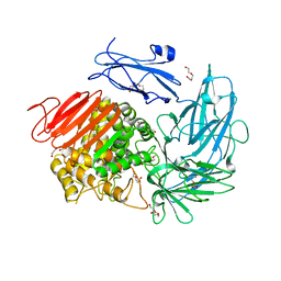
5Z59
 
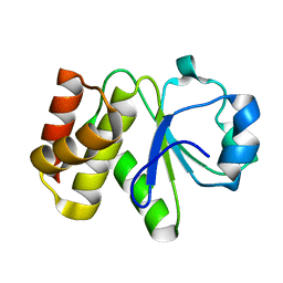 | | Crystal structure of Tk-PTP in the inactive form | | 分子名称: | Protein-tyrosine phosphatase | | 著者 | Ku, B, Yun, H.Y, Kim, S.J. | | 登録日 | 2018-01-17 | | 公開日 | 2018-06-27 | | 最終更新日 | 2023-11-22 | | 実験手法 | X-RAY DIFFRACTION (1.703 Å) | | 主引用文献 | Structural study reveals the temperature-dependent conformational flexibility of Tk-PTP, a protein tyrosine phosphatase from Thermococcus kodakaraensis KOD1
PLoS ONE, 13, 2018
|
|
3P2F
 | |
3P2H
 | |
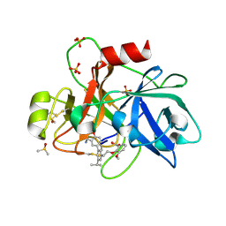
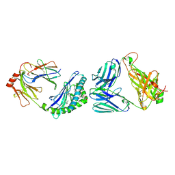
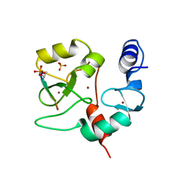
5Z5B
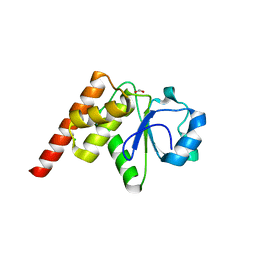 | | Crystal structure of Tk-PTP in the G95A mutant form | | 分子名称: | CHLORIDE ION, FORMIC ACID, Protein-tyrosine phosphatase | | 著者 | Ku, B, Yun, H.Y, Kim, S.J. | | 登録日 | 2018-01-17 | | 公開日 | 2018-06-27 | | 最終更新日 | 2023-11-22 | | 実験手法 | X-RAY DIFFRACTION (2.3 Å) | | 主引用文献 | Structural study reveals the temperature-dependent conformational flexibility of Tk-PTP, a protein tyrosine phosphatase from Thermococcus kodakaraensis KOD1
PLoS ONE, 13, 2018
|
|
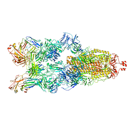
6YEJ
 | | Cryo-EM structure of the Full-length disease type human Huntingtin | | 分子名称: | Huntingtin | | 著者 | Tame, G, Jung, T, Dal Perraro, M, Hebert, H, Song, J. | | 登録日 | 2020-03-24 | | 公開日 | 2020-12-16 | | 実験手法 | ELECTRON MICROSCOPY (18.200001 Å) | | 主引用文献 | The Polyglutamine Expansion at the N-Terminal of Huntingtin Protein Modulates the Dynamic Configuration and Phosphorylation of the C-Terminal HEAT Domain.
Structure, 28, 2020
|
|
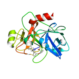
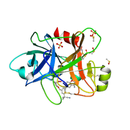
5Z5A
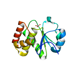 | | Crystal structure of Tk-PTP in the active form | | 分子名称: | Protein-tyrosine phosphatase, VANADATE ION | | 著者 | Ku, B, Yun, H.Y, Kim, S.J. | | 登録日 | 2018-01-17 | | 公開日 | 2018-07-04 | | 最終更新日 | 2023-11-22 | | 実験手法 | X-RAY DIFFRACTION (1.8 Å) | | 主引用文献 | Structural study reveals the temperature-dependent conformational flexibility of Tk-PTP, a protein tyrosine phosphatase from Thermococcus kodakaraensis KOD1
PLoS ONE, 13, 2018
|
|
7EC1
 | | Crystal structure of SdgB (ligand-free form) | | 分子名称: | GLYCEROL, Glycosyl transferase, group 1 family protein, ... | | 著者 | Kim, D.-G, Baek, I, Lee, Y, Kim, H.S. | | 登録日 | 2021-03-11 | | 公開日 | 2021-03-24 | | 最終更新日 | 2024-05-29 | | 実験手法 | X-RAY DIFFRACTION (1.85 Å) | | 主引用文献 | Structural basis for SdgB- and SdgA-mediated glycosylation of staphylococcal adhesive proteins.
Acta Crystallogr D Struct Biol, 77, 2021
|
|
7EC6
 | | Crystal structure of SdgB (complexed with peptides) | | 分子名称: | ASP-SER-ASP, Glycosyl transferase, group 1 family protein | | 著者 | Kim, D.-G, Baek, I, Lee, Y, Kim, H.S. | | 登録日 | 2021-03-11 | | 公開日 | 2022-03-16 | | 最終更新日 | 2023-11-29 | | 実験手法 | X-RAY DIFFRACTION (1.9 Å) | | 主引用文献 | Structural basis for SdgB- and SdgA-mediated glycosylation of staphylococcal adhesive proteins.
Acta Crystallogr D Struct Biol, 77, 2021
|
|
7EC7
 | | Crystal structure of SdgB (complexed with phosphate ions) | | 分子名称: | Glycosyl transferase, group 1 family protein, PHOSPHATE ION | | 著者 | Kim, D.-G, Baek, I, Lee, Y, Kim, H.S. | | 登録日 | 2021-03-11 | | 公開日 | 2022-03-16 | | 最終更新日 | 2023-11-29 | | 実験手法 | X-RAY DIFFRACTION (3.2 Å) | | 主引用文献 | Structural basis for SdgB- and SdgA-mediated glycosylation of staphylococcal adhesive proteins.
Acta Crystallogr D Struct Biol, 77, 2021
|
|
7EC3
 | | Crystal structure of SdgB (complexed with UDP, GlcNAc, and Glycosylated peptide) | | 分子名称: | 2-acetamido-2-deoxy-alpha-D-glucopyranose-(1-35)-[2-acetamido-2-deoxy-alpha-D-galactopyranose-(1-65)]5,6-DIHYDRO-BENZO[H]CINNOLIN-3-YLAMINE, 2-acetamido-2-deoxy-beta-D-glucopyranose, Glycosyl transferase, ... | | 著者 | Kim, D.-G, Baek, I, Lee, Y, Kim, H.S. | | 登録日 | 2021-03-11 | | 公開日 | 2022-03-16 | | 最終更新日 | 2023-11-29 | | 実験手法 | X-RAY DIFFRACTION (2.5 Å) | | 主引用文献 | Structural basis for SdgB- and SdgA-mediated glycosylation of staphylococcal adhesive proteins.
Acta Crystallogr D Struct Biol, 77, 2021
|
|
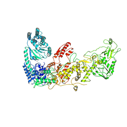
6TS4
 | | Coagulation factor XI protease domain in complex with active site inhibitor | | 分子名称: | 2-[2-[[3-[3-(aminomethyl)phenyl]phenyl]carbonylamino]phenyl]ethanoic acid, Coagulation factor XI, DIMETHYL SULFOXIDE, ... | | 著者 | Renatus, M, Schiering, N. | | 登録日 | 2019-12-19 | | 公開日 | 2020-07-08 | | 最終更新日 | 2020-08-26 | | 実験手法 | X-RAY DIFFRACTION (1.17 Å) | | 主引用文献 | Structure-Based Design and Preclinical Characterization of Selective and Orally Bioavailable Factor XIa Inhibitors: Demonstrating the Power of an Integrated S1 Protease Family Approach.
J.Med.Chem., 63, 2020
|
|
7YC5
 | |
6TS7
 | | Coagulation factor XI protease domain in complex with active site inhibitor | | 分子名称: | 2-[2-[[3-(1,2,3,4-tetrahydroisoquinolin-7-yl)phenyl]methoxy]phenyl]ethanoic acid, Coagulation factor XI | | 著者 | Renatus, M, Schiering, N. | | 登録日 | 2019-12-20 | | 公開日 | 2020-07-08 | | 最終更新日 | 2020-08-26 | | 実験手法 | X-RAY DIFFRACTION (2.63 Å) | | 主引用文献 | Structure-Based Design and Preclinical Characterization of Selective and Orally Bioavailable Factor XIa Inhibitors: Demonstrating the Power of an Integrated S1 Protease Family Approach.
J.Med.Chem., 63, 2020
|
|
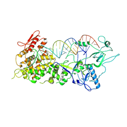
6TS5
 | | Coagulation factor XI protease domain in complex with active site inhibitor | | 分子名称: | 2-[2-[3-[(3~{S})-3-azanyl-2,3-dihydro-1-benzofuran-5-yl]-5-propan-2-yl-phenyl]ethoxy]-3-methoxy-benzoic acid, Coagulation factor XI, DIMETHYL SULFOXIDE, ... | | 著者 | Renatus, M, Schiering, N. | | 登録日 | 2019-12-20 | | 公開日 | 2020-07-08 | | 最終更新日 | 2024-05-01 | | 実験手法 | X-RAY DIFFRACTION (1.29 Å) | | 主引用文献 | Structure-Based Design and Preclinical Characterization of Selective and Orally Bioavailable Factor XIa Inhibitors: Demonstrating the Power of an Integrated S1 Protease Family Approach.
J.Med.Chem., 63, 2020
|
|
3SH4
 | |
6XNG
 | | MHC-like protein complex structure | | 分子名称: | (3R)-N-[(2S,3R)-1-(alpha-D-galactopyranosyloxy)-3-hydroxyheptadecan-2-yl]-3-hydroxyheptadecanamide, 2-acetamido-2-deoxy-beta-D-glucopyranose-(1-4)-2-acetamido-2-deoxy-beta-D-glucopyranose, Antigen-presenting glycoprotein CD1d1, ... | | 著者 | Thirunavukkarasu, P, Le Nours, J, Rossjohn, J. | | 登録日 | 2020-07-02 | | 公開日 | 2021-11-10 | | 最終更新日 | 2023-10-18 | | 実験手法 | X-RAY DIFFRACTION (2.79 Å) | | 主引用文献 | Host immunomodulatory lipids created by symbionts from dietary amino acids.
Nature, 600, 2021
|
|
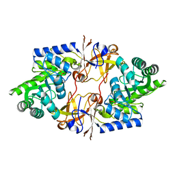
6TS6
 | | Coagulation factor XI protease domain in complex with active site inhibitor | | 分子名称: | 2-[2-[[3-[(3~{S})-3-azanyl-2,3-dihydro-1-benzofuran-5-yl]-5-(2-cyanopropan-2-yl)phenyl]methoxy]phenyl]ethanoic acid, Coagulation factor XI, DIMETHYL SULFOXIDE, ... | | 著者 | Renatus, M, Schiering, N. | | 登録日 | 2019-12-20 | | 公開日 | 2020-07-08 | | 最終更新日 | 2020-08-26 | | 実験手法 | X-RAY DIFFRACTION (1.33 Å) | | 主引用文献 | Structure-Based Design and Preclinical Characterization of Selective and Orally Bioavailable Factor XIa Inhibitors: Demonstrating the Power of an Integrated S1 Protease Family Approach.
J.Med.Chem., 63, 2020
|
|
6USY
 | | COAGULATION FACTOR XI CATALYTIC DOMAIN (C123S) IN COMPLEX WITH NVP-XIV936 | | 分子名称: | 1-[(2S)-2-{3-[(3S)-3-amino-2,3-dihydro-1-benzofuran-5-yl]-5-(propan-2-yl)phenyl}-2-hydroxyethyl]-1H-indole-7-carboxylic acid, Coagulation factor XIa light chain | | 著者 | Weihofen, W.A, Clark, K, Nunes, S. | | 登録日 | 2019-10-28 | | 公開日 | 2020-07-01 | | 最終更新日 | 2023-10-11 | | 実験手法 | X-RAY DIFFRACTION (1.26 Å) | | 主引用文献 | Structure-Based Design and Preclinical Characterization of Selective and Orally Bioavailable Factor XIa Inhibitors: Demonstrating the Power of an Integrated S1 Protease Family Approach.
J.Med.Chem., 63, 2020
|
|
3SH5
 | |
6T7P
 | | human plasmakallikrein protease domain in complex with active site directed inhibitor | | 分子名称: | (2~{S},4~{R})-1-[[(3~{S})-3-azanyl-2,3-dihydro-1-benzofuran-6-yl]carbonyl]-~{N}-(3-chlorophenyl)-4-phenyl-pyrrolidine-2-carboxamide, DIMETHYL SULFOXIDE, GLUTATHIONE, ... | | 著者 | Renatus, M. | | 登録日 | 2019-10-22 | | 公開日 | 2020-07-08 | | 最終更新日 | 2024-01-24 | | 実験手法 | X-RAY DIFFRACTION (1.416 Å) | | 主引用文献 | Structure-Based Design and Preclinical Characterization of Selective and Orally Bioavailable Factor XIa Inhibitors: Demonstrating the Power of an Integrated S1 Protease Family Approach.
J.Med.Chem., 63, 2020
|
|
7XW3
 | |
7XW2
 | |
7DIB
 | |
6I60
 | |
4QBS
 | |


