6O9C
 
 | |
3E37
 | |
3E27
 | |
3E32
 | |
3E34
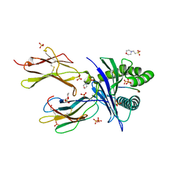
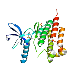
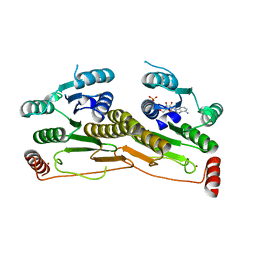
 | | Protein farnesyltransferase complexed with FPP and ethylenediamine-scaffold inhibitor 10 | | 分子名称: | 3-{2'-[{[1-(tert-butoxycarbonyl)piperidin-4-yl]methyl}(2-{(4-cyanophenyl)[(1-methyl-1H-imidazol-5-yl)methyl]amino}ethyl)sulfamoyl]biphenyl-3-yl}propanoic acid, FARNESYL DIPHOSPHATE, Protein farnesyltransferase subunit beta, ... | | 著者 | Hast, M.A, Beese, L.S. | | 登録日 | 2008-08-06 | | 公開日 | 2009-03-10 | | 最終更新日 | 2024-02-21 | | 実験手法 | X-RAY DIFFRACTION (2.05 Å) | | 主引用文献 | Structural basis for binding and selectivity of antimalarial and anticancer ethylenediamine inhibitors to protein farnesyltransferase.
Chem.Biol., 16, 2009
|
|
3E30
 | |
3F4C
 | | Crystal structure of organophosphorus hydrolase from Geobacillus stearothermophilus strain 10, with glycerol bound | | 分子名称: | COBALT (II) ION, GLYCEROL, Organophosphorus hydrolase | | 著者 | Hawwa, R, Aikens, J, Turner, R.J, Santarsiero, B, Mesecar, A. | | 登録日 | 2008-10-31 | | 公開日 | 2009-09-15 | | 最終更新日 | 2023-11-15 | | 実験手法 | X-RAY DIFFRACTION (2.07 Å) | | 主引用文献 | Structural basis for thermostability revealed through the identification and characterization of a highly thermostable phosphotriesterase-like lactonase from Geobacillus stearothermophilus.
Arch.Biochem.Biophys., 488, 2009
|
|
3FZO
 | | Crystal Structure of PYK2-Apo, Proline-rich Tyrosine Kinase | | 分子名称: | Protein tyrosine kinase 2 beta | | 著者 | Han, S. | | 登録日 | 2009-01-26 | | 公開日 | 2009-03-31 | | 最終更新日 | 2023-09-06 | | 実験手法 | X-RAY DIFFRACTION (2.2 Å) | | 主引用文献 | Structural characterization of proline-rich tyrosine kinase 2 (PYK2) reveals a unique (DFG-out) conformation and enables inhibitor design.
J.Biol.Chem., 284, 2009
|
|
3FZR
 | | Crystal structure of PYK2 complexed with PF-431396 | | 分子名称: | N-methyl-N-{2-[({2-[(2-oxo-2,3-dihydro-1H-indol-5-yl)amino]-5-(trifluoromethyl)pyrimidin-4-yl}amino)methyl]phenyl}methanesulfonamide, PHOSPHATE ION, Protein tyrosine kinase 2 beta | | 著者 | Han, S. | | 登録日 | 2009-01-26 | | 公開日 | 2009-03-31 | | 最終更新日 | 2024-02-21 | | 実験手法 | X-RAY DIFFRACTION (2.7 Å) | | 主引用文献 | Structural characterization of proline-rich tyrosine kinase 2 (PYK2) reveals a unique (DFG-out) conformation and enables inhibitor design.
J.Biol.Chem., 284, 2009
|
|
3FZP
 | | Crystal structure of PYK2 complexed with ATPgS | | 分子名称: | PHOSPHOTHIOPHOSPHORIC ACID-ADENYLATE ESTER, Protein tyrosine kinase 2 beta, SULFATE ION | | 著者 | Han, S. | | 登録日 | 2009-01-26 | | 公開日 | 2009-03-31 | | 最終更新日 | 2024-02-21 | | 実験手法 | X-RAY DIFFRACTION (2.1 Å) | | 主引用文献 | Structural characterization of proline-rich tyrosine kinase 2 (PYK2) reveals a unique (DFG-out) conformation and enables inhibitor design.
J.Biol.Chem., 284, 2009
|
|
3FZS
 | | Crystal Structure of PYK2 complexed with BIRB796 | | 分子名称: | 1-(5-TERT-BUTYL-2-P-TOLYL-2H-PYRAZOL-3-YL)-3-[4-(2-MORPHOLIN-4-YL-ETHOXY)-NAPHTHALEN-1-YL]-UREA, Protein tyrosine kinase 2 beta | | 著者 | Han, S. | | 登録日 | 2009-01-26 | | 公開日 | 2009-03-31 | | 最終更新日 | 2024-02-21 | | 実験手法 | X-RAY DIFFRACTION (1.75 Å) | | 主引用文献 | Structural characterization of proline-rich tyrosine kinase 2 (PYK2) reveals a unique (DFG-out) conformation and enables inhibitor design.
J.Biol.Chem., 284, 2009
|
|
3FZT
 | | Crystal structure of PYK2 complexed with PF-4618433 | | 分子名称: | 1-[5-tert-butyl-2-(4-methylphenyl)-1,2-dihydro-3H-pyrazol-3-ylidene]-3-{3-[(pyridin-3-yloxy)methyl]-1H-pyrazol-5-yl}urea, Protein tyrosine kinase 2 beta | | 著者 | Han, S. | | 登録日 | 2009-01-26 | | 公開日 | 2009-03-31 | | 最終更新日 | 2024-02-21 | | 実験手法 | X-RAY DIFFRACTION (1.95 Å) | | 主引用文献 | Structural characterization of proline-rich tyrosine kinase 2 (PYK2) reveals a unique (DFG-out) conformation and enables inhibitor design.
J.Biol.Chem., 284, 2009
|
|
3GE3
 | | Crystal Structure of the reduced Toluene 4-Monooxygenase HD T201A mutant complex | | 分子名称: | ACETATE ION, CHLORIDE ION, FE (III) ION, ... | | 著者 | Elsen, N.L, Bailey, L.J, Fox, B.G. | | 登録日 | 2009-02-24 | | 公開日 | 2009-07-28 | | 最終更新日 | 2023-09-06 | | 実験手法 | X-RAY DIFFRACTION (1.52 Å) | | 主引用文献 | Role for threonine 201 in the catalytic cycle of the soluble diiron hydroxylase toluene 4-monooxygenase.
Biochemistry, 48, 2009
|
|
3GAB
 | | C-terminal domain of Bacillus subtilis MutL crystal form I | | 分子名称: | DNA mismatch repair protein mutL | | 著者 | Guarne, A, Pillon, M.C, Lorenowicz, J.J, Mitchell, R.R, Chung, Y.S, Friedhoff, P. | | 登録日 | 2009-02-17 | | 公開日 | 2010-07-21 | | 最終更新日 | 2024-02-21 | | 実験手法 | X-RAY DIFFRACTION (2.5 Å) | | 主引用文献 | Structure of the endonuclease domain of MutL: unlicensed to cut.
Mol.Cell, 39, 2010
|
|
3E33
 | |
3DLK
 | | Crystal Structure of an engineered form of the HIV-1 Reverse Transcriptase, RT69A | | 分子名称: | Reverse transcriptase/ribonuclease H, SULFATE ION, p51 RT | | 著者 | Ho, W.C, Bauman, J.D, Himmel, D.M, Das, K, Arnold, E. | | 登録日 | 2008-06-27 | | 公開日 | 2008-10-07 | | 最終更新日 | 2023-08-30 | | 実験手法 | X-RAY DIFFRACTION (1.85 Å) | | 主引用文献 | Crystal engineering of HIV-1 reverse transcriptase for structure-based drug design.
Nucleic Acids Res., 36, 2008
|
|
3H8V
 | | Human Ubiquitin-activating Enzyme 5 in Complex with ATP | | 分子名称: | ADENOSINE-5'-TRIPHOSPHATE, Ubiquitin-like modifier-activating enzyme 5, ZINC ION | | 著者 | Walker, J.R, Bacik, J.P, Rastgoo, N, Weigelt, J, Bountra, C, Edwards, A.M, Arrowsmith, C.H, Bochkarev, A, Dhe-Paganon, S, Structural Genomics Consortium (SGC) | | 登録日 | 2009-04-29 | | 公開日 | 2009-05-26 | | 最終更新日 | 2023-09-06 | | 実験手法 | X-RAY DIFFRACTION (2 Å) | | 主引用文献 | Crystal structure of the human ubiquitin-activating enzyme 5 (UBA5) bound to ATP: mechanistic insights into a minimalistic E1 enzyme.
J.Biol.Chem., 285, 2010
|
|
3GU1
 | | Y97W mutant in organophosphorus hydrolase from Deinococcus radiodurans | | 分子名称: | COBALT (II) ION, GLYCEROL, Organophosphorus hydrolase | | 著者 | Hawwa, R, Larsen, S, Ratia, K, Mesecar, A. | | 登録日 | 2009-03-28 | | 公開日 | 2009-06-30 | | 最終更新日 | 2021-10-13 | | 実験手法 | X-RAY DIFFRACTION (2 Å) | | 主引用文献 | Structure-based and random mutagenesis approaches increase the organophosphate-degrading activity of a phosphotriesterase homologue from Deinococcus radiodurans.
J.Mol.Biol., 393, 2009
|
|
3GTH
 | | D71G/E101G/M234I mutant in organophosphorus hydrolase from Deinococcus radiodurans | | 分子名称: | COBALT (II) ION, FORMIC ACID, Organophosphorus hydrolase | | 著者 | Hawwa, R, Larsen, S, Ratia, K, Mesecar, A. | | 登録日 | 2009-03-27 | | 公開日 | 2009-06-30 | | 最終更新日 | 2021-10-20 | | 実験手法 | X-RAY DIFFRACTION (1.98 Å) | | 主引用文献 | Structure-based and random mutagenesis approaches increase the organophosphate-degrading activity of a phosphotriesterase homologue from Deinococcus radiodurans.
J.Mol.Biol., 393, 2009
|
|
3GU2
 | | Y97L/G100-/E101- mutant in organophosphorus hydrolase | | 分子名称: | COBALT (II) ION, Organophosphorus hydrolase | | 著者 | Hawwa, R, Larsen, S, Ratia, K, Mesecar, A. | | 登録日 | 2009-03-28 | | 公開日 | 2009-06-30 | | 最終更新日 | 2021-10-13 | | 実験手法 | X-RAY DIFFRACTION (2 Å) | | 主引用文献 | Structure-based and random mutagenesis approaches increase the organophosphate-degrading activity of a phosphotriesterase homologue from Deinococcus radiodurans.
J.Mol.Biol., 393, 2009
|
|
3GU9
 | | R228A mutation in organophosphorus hydrolase from Deinococcus radiodurans | | 分子名称: | COBALT (II) ION, Organophosphorus hydrolase | | 著者 | Hawwa, R, Larsen, S, Ratia, K, Mesecar, A. | | 登録日 | 2009-03-28 | | 公開日 | 2009-06-30 | | 最終更新日 | 2021-10-13 | | 実験手法 | X-RAY DIFFRACTION (2.06 Å) | | 主引用文献 | Structure-based and random mutagenesis approaches increase the organophosphate-degrading activity of a phosphotriesterase homologue from Deinococcus radiodurans.
J.Mol.Biol., 393, 2009
|
|
3GX2
 | |
3GX6
 | |
3F4D
 | | Crystal structure of organophosphorus hydrolase from Geobacillus stearothermophilus strain 10 | | 分子名称: | COBALT (II) ION, Organophosphorus hydrolase | | 著者 | Hawwa, R, Aikens, J, Turner, R.J, Santarsiero, B, Mesecar, A. | | 登録日 | 2008-10-31 | | 公開日 | 2009-08-18 | | 最終更新日 | 2023-11-15 | | 実験手法 | X-RAY DIFFRACTION (2.36 Å) | | 主引用文献 | Structural basis for thermostability revealed through the identification and characterization of a highly thermostable phosphotriesterase-like lactonase from Geobacillus stearothermophilus.
Arch.Biochem.Biophys., 488, 2009
|
|
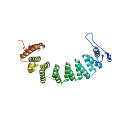
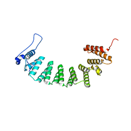
3GK1
 | | X-ray structure of bovine SBi132,Ca(2+)-S100B | | 分子名称: | 2-[(5-hex-1-yn-1-ylfuran-2-yl)carbonyl]-N-methylhydrazinecarbothioamide, CACODYLATE ION, CALCIUM ION, ... | | 著者 | Charpentier, T.H, Weber, D.J, Toth, E.A. | | 登録日 | 2009-03-09 | | 公開日 | 2009-06-09 | | 最終更新日 | 2023-09-06 | | 実験手法 | X-RAY DIFFRACTION (2.1 Å) | | 主引用文献 | Small molecules bound to unique sites in the target protein binding cleft of calcium-bound S100B as characterized by nuclear magnetic resonance and X-ray crystallography.
Biochemistry, 48, 2009
|
|


