1QUV
 
 | |
2OUJ
 | |
3VSO
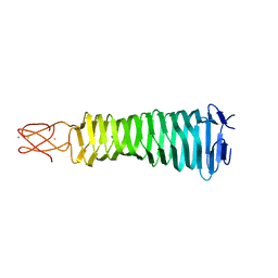
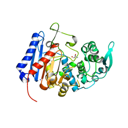
 | | Human PPAR gamma ligand binding domain in complex with a gamma selective agonist mekt21 | | 分子名称: | (2R)-2-benzyl-3-[4-propoxy-3-({[4-(pyrimidin-2-yl)benzoyl]amino}methyl)phenyl]propanoic acid, Peroxisome proliferator-activated receptor gamma | | 著者 | Oyama, T, Waku, T, Ohashi, M, Morikawa, K, Miyachi, H. | | 登録日 | 2012-04-30 | | 公開日 | 2013-05-01 | | 最終更新日 | 2023-11-08 | | 実験手法 | X-RAY DIFFRACTION (2 Å) | | 主引用文献 | Design and synthesis of a series of alpha-benzyl phenylpropanoic acid-type peroxisome proliferator-activated receptor (PPAR) gamma partial agonists with improved aqueous solubility
Bioorg.Med.Chem., 21, 2013
|
|
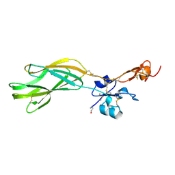
2OUH
 | | Crystal structure of the Thrombospondin-1 N-terminal domain in complex with fractionated Heparin DP10 | | 分子名称: | SULFATE ION, Thrombospondin-1 | | 著者 | Tan, K, Joachimiak, A, Wang, J, Lawler, J. | | 登録日 | 2007-02-11 | | 公開日 | 2008-01-08 | | 最終更新日 | 2024-10-09 | | 実験手法 | X-RAY DIFFRACTION (2.4 Å) | | 主引用文献 | Heparin-induced cis- and trans-Dimerization Modes of the Thrombospondin-1 N-terminal Domain.
J.Biol.Chem., 283, 2008
|
|
7T1V
 | |
2ES3
 | |
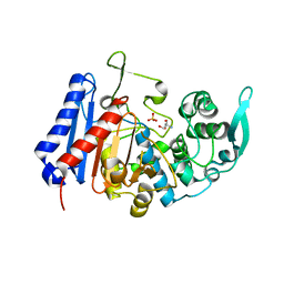
2YZ3
 | | Crystallographic Investigation of Inhibition Mode of the VIM-2 Metallo-beta-lactamase from Pseudomonas aeruginosa with Mercaptocarboxylate Inhibitor | | 分子名称: | (S)-2-(MERCAPTOMETHYL)-5-PHENYLPENTANOIC ACID, Metallo-beta-lactamase, SULFATE ION, ... | | 著者 | Yamaguchi, Y, Yamagata, Y, Arakawa, Y, Kurosaki, H. | | 登録日 | 2007-05-02 | | 公開日 | 2008-03-11 | | 最終更新日 | 2024-03-13 | | 実験手法 | X-RAY DIFFRACTION (2.3 Å) | | 主引用文献 | Crystallographic investigation of the inhibition mode of a VIM-2 metallo-beta-lactamase from Pseudomonas aeruginosa by a mercaptocarboxylate inhibitor.
J.Med.Chem., 50, 2007
|
|
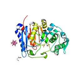
3AQJ
 | | Crystal Structure of a C-terminal domain of the bacteriophage P2 tail spike protein, gpV | | 分子名称: | Baseplate assembly protein V, CALCIUM ION, CHLORIDE ION, ... | | 著者 | Takeda, S, Yamashita, E, Nakagawa, A. | | 登録日 | 2010-11-06 | | 公開日 | 2011-08-10 | | 最終更新日 | 2024-03-13 | | 実験手法 | X-RAY DIFFRACTION (1.27 Å) | | 主引用文献 | The host-binding domain of the P2 phage tail spike reveals a trimeric iron-binding structure
Acta Crystallogr.,Sect.F, 67, 2011
|
|
5E6V
 | |
7U1B
 | |
7U1C
 | | Structure of EstG crystalized with SO4 and Tris | | 分子名称: | 2-AMINO-2-HYDROXYMETHYL-PROPANE-1,3-DIOL, Beta-lactamase domain-containing protein, SODIUM ION, ... | | 著者 | Gabelli, S.B, Chen, Z. | | 登録日 | 2022-02-20 | | 公開日 | 2023-01-11 | | 最終更新日 | 2023-10-25 | | 実験手法 | X-RAY DIFFRACTION (2.09 Å) | | 主引用文献 | EstG is a novel esterase required for cell envelope integrity in Caulobacter.
Curr.Biol., 33, 2023
|
|
7UDA
 | | Structure of the EstG | | 分子名称: | 2-AMINO-2-HYDROXYMETHYL-PROPANE-1,3-DIOL, Beta-lactamase domain-containing protein, SODIUM ION | | 著者 | Chen, Z, Gabelli, S.B. | | 登録日 | 2022-03-18 | | 公開日 | 2023-01-11 | | 最終更新日 | 2023-10-25 | | 実験手法 | X-RAY DIFFRACTION (2.47 Å) | | 主引用文献 | EstG is a novel esterase required for cell envelope integrity in Caulobacter.
Curr.Biol., 33, 2023
|
|
7UFG
 | | Cryo-EM structure of PAPP-A in complex with IGFBP5 | | 分子名称: | Insulin-like growth factor-binding protein 5, Pappalysin-1, ZINC ION | | 著者 | Judge, R.A, Jain, R, Hao, Q, Ouch, C, Sridar, J, Smith, C.L, Wang, J.C.K, Eaton, D. | | 登録日 | 2022-03-22 | | 公開日 | 2022-09-28 | | 実験手法 | ELECTRON MICROSCOPY (3.28 Å) | | 主引用文献 | Structure of the PAPP-ABP5 complex reveals mechanism of substrate recognition
Nat Commun, 13, 2022
|
|
7UQ0
 | | Putative periplasmic iron siderophore binding protein FecB (Rv3044) from Mycobacterium tuberculosis | | 分子名称: | CITRIC ACID, GLYCEROL, PENTAETHYLENE GLYCOL, ... | | 著者 | Chao, A, Cuthbert, B.J, Goulding, C.W. | | 登録日 | 2022-04-18 | | 公開日 | 2022-10-05 | | 最終更新日 | 2023-11-08 | | 実験手法 | X-RAY DIFFRACTION (2 Å) | | 主引用文献 | Differentiating the roles of Mycobacterium tuberculosis substrate binding proteins, FecB and FecB2, in iron uptake.
Plos Pathog., 19, 2023
|
|
6IQN
 | | Crystal structure of TrkA kinase with ligand | | 分子名称: | 4-[[4-azanyl-3-(4-cyclohexylpiperazin-1-yl)-9,10-bis(oxidanylidene)anthracen-1-yl]amino]benzoic acid, High affinity nerve growth factor receptor | | 著者 | Noritaka, F. | | 登録日 | 2018-11-08 | | 公開日 | 2020-01-22 | | 最終更新日 | 2023-11-22 | | 実験手法 | X-RAY DIFFRACTION (2.54 Å) | | 主引用文献 | An isoform-selective inhibitor of tropomyosin receptor kinase A behaves as molecular glue.
Bioorg.Med.Chem.Lett., 30, 2020
|
|
2KLN
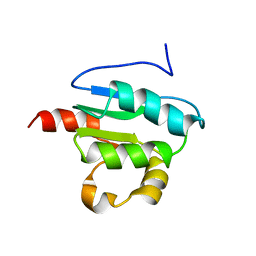 | | Solution Structure of STAS domain of RV1739c from M. tuberculosis | | 分子名称: | PROBABLE SULPHATE-TRANSPORT TRANSMEMBRANE PROTEIN, COG0659 | | 著者 | Sharma, A.K, Ye, L, Zolotarev, A.S, Alper, S.L, Rigby, A.C. | | 登録日 | 2009-07-06 | | 公開日 | 2010-12-15 | | 最終更新日 | 2024-05-01 | | 実験手法 | SOLUTION NMR | | 主引用文献 | Solution Structure of the Guanine Nucleotide-binding STAS Domain of SLC26-related SulP Protein Rv1739c from Mycobacterium tuberculosis.
J.Biol.Chem., 286, 2011
|
|
8I4O
 | | Design of a split green fluorescent protein for sensing and tracking an beta-amyloid | | 分子名称: | Beta-amyloid, Split Green flourescent protein | | 著者 | Taegeun, Y, Jinsu, L, Jungmin, Y, Jungmin, C, Wondo, H, Song, J.J, Haksung, K. | | 登録日 | 2023-01-20 | | 公開日 | 2023-11-29 | | 最終更新日 | 2023-12-13 | | 実験手法 | X-RAY DIFFRACTION (3.1 Å) | | 主引用文献 | Engineering of a Fluorescent Protein for a Sensing of an Intrinsically Disordered Protein through Transition in the Chromophore State.
Jacs Au, 3, 2023
|
|
3WPN
 | |


