Loading
PDBj
メニューPDBj@FacebookPDBj@TwitterPDBj@YouTubewwPDB FoundationwwPDB
RCSB PDBPDBeBMRBAdv. SearchSearch help
Search by PDB author
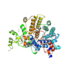
5KHJ
DownloadVisualize
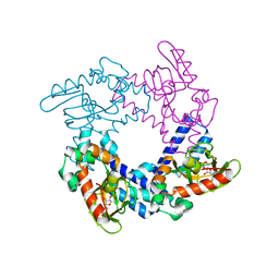
BU of 5khj by Molmil
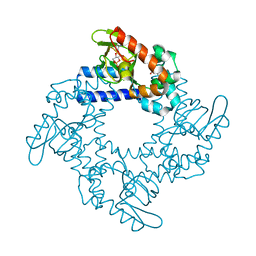
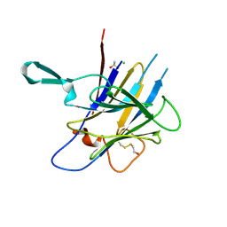
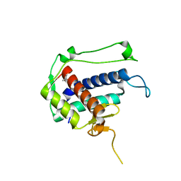
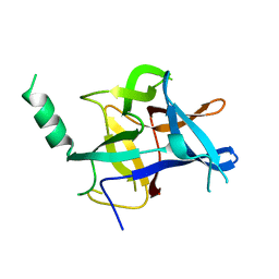
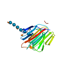
HCN2 CNBD in complex with uridine-3', 5'-cyclic monophosphate (cUMP)
分子名称: Potassium/sodium hyperpolarization-activated cyclic nucleotide-gated channel 2, Uridine-3',5'-cyclic monophosphate
著者Ng, L.C.T, Putrenko, I, Baronas, V, Van Petegem, F, Accili, E.A.
登録日2016-06-14
公開日2016-09-14
最終更新日2023-09-27
実験手法X-RAY DIFFRACTION (2.01 Å)
主引用文献Cyclic Purine and Pyrimidine Nucleotides Bind to the HCN2 Ion Channel and Variably Promote C-Terminal Domain Interactions and Opening.
Structure, 24, 2016
5KHK
DownloadVisualize
BU of 5khk by Molmil
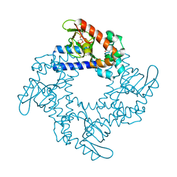
HCN2 CNBD in complex with 2-aminopurine riboside-3', 5'-cyclic monophosphate (2-NH2-cPuMP)
分子名称: 2-Aminopurine riboside-3',5'-cyclic monophosphate, Potassium/sodium hyperpolarization-activated cyclic nucleotide-gated channel 2
著者Ng, L.C.T, Putrenko, I, Baronas, V, Van Petegem, F, Accili, E.A.
登録日2016-06-14
公開日2016-09-14
最終更新日2023-09-27
実験手法X-RAY DIFFRACTION (2.07 Å)
主引用文献Cyclic Purine and Pyrimidine Nucleotides Bind to the HCN2 Ion Channel and Variably Promote C-Terminal Domain Interactions and Opening.
Structure, 24, 2016
5KHG
DownloadVisualize
BU of 5khg by Molmil
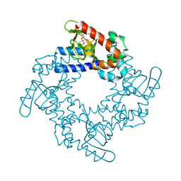
HCN2 CNBD in complex with cytidine-3', 5'-cyclic monophosphate (cCMP)
分子名称: 4-amino-1-[(2S,4aR,6R,7R,7aS)-2,7-dihydroxy-2-oxidotetrahydro-4H-furo[3,2-d][1,3,2]dioxaphosphinin-6-yl]pyrimidin-2(1H)-one, Potassium/sodium hyperpolarization-activated cyclic nucleotide-gated channel 2
著者Ng, L.C.T, Putrenko, I, Baronas, V, Van Petegem, F, Accili, E.A.
登録日2016-06-14
公開日2016-09-14
最終更新日2023-09-27
実験手法X-RAY DIFFRACTION (2.241 Å)
主引用文献Cyclic Purine and Pyrimidine Nucleotides Bind to the HCN2 Ion Channel and Variably Promote C-Terminal Domain Interactions and Opening.
Structure, 24, 2016
5KHH
DownloadVisualize
BU of 5khh by Molmil
HCN2 CNBD in complex with inosine-3', 5'-cyclic monophosphate (cIMP)
分子名称: Inosine-3',5'-cyclic monophosphate, Potassium/sodium hyperpolarization-activated cyclic nucleotide-gated channel 2
著者Ng, L.C.T, Putrenko, I, Baronas, V, Van Petegem, F, Accili, E.A.
登録日2016-06-14
公開日2016-09-14
最終更新日2023-09-27
実験手法X-RAY DIFFRACTION (1.77 Å)
主引用文献Cyclic Purine and Pyrimidine Nucleotides Bind to the HCN2 Ion Channel and Variably Promote C-Terminal Domain Interactions and Opening.
Structure, 24, 2016
6DLG
DownloadVisualize
BU of 6dlg by Molmil
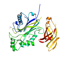
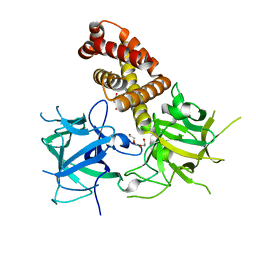
Crystal structure of a SHIP1 surface entropy reduction mutant
分子名称: ISOPROPYL ALCOHOL, Phosphatidylinositol 3,4,5-trisphosphate 5-phosphatase 1
著者Gardill, B.R, Cheung, S.T, Mui, A.L, Van Petegem, F.
登録日2018-06-01
公開日2019-06-05
最終更新日2024-03-13
実験手法X-RAY DIFFRACTION (1.499 Å)
主引用文献Interleukin-10 and Small Molecule SHIP1 Allosteric Regulators Trigger Anti-Inflammatory Effects Through SHIP1/STAT3 Complexes
Biorxiv, 2020
5C33
DownloadVisualize
BU of 5c33 by Molmil
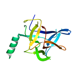
Crystal Structure of Mouse Ryanodine Receptor 2 SPRY1 Domain
分子名称: CHLORIDE ION, ISOPROPYL ALCOHOL, Ryanodine receptor 2
著者Yuchi, Z, Van Petegem, F.
登録日2015-06-16
公開日2015-08-05
最終更新日2019-12-04
実験手法X-RAY DIFFRACTION (1.21 Å)
主引用文献Crystal structures of ryanodine receptor SPRY1 and tandem-repeat domains reveal a critical FKBP12 binding determinant.
Nat Commun, 6, 2015
5C30
DownloadVisualize
BU of 5c30 by Molmil
Crystal Structure of Rabbit Ryanodine Receptor 1 Repeat12 Domain
分子名称: ISOPROPYL ALCOHOL, Ryanodine receptor 1
著者Yuchi, Z, Van Petegem, F.
登録日2015-06-16
公開日2015-08-05
最終更新日2024-03-06
実験手法X-RAY DIFFRACTION (1.55 Å)
主引用文献Crystal structures of ryanodine receptor SPRY1 and tandem-repeat domains reveal a critical FKBP12 binding determinant.
Nat Commun, 6, 2015
4KEI
DownloadVisualize
BU of 4kei by Molmil
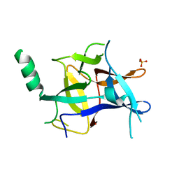
Crystal structure of mouse Ryanodine Receptor 2 (1-217) disease mutant P164S
分子名称: Ryanodine receptor 2
著者Kimlicka, L, Van Petegem, F.
登録日2013-04-25
公開日2013-09-04
最終更新日2023-09-20
実験手法X-RAY DIFFRACTION (2.406 Å)
主引用文献Type 2 Ryanodine Receptor Domain A Contains a Unique and Dynamic alpha-Helix That Transitions to a beta-Strand in a Mutant Linked with a Heritable Cardiomyopathy.
J.Mol.Biol., 425, 2013
5DZG
DownloadVisualize
BU of 5dzg by Molmil
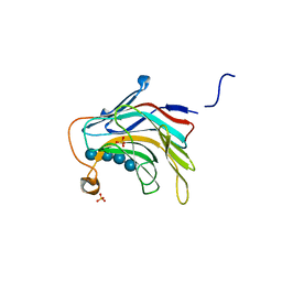
Crystal Structure of the catalytic nucleophile mutant of VvEG16 in complex with a xyloglucan tetradecasaccharide
分子名称: VvEG16, endo-glucanase, alpha-D-xylopyranose-(1-6)-beta-D-glucopyranose-(1-4)-[alpha-D-xylopyranose-(1-6)]beta-D-glucopyranose-(1-4)-[alpha-D-xylopyranose-(1-6)]beta-D-glucopyranose-(1-4)-alpha-D-glucopyranose, ...
著者McGregor, N.G.S, Tung, C.C, Van Petegem, F, Brumer, H.
登録日2015-09-25
公開日2016-09-21
最終更新日2023-09-27
実験手法X-RAY DIFFRACTION (1.79 Å)
主引用文献Crystallographic insight into the evolutionary origins of xyloglucan endotransglycosylases and endohydrolases.
Plant J., 89, 2017
4KEK
DownloadVisualize
BU of 4kek by Molmil
Crystal structure of mouse Ryanodine Receptor 2 (1-217) disease mutant R176Q
分子名称: Ryanodine receptor 2, SULFATE ION
著者Kimlicka, L, Van Petegem, F.
登録日2013-04-25
公開日2013-09-04
最終更新日2023-09-20
実験手法X-RAY DIFFRACTION (2.146 Å)
主引用文献Type 2 Ryanodine Receptor Domain A Contains a Unique and Dynamic alpha-Helix That Transitions to a beta-Strand in a Mutant Linked with a Heritable Cardiomyopathy.
J.Mol.Biol., 425, 2013
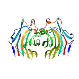
5DZE
DownloadVisualize
BU of 5dze by Molmil
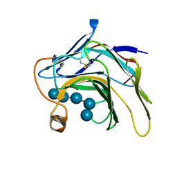
Crystal Structure of the catalytic nucleophile mutant of VvEG16 in complex with cellotetraose
分子名称: beta-D-glucopyranose, beta-D-glucopyranose-(1-4)-beta-D-glucopyranose-(1-4)-beta-D-glucopyranose-(1-4)-alpha-D-glucopyranose, endo-glucanase
著者McGregor, N.G.S, Tung, C.C, Van Petegem, F, Brumer, H.
登録日2015-09-25
公開日2016-09-21
最終更新日2020-07-29
実験手法X-RAY DIFFRACTION (0.97 Å)
主引用文献Crystallographic insight into the evolutionary origins of xyloglucan endotransglycosylases and endohydrolases.
Plant J., 89, 2017
4KEJ
DownloadVisualize
BU of 4kej by Molmil
Crystal structure of mouse Ryanodine Receptor 2 (1-217) disease mutant R169Q
分子名称: Ryanodine receptor 2
著者Kimlicka, L, Van Petegem, F.
登録日2013-04-25
公開日2013-09-04
最終更新日2023-09-20
実験手法X-RAY DIFFRACTION (2.553 Å)
主引用文献Type 2 Ryanodine Receptor Domain A Contains a Unique and Dynamic alpha-Helix That Transitions to a beta-Strand in a Mutant Linked with a Heritable Cardiomyopathy.
J.Mol.Biol., 425, 2013
4L4I
DownloadVisualize
BU of 4l4i by Molmil
Crystal structure of mouse Ryanodine Receptor isoform 2 (RyR2) 1-547 disease mutant R420Q
分子名称: GLYCEROL, Ryanodine receptor 2
著者Kimlicka, L, Van Petegem, F.
登録日2013-06-07
公開日2013-07-24
最終更新日2023-09-20
実験手法X-RAY DIFFRACTION (2.15 Å)
主引用文献Crystal structure of ryanodine receptor disease mutant
To be Published
4L4H
DownloadVisualize
BU of 4l4h by Molmil
Crystal structure of mouse Ryanodine Receptor isoform 2 (RyR2) 1-547
分子名称: CHLORIDE ION, DI(HYDROXYETHYL)ETHER, Ryanodine receptor 2, ...
著者Kimlicka, L, Tung, C.C, Van Petegem, F.
登録日2013-06-07
公開日2013-07-24
最終更新日2023-09-20
実験手法X-RAY DIFFRACTION (2 Å)
主引用文献Crystal structure of a ryanodine receptor domain
To be Published
5DZF
DownloadVisualize
BU of 5dzf by Molmil
Crystal Structure of the catalytic nucleophile mutant of VvEG16 in complex with a mixed-linkage glucan octasaccharide
分子名称: SULFATE ION, beta-D-glucopyranose, beta-D-glucopyranose-(1-3)-beta-D-glucopyranose-(1-4)-beta-D-glucopyranose-(1-4)-alpha-D-glucopyranose, ...
著者McGregor, N.G.S, Tung, C.C, Van Petegem, F, Brumer, H.
登録日2015-09-25
公開日2016-09-21
最終更新日2023-09-27
実験手法X-RAY DIFFRACTION (1.65 Å)
主引用文献Crystallographic insight into the evolutionary origins of xyloglucan endotransglycosylases and endohydrolases.
Plant J., 89, 2017
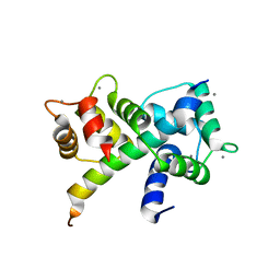
6U3A
DownloadVisualize
BU of 6u3a by Molmil
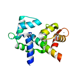
1.65 Angstrom crystal structure of the N97S Ca-CaM:CaV1.2 IQ domain complex
分子名称: CALCIUM ION, Calmodulin-1, SODIUM ION, ...
著者Wang, K, Van Petegem, F.
登録日2019-08-21
公開日2020-02-19
最終更新日2023-10-11
実験手法X-RAY DIFFRACTION (1.65 Å)
主引用文献Arrhythmia mutations in calmodulin can disrupt cooperativity of Ca2+binding and cause misfolding.
J. Physiol. (Lond.), 598, 2020
6U39
DownloadVisualize
BU of 6u39 by Molmil
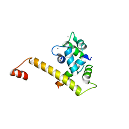
2.4 Angstrom crystal structure of the D129G Ca-CaM:CaV1.2 IQ domain complex
分子名称: CALCIUM ION, Calmodulin-1, Voltage-dependent L-type calcium channel subunit alpha-1C
著者Wang, K, Van Petegem, F.
登録日2019-08-21
公開日2020-02-19
最終更新日2023-10-11
実験手法X-RAY DIFFRACTION (2.4 Å)
主引用文献Arrhythmia mutations in calmodulin can disrupt cooperativity of Ca2+binding and cause misfolding.
J. Physiol. (Lond.), 598, 2020
6E60
DownloadVisualize
BU of 6e60 by Molmil
Bacteroides ovatus mixed-linkage glucan utilization locus (MLGUL) SGBP-A
分子名称: 1,2-ETHANEDIOL, MAGNESIUM ION, mixed-linkage glucan utilization locus (MLGUL) SGBP-B
著者Tamura, K, Gardill, B.R, Brumer, H, Van Petegem, F.
登録日2018-07-23
公開日2019-05-15
最終更新日2024-04-03
実験手法X-RAY DIFFRACTION (1.5 Å)
主引用文献Surface glycan-binding proteins are essential for cereal beta-glucan utilization by the human gut symbiont Bacteroides ovatus.
Cell.Mol.Life Sci., 76, 2019
6U3B
DownloadVisualize
BU of 6u3b by Molmil
1.7 Angstrom crystal structure of the Q135P Ca-CaM:CaV1.2 IQ domain complex
分子名称: CALCIUM ION, Calmodulin-1, Voltage-dependent L-type calcium channel subunit alpha-1C
著者Wang, K, Van Petegem, F.
登録日2019-08-21
公開日2020-02-19
最終更新日2023-10-11
実験手法X-RAY DIFFRACTION (1.7 Å)
主引用文献Arrhythmia mutations in calmodulin can disrupt cooperativity of Ca2+binding and cause misfolding.
J. Physiol. (Lond.), 598, 2020
6VHO
DownloadVisualize
BU of 6vho by Molmil
Glycoside hydrolase family 16 endo-glucanase from Bacteroides ovatus in complex with G4G4G3G-NHCOCH2Br
分子名称: Glycosyl hydrolase family 16, beta-D-glucopyranose-(1-4)-beta-D-glucopyranose-(1-3)-N-acetyl-beta-D-glucopyranosylamine
著者Tamura, K, Brumer, H, van Petegem, F.
登録日2020-01-10
公開日2021-01-13
最終更新日2023-10-11
実験手法X-RAY DIFFRACTION (2.148 Å)
主引用文献Orthogonal Active-Site Labels for Mixed-Linkage endo-beta-Glucanases.
Acs Chem.Biol., 16, 2021
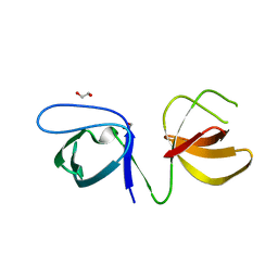
6UY7
DownloadVisualize
BU of 6uy7 by Molmil
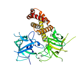
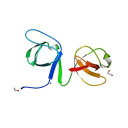
Crystal structure of the STAC3 tandem SH3 domains - P269R
分子名称: 1,2-ETHANEDIOL, SH3 and cysteine-rich domain-containing protein 3
著者Rufenach, B, Van Petegem, F.
登録日2019-11-12
公開日2020-06-17
最終更新日2023-10-11
実験手法X-RAY DIFFRACTION (2.105 Å)
主引用文献Multiple Sequence Variants in STAC3 Affect Interactions with CaV1.1 and Excitation-Contraction Coupling.
Structure, 28, 2020
6UY9
DownloadVisualize
BU of 6uy9 by Molmil
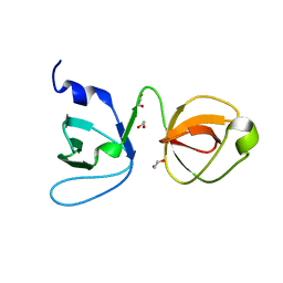
Crystal structure of the STAC3 tandem SH3 domains - P269R, W284S
分子名称: 1,2-ETHANEDIOL, SH3 and cysteine-rich domain-containing protein 3, SODIUM ION
著者Rufenach, B, Van Petegem, F.
登録日2019-11-12
公開日2020-06-17
最終更新日2023-10-11
実験手法X-RAY DIFFRACTION (1.6 Å)
主引用文献Multiple Sequence Variants in STAC3 Affect Interactions with CaV1.1 and Excitation-Contraction Coupling.
Structure, 28, 2020
6UY8
DownloadVisualize
BU of 6uy8 by Molmil
Crystal structure of the STAC3 tandem SH3 domains - P269R, K329N
分子名称: 1,2-ETHANEDIOL, CHLORIDE ION, SH3 and cysteine-rich domain-containing protein 3
著者Rufenach, B, Van Petegem, F.
登録日2019-11-12
公開日2020-06-17
最終更新日2023-10-11
実験手法X-RAY DIFFRACTION (1.649 Å)
主引用文献Multiple Sequence Variants in STAC3 Affect Interactions with CaV1.1 and Excitation-Contraction Coupling.
Structure, 28, 2020
6W1N
DownloadVisualize
BU of 6w1n by Molmil
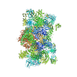
Pig Ryanodine Receptor (WT) in 5mM EGTA condition
分子名称: Peptidyl-prolyl cis-trans isomerase FKBP1B, Ryanodine Receptor, ZINC ION
著者Woll, K.W, Haji-Ghassemi, O, Van Petegem, F.
登録日2020-03-04
公開日2021-01-06
最終更新日2024-03-06
実験手法ELECTRON MICROSCOPY (4 Å)
主引用文献Pathological conformations of disease mutant Ryanodine Receptors revealed by cryo-EM.
Nat Commun, 12, 2021
6X34
DownloadVisualize
BU of 6x34 by Molmil
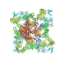
Pig R615C RyR1 EGTA (all classes, open)
分子名称: Peptidyl-prolyl cis-trans isomerase FKBP1B, Ryanodine Receptor, ZINC ION
著者Woll, K.W, Haji-Ghassemi, O, Van Petegem, F.
登録日2020-05-21
公開日2021-01-13
最終更新日2024-03-06
実験手法ELECTRON MICROSCOPY (4.7 Å)
主引用文献Pathological conformations of disease mutant Ryanodine Receptors revealed by cryo-EM.
Nat Commun, 12, 2021

221051

件を2024-06-12に公開中

PDB statisticsPDBj update infoContact PDBjnumon