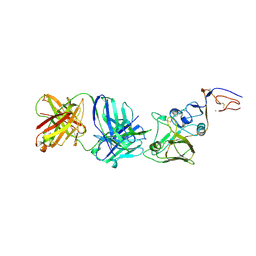
4E58
 
 | | Crystal structure of GCC(LCG)CCGC duplex containing LNA residue | | 分子名称: | RNA duplex containing CCG repeats, SULFATE ION | | 著者 | Kiliszek, A, Kierzek, R, Krzyzosiak, W.J, Rypniewski, R. | | 登録日 | 2012-03-14 | | 公開日 | 2012-12-12 | | 最終更新日 | 2023-09-13 | | 実験手法 | X-RAY DIFFRACTION (1.952 Å) | | 主引用文献 | Crystallographic characterization of CCG repeats.
Nucleic Acids Res., 40, 2012
|
|
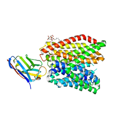
5HES
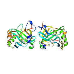
 | | Human leucine zipper- and sterile alpha motif-containing kinase (ZAK, MLT, HCCS-4, MRK, AZK, MLTK) in complex with vemurafenib | | 分子名称: | 1,2-ETHANEDIOL, Mitogen-activated protein kinase kinase kinase MLT, N-(3-{[5-(4-chlorophenyl)-1H-pyrrolo[2,3-b]pyridin-3-yl]carbonyl}-2,4-difluorophenyl)propane-1-sulfonamide | | 著者 | Mathea, S, Salah, E, Abdul Azeez, K.R, Tallant, C, Szklarz, M, Chaikuad, A, Shrestha, B, Sorrell, F.J, Elkins, J.M, Shrestha, L, Burgess-Brown, N, von Delft, F, Arrowsmith, C.H, Edwards, A.M, Bountra, C, Knapp, S. | | 登録日 | 2016-01-06 | | 公開日 | 2016-03-30 | | 最終更新日 | 2024-01-10 | | 実験手法 | X-RAY DIFFRACTION (2.14 Å) | | 主引用文献 | Structure of the Human Protein Kinase ZAK in Complex with Vemurafenib.
Acs Chem.Biol., 11, 2016
|
|
7R0J
 | | Structure of the V2 receptor Cter-arrestin2-ScFv30 complex | | 分子名称: | Arrestin2, ScFv30, V2R Cter | | 著者 | Bous, J, Fouillen, A, Trapani, S, Granier, S, Mouillac, B, Bron, P. | | 登録日 | 2022-02-02 | | 公開日 | 2022-09-14 | | 実験手法 | ELECTRON MICROSCOPY (4.23 Å) | | 主引用文献 | Structure of the vasopressin hormone-V2 receptor-beta-arrestin1 ternary complex.
Sci Adv, 8, 2022
|
|
7R0C
 | | Structure of the AVP-V2R-arrestin2-ScFv30 complex | | 分子名称: | AVP, Arrestin2, ScFv30, ... | | 著者 | Bous, J, Fouillen, A, Trapani, S, Granier, S, Mouillac, B, Bron, P. | | 登録日 | 2022-02-01 | | 公開日 | 2022-09-14 | | 実験手法 | ELECTRON MICROSCOPY (4.73 Å) | | 主引用文献 | Structure of the vasopressin hormone-V2 receptor-beta-arrestin1 ternary complex.
Sci Adv, 8, 2022
|
|
6FE0
 | |
6FE2
 | |
6FE1
 | |
7A05
 | | NMR structure of D3-D4 domains of Vibrio vulnificus ribosomal protein S1 | | 分子名称: | 30S ribosomal protein S1 | | 著者 | Qureshi, N.S, Matzel, T, Cetiner, E.C, Schnieders, S, Jonker, H.R.A, Schwalbe, H, Fuertig, B. | | 登録日 | 2020-08-06 | | 公開日 | 2021-06-23 | | 最終更新日 | 2024-01-17 | | 実験手法 | SOLUTION NMR | | 主引用文献 | NMR structure of the Vibrio vulnificus ribosomal protein S1 domains D3 and D4 provides insights into molecular recognition of single-stranded RNAs.
Nucleic Acids Res., 49, 2021
|
|
6R8N
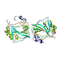
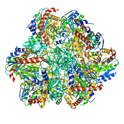 | | STRUCTURE DETERMINATION OF THE TETRAHEDRAL AMINOPEPTIDASE TET2 FROM P. HORIKOSHII BY USE OF COMBINED SOLID-STATE NMR, SOLUTION-STATE NMR AND EM DATA 4.1 A, FOLLOWED BY REAL_SPACE_REFINEMENT AT 4.1 A | | 分子名称: | Tetrahedral aminopeptidase, ZINC ION | | 著者 | Colletier, J.-P, Gauto, D, Estrozi, L, Favier, A, Effantin, G, Schoehn, G, Boisbouvier, J, Schanda, P. | | 登録日 | 2019-04-02 | | 公開日 | 2019-08-14 | | 最終更新日 | 2023-09-13 | | 実験手法 | ELECTRON MICROSCOPY (4.1 Å), SOLUTION NMR | | 主引用文献 | Integrated NMR and cryo-EM atomic-resolution structure determination of a half-megadalton enzyme complex.
Nat Commun, 10, 2019
|
|
6G9U
 | |
6G98
 | |
1GIG
 | |
5NFO
 | |
5NFN
 | |
7BBV
 | | Pectate lyase B from Verticillium dahliae | | 分子名称: | CALCIUM ION, DI(HYDROXYETHYL)ETHER, Pectate lyase B, ... | | 著者 | Safran, J, Habrylo, O, Bouckaert, J, Pau Roblot, C, Senechal, F, Pelloux, J. | | 登録日 | 2020-12-18 | | 公開日 | 2022-07-13 | | 最終更新日 | 2024-02-07 | | 実験手法 | X-RAY DIFFRACTION (1.2 Å) | | 主引用文献 | The specificity of pectate lyase VdPelB from Verticilium dahliae is highlighted by structural, dynamical and biochemical characterizations.
Int.J.Biol.Macromol., 231, 2023
|
|
1X94
 | |
2VIU
 | | INFLUENZA VIRUS HEMAGGLUTININ | | 分子名称: | 2-acetamido-2-deoxy-beta-D-glucopyranose, HEMAGGLUTININ, beta-D-mannopyranose-(1-4)-2-acetamido-2-deoxy-beta-D-glucopyranose-(1-4)-2-acetamido-2-deoxy-beta-D-glucopyranose | | 著者 | Bizebard, T, Fleury, D, Gigant, B, Wharton, S.A, Skehel, J.J, Knossow, M. | | 登録日 | 1997-12-22 | | 公開日 | 1998-04-29 | | 最終更新日 | 2021-11-03 | | 実験手法 | X-RAY DIFFRACTION (2.5 Å) | | 主引用文献 | Antigen distortion allows influenza virus to escape neutralization.
Nat.Struct.Biol., 5, 1998
|
|
2VIS
 | | INFLUENZA VIRUS HEMAGGLUTININ, (ESCAPE) MUTANT WITH THR 131 REPLACED BY ILE, COMPLEXED WITH A NEUTRALIZING ANTIBODY | | 分子名称: | 2-acetamido-2-deoxy-beta-D-glucopyranose, HEMAGGLUTININ, IMMUNOGLOBULIN (IGG1, ... | | 著者 | Bizebard, T, Fleury, D, Gigant, B, Wharton, S.A, Skehel, J.J, Knossow, M. | | 登録日 | 1997-12-22 | | 公開日 | 1998-04-29 | | 最終更新日 | 2021-11-03 | | 実験手法 | X-RAY DIFFRACTION (3.25 Å) | | 主引用文献 | Antigen distortion allows influenza virus to escape neutralization.
Nat.Struct.Biol., 5, 1998
|
|
2VIT
 | | INFLUENZA VIRUS HEMAGGLUTININ, MUTANT WITH THR 155 REPLACED BY ILE, COMPLEXED WITH A NEUTRALIZING ANTIBODY | | 分子名称: | HEMAGGLUTININ, IMMUNOGLOBULIN (IGG1, LAMBDA), ... | | 著者 | Bizebard, T, Fleury, D, Gigant, B, Wharton, S.A, Skehel, J.J, Knossow, M. | | 登録日 | 1997-12-22 | | 公開日 | 1998-04-29 | | 最終更新日 | 2021-11-03 | | 実験手法 | X-RAY DIFFRACTION (3.25 Å) | | 主引用文献 | Antigen distortion allows influenza virus to escape neutralization.
Nat.Struct.Biol., 5, 1998
|
|
6GS4
 | | Crystal structure of peptide transporter DtpA-nanobody in complex with valganciclovir | | 分子名称: | DODECYL-BETA-D-MALTOSIDE, Dipeptide and tripeptide permease A, [(2~{S})-2-[(2-azanyl-6-oxidanylidene-3~{H}-purin-9-yl)methoxy]-3-oxidanyl-propyl] (2~{S})-2-azanyl-3-methyl-butanoate, ... | | 著者 | Ural-Blimke, Y, Flayhan, A, Quistgaard, E.M, Loew, C. | | 登録日 | 2018-06-13 | | 公開日 | 2019-01-30 | | 最終更新日 | 2024-01-17 | | 実験手法 | X-RAY DIFFRACTION (2.645 Å) | | 主引用文献 | Structure of Prototypic Peptide Transporter DtpA from E. coli in Complex with Valganciclovir Provides Insights into Drug Binding of Human PepT1.
J. Am. Chem. Soc., 141, 2019
|
|
6GS7
 | | Crystal structure of peptide transporter DtpA-nanobody in glycine buffer | | 分子名称: | Dipeptide and tripeptide permease A, nanobody | | 著者 | Ural-Blimke, Y, Flayhan, A, Quistgaard, E.M, Loew, C. | | 登録日 | 2018-06-13 | | 公開日 | 2019-01-30 | | 最終更新日 | 2024-01-17 | | 実験手法 | X-RAY DIFFRACTION (3.3 Å) | | 主引用文献 | Structure of Prototypic Peptide Transporter DtpA from E. coli in Complex with Valganciclovir Provides Insights into Drug Binding of Human PepT1.
J. Am. Chem. Soc., 141, 2019
|
|
6GS1
 | | Crystal structure of peptide transporter DtpA-nanobody in MES buffer | | 分子名称: | Dipeptide and tripeptide permease A, Nanobody 00 | | 著者 | Ural-Blimke, Y, Flayhan, A, Loew, C, Quistgaard, E.M. | | 登録日 | 2018-06-13 | | 公開日 | 2019-01-30 | | 最終更新日 | 2024-01-17 | | 実験手法 | X-RAY DIFFRACTION (3.29 Å) | | 主引用文献 | Structure of Prototypic Peptide Transporter DtpA from E. coli in Complex with Valganciclovir Provides Insights into Drug Binding of Human PepT1.
J. Am. Chem. Soc., 141, 2019
|
|
2EA2
 | | h-MetAP2 complexed with A773812 | | 分子名称: | 3-ETHYL-6-{[(4-FLUOROPHENYL)SULFONYL]AMINO}-2-METHYLBENZOIC ACID, MANGANESE (II) ION, Methionine aminopeptidase 2 | | 著者 | Park, C.H. | | 登録日 | 2007-01-30 | | 公開日 | 2008-02-05 | | 最終更新日 | 2023-10-25 | | 実験手法 | X-RAY DIFFRACTION (2.5 Å) | | 主引用文献 | Lead optimization of methionine aminopeptidase-2 (MetAP2) inhibitors containing sulfonamides of 5,6-disubstituted anthranilic acids
Bioorg.Med.Chem.Lett., 17, 2007
|
|
2EA4
 | | h-MetAP2 complexed with A797859 | | 分子名称: | 2-(2-AMINOETHOXY)-3-ETHYL-6-{[(4-FLUOROPHENYL)SULFONYL]AMINO}BENZOIC ACID, MANGANESE (II) ION, Methionine aminopeptidase 2 | | 著者 | Park, C.H. | | 登録日 | 2007-01-30 | | 公開日 | 2008-02-05 | | 最終更新日 | 2023-10-25 | | 実験手法 | X-RAY DIFFRACTION (2.35 Å) | | 主引用文献 | Lead optimization of methionine aminopeptidase-2 (MetAP2) inhibitors containing sulfonamides of 5,6-disubstituted anthranilic acids
Bioorg.Med.Chem.Lett., 17, 2007
|
|
1YW7
 | | h-MetAP2 complexed with A444148 | | 分子名称: | 5-METHYL-2-[(PHENYLSULFONYL)AMINO]BENZOIC ACID, MANGANESE (II) ION, Methionine aminopeptidase 2 | | 著者 | Park, C.H. | | 登録日 | 2005-02-17 | | 公開日 | 2006-02-21 | | 最終更新日 | 2011-07-13 | | 実験手法 | X-RAY DIFFRACTION (1.85 Å) | | 主引用文献 | Discovery and optimization of anthranilic acid sulfonamides as inhibitors of methionine aminopeptidase-2: a structural basis for the reduction of albumin binding.
J.Med.Chem., 49, 2006
|
|


