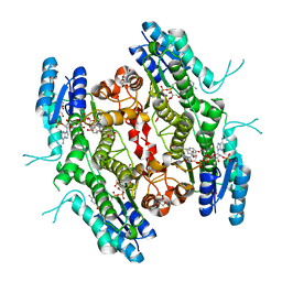
5JCX
 
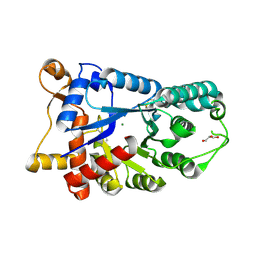
 | | Trypanosoma brucei PTR1 in complex with inhibitor NP-29 | | 分子名称: | 3,5,7-trihydroxy-2-(2-hydroxyphenyl)-4H-1-benzopyran-4-one, ACETATE ION, GLYCEROL, ... | | 著者 | Landi, G, Pozzi, C, Di Pisa, F, Dello Iacono, L, Mangani, S. | | 登録日 | 2016-04-15 | | 公開日 | 2016-08-17 | | 最終更新日 | 2024-01-10 | | 実験手法 | X-RAY DIFFRACTION (1.43 Å) | | 主引用文献 | Profiling of Flavonol Derivatives for the Development of Antitrypanosomatidic Drugs.
J.Med.Chem., 59, 2016
|
|
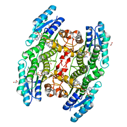
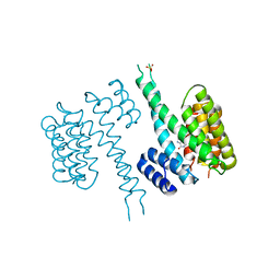
5JDI
 | | Trypanosoma brucei PTR1 in complex with cofactor and inhibitor NMT-H024 (compound 2) | | 分子名称: | 3,6-dihydroxy-2-(3-hydroxyphenyl)-4H-1-benzopyran-4-one, ACETATE ION, NADP NICOTINAMIDE-ADENINE-DINUCLEOTIDE PHOSPHATE, ... | | 著者 | Landi, G, Pozzi, C, Di Pisa, F, Dello lacono, L, Mangani, S. | | 登録日 | 2016-04-16 | | 公開日 | 2016-08-17 | | 最終更新日 | 2024-01-10 | | 実験手法 | X-RAY DIFFRACTION (1.38 Å) | | 主引用文献 | Profiling of Flavonol Derivatives for the Development of Antitrypanosomatidic Drugs.
J.Med.Chem., 59, 2016
|
|
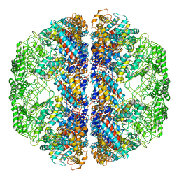
5A1A
 | | 2.2 A resolution cryo-EM structure of beta-galactosidase in complex with a cell-permeant inhibitor | | 分子名称: | 2-phenylethyl 1-thio-beta-D-galactopyranoside, BETA-GALACTOSIDASE, MAGNESIUM ION, ... | | 著者 | Bartesaghi, A, Merk, A, Banerjee, S, Matthies, D, Wu, X, Milne, J, Subramaniam, S. | | 登録日 | 2015-04-29 | | 公開日 | 2015-05-06 | | 最終更新日 | 2024-05-08 | | 実験手法 | ELECTRON MICROSCOPY (2.2 Å) | | 主引用文献 | 2.2 A Resolution Cryo-Em Structure of Beta-Galactosidase in Complex with a Cell-Permeant Inhibitor
Science, 348, 2015
|
|
1D8I
 | |
3IZI
 | | Mm-cpn rls with ATP | | 分子名称: | Chaperonin | | 著者 | Douglas, N.R, Reissmann, S, Zhang, J, Chen, B, Jakana, J, Kumar, R, Chiu, W, Frydman, J. | | 登録日 | 2010-10-29 | | 公開日 | 2011-02-02 | | 最終更新日 | 2024-02-21 | | 実験手法 | ELECTRON MICROSCOPY (6.7 Å) | | 主引用文献 | Dual Action of ATP Hydrolysis Couples Lid Closure to Substrate Release into the Group II Chaperonin Chamber.
Cell(Cambridge,Mass.), 144, 2011
|
|
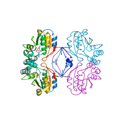
1Q0T
 | | Ternary Structure of T4DAM with AdoHcy and DNA | | 分子名称: | 5'-D(*AP*CP*AP*GP*GP*AP*TP*CP*CP*TP*GP*T)-3', DNA adenine methylase, IODIDE ION, ... | | 著者 | Yang, Z, Horton, J.R, Zhou, L, Zhang, X.J, Dong, A, Zhang, X, Schlagman, S.L, Kossykh, V, Hattman, S, Cheng, X. | | 登録日 | 2003-07-17 | | 公開日 | 2003-09-23 | | 最終更新日 | 2024-02-14 | | 実験手法 | X-RAY DIFFRACTION (3.1 Å) | | 主引用文献 | Structure of the bacteriophage T4 DNA adenine methyltransferase
Nat.Struct.Biol., 10, 2003
|
|
6TBX
 | | Trypanosoma brucei PTR1 (TbPTR1) in complex with a tricyclic-based inhibitor | | 分子名称: | 2,4-bis(azanyl)-9~{H}-pyrimido[4,5-b]indol-6-ol, ACETATE ION, GLYCEROL, ... | | 著者 | Landi, G, Tassone, G, Pozzi, C, Mangani, S. | | 登録日 | 2019-11-04 | | 公開日 | 2020-06-03 | | 最終更新日 | 2024-01-24 | | 実験手法 | X-RAY DIFFRACTION (1.3 Å) | | 主引用文献 | High-resolution crystal structure of Trypanosoma brucei pteridine reductase 1 in complex with an innovative tricyclic-based inhibitor.
Acta Crystallogr D Struct Biol, 76, 2020
|
|
3E2V
 | | Crystal structure of an uncharacterized amidohydrolase from Saccharomyces cerevisiae | | 分子名称: | 3'-5'-exonuclease, GLYCEROL, MAGNESIUM ION | | 著者 | Bonanno, J.B, Dickey, M, Bain, K.T, Hu, S, Romero, R, Smith, D, Wasserman, S, Sauder, J.M, Burley, S.K, Almo, S.C, New York SGX Research Center for Structural Genomics (NYSGXRC) | | 登録日 | 2008-08-06 | | 公開日 | 2008-08-26 | | 最終更新日 | 2021-02-10 | | 実験手法 | X-RAY DIFFRACTION (1.5 Å) | | 主引用文献 | Crystal structure of an uncharacterized amidohydrolase from Saccharomyces cerevisiae
To be Published
|
|
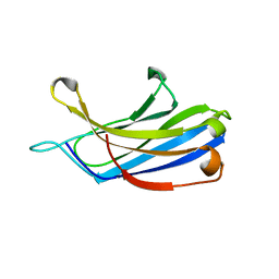
5DZX
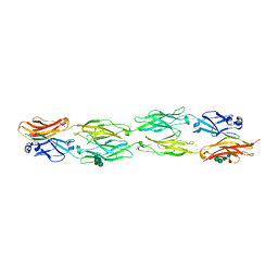 | | Protocadherin beta 6 extracellular cadherin domains 1-4 | | 分子名称: | 2-acetamido-2-deoxy-beta-D-glucopyranose-(1-4)-2-acetamido-2-deoxy-beta-D-glucopyranose, CALCIUM ION, Protocadherin beta 6, ... | | 著者 | Goodman, K.M, Mannepalli, S, Bahna, F, Honig, B, Shapiro, L. | | 登録日 | 2015-09-26 | | 公開日 | 2016-05-04 | | 最終更新日 | 2023-09-27 | | 実験手法 | X-RAY DIFFRACTION (2.879 Å) | | 主引用文献 | Structural Basis of Diverse Homophilic Recognition by Clustered alpha- and beta-Protocadherins.
Neuron, 90, 2016
|
|
3LOS
 | | Atomic Model of Mm-cpn in the Closed State | | 分子名称: | Chaperonin | | 著者 | Zhang, J, Baker, M.L, Schroeder, G, Douglas, N.R, Reissmann, S, Jakana, J, Dougherty, M, Fu, C.J, Levitt, M, Ludtke, S.J, Frydman, J, Chiu, W. | | 登録日 | 2010-02-04 | | 公開日 | 2010-03-16 | | 最終更新日 | 2018-07-18 | | 実験手法 | ELECTRON MICROSCOPY (4.3 Å) | | 主引用文献 | Mechanism of folding chamber closure in a group II chaperonin
Nature, 463, 2010
|
|
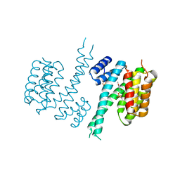
5L42
 | | Leishmania major Pteridine reductase 1 (PTR1) in complex with compound 3 | | 分子名称: | (2~{R})-2-[3,4-bis(oxidanyl)phenyl]-6-oxidanyl-2,3-dihydrochromen-4-one, 1,2-ETHANEDIOL, ACETATE ION, ... | | 著者 | Dello Iacono, L, Di Pisa, F, Pozzi, C, Landi, G, Mangani, S. | | 登録日 | 2016-05-24 | | 公開日 | 2017-05-03 | | 最終更新日 | 2024-01-10 | | 実験手法 | X-RAY DIFFRACTION (2.1 Å) | | 主引用文献 | Chroman-4-One Derivatives Targeting Pteridine Reductase 1 and Showing Anti-Parasitic Activity.
Molecules, 22, 2017
|
|
6TLF
 | |
2B82
 | | Crystal structure of AphA class B acid phosphatase/phosphotransferase ternary complex with adenosine and phosphate bound to the catalytic metal at 1.2 A resolution | | 分子名称: | ADENOSINE, MAGNESIUM ION, PHOSPHATE ION, ... | | 著者 | Calderone, V, Forleo, C, Benvenuti, M, Thaller, M.C, Rossolini, G.M, Mangani, S. | | 登録日 | 2005-10-06 | | 公開日 | 2005-11-29 | | 最終更新日 | 2023-08-23 | | 実験手法 | X-RAY DIFFRACTION (1.25 Å) | | 主引用文献 | A structure-based proposal for the catalytic mechanism of the bacterial acid phosphatase AphA belonging to the DDDD superfamily of phosphohydrolases
J.Mol.Biol., 355, 2006
|
|
6TM7
 | |
1AVN
 | | HUMAN CARBONIC ANHYDRASE II COMPLEXED WITH THE HISTAMINE ACTIVATOR | | 分子名称: | AZIDE ION, CARBONIC ANHYDRASE II, HISTAMINE, ... | | 著者 | Briganti, F, Mangani, S, Orioli, P, Scozzafava, A, Vernaglione, G, Supuran, C.T. | | 登録日 | 1997-09-17 | | 公開日 | 1997-12-24 | | 最終更新日 | 2024-02-07 | | 実験手法 | X-RAY DIFFRACTION (2 Å) | | 主引用文献 | Carbonic anhydrase activators: X-ray crystallographic and spectroscopic investigations for the interaction of isozymes I and II with histamine.
Biochemistry, 36, 1997
|
|
1Q3A
 | | Crystal structure of the catalytic domain of human matrix metalloproteinase 10 | | 分子名称: | CALCIUM ION, N-ISOBUTYL-N-[4-METHOXYPHENYLSULFONYL]GLYCYL HYDROXAMIC ACID, Stromelysin-2, ... | | 著者 | Calderone, V, Bertini, I, Fragai, M, Luchinat, C, Mangani, S, Terni, B. | | 登録日 | 2003-07-29 | | 公開日 | 2004-04-06 | | 最終更新日 | 2023-08-16 | | 実験手法 | X-RAY DIFFRACTION (2.1 Å) | | 主引用文献 | Crystal structure of the catalytic domain of human matrix metalloproteinase 10.
J.Mol.Biol., 336, 2004
|
|
1KXT
 | | Camelid VHH Domains in Complex with Porcine Pancreatic alpha-Amylase | | 分子名称: | ALPHA-AMYLASE, PANCREATIC, CALCIUM ION, ... | | 著者 | Desmyter, A, Spinelli, S, Payan, F, Lauwereys, M, Wyns, L, Muyldermans, S, Cambillau, C. | | 登録日 | 2002-02-01 | | 公開日 | 2002-06-19 | | 最終更新日 | 2023-08-16 | | 実験手法 | X-RAY DIFFRACTION (2 Å) | | 主引用文献 | Three camelid VHH domains in complex with porcine pancreatic alpha-amylase. Inhibition and versatility of binding topology.
J.Biol.Chem., 277, 2002
|
|
3DHU
 | | Crystal structure of an alpha-amylase from Lactobacillus plantarum | | 分子名称: | Alpha-amylase | | 著者 | Bonanno, J.B, Dickey, M, Bain, K.T, Iizuka, M, Ozyurt, S, Smith, D, Wasserman, S, Sauder, J.M, Burley, S.K, Almo, S.C, New York SGX Research Center for Structural Genomics (NYSGXRC) | | 登録日 | 2008-06-18 | | 公開日 | 2008-08-12 | | 最終更新日 | 2024-02-21 | | 実験手法 | X-RAY DIFFRACTION (2 Å) | | 主引用文献 | Crystal structure of an alpha-amylase from Lactobacillus plantarum
To be Published
|
|
1BV3
 | | HUMAN CARBONIC ANHYDRASE II COMPLEXED WITH UREA | | 分子名称: | 4-(HYDROXYMERCURY)BENZOIC ACID, PROTEIN (CARBONIC ANHYDRASE II), UREA, ... | | 著者 | Briganti, F, Mangani, S, Scozzafava, A, Vernaglione, G, Supuran, C.T. | | 登録日 | 1998-09-22 | | 公開日 | 1999-09-28 | | 最終更新日 | 2024-02-07 | | 実験手法 | X-RAY DIFFRACTION (1.85 Å) | | 主引用文献 | Carbonic anhydrase catalyzes cyanamide hydration to urea: is it mimicking the physiological reaction?
J.Biol.Inorg.Chem., 4, 1999
|
|
3IZJ
 | | Mm-cpn rls with ATP and AlFx | | 分子名称: | Chaperonin | | 著者 | Douglas, N.R, Reissmann, S, Zhang, J, Chen, B, Jakana, J, Kumar, R, Chiu, W, Frydman, J. | | 登録日 | 2010-10-29 | | 公開日 | 2011-02-02 | | 最終更新日 | 2024-02-21 | | 実験手法 | ELECTRON MICROSCOPY (6.9 Å) | | 主引用文献 | Dual Action of ATP Hydrolysis Couples Lid Closure to Substrate Release into the Group II Chaperonin Chamber.
Cell(Cambridge,Mass.), 144, 2011
|
|
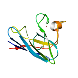
3FDW
 | | Crystal structure of a C2 domain from human synaptotagmin-like protein 4 | | 分子名称: | Synaptotagmin-like protein 4 | | 著者 | Bonanno, J.B, Rutter, M, Bain, K.T, Miller, S, Romero, R, Wasserman, S, Sauder, J.M, Burley, S.K, Almo, S.C, New York SGX Research Center for Structural Genomics (NYSGXRC) | | 登録日 | 2008-11-26 | | 公開日 | 2008-12-23 | | 最終更新日 | 2023-12-27 | | 実験手法 | X-RAY DIFFRACTION (2.2 Å) | | 主引用文献 | Crystal structure of a C2 domain from human synaptotagmin-like protein 4
To be Published
|
|
5DZY
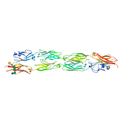 | | Protocadherin beta 8 extracellular cadherin domains 1-4 | | 分子名称: | 2-acetamido-2-deoxy-beta-D-glucopyranose, CALCIUM ION, Pcdhb8 protein, ... | | 著者 | Goodman, K.M, Bahna, F, Mannepalli, S, Honig, B, Shapiro, L. | | 登録日 | 2015-09-26 | | 公開日 | 2016-05-04 | | 最終更新日 | 2023-09-27 | | 実験手法 | X-RAY DIFFRACTION (2.9 Å) | | 主引用文献 | Structural Basis of Diverse Homophilic Recognition by Clustered alpha- and beta-Protocadherins.
Neuron, 90, 2016
|
|
1OSC
 | | Crystal structure of rat CUTA1 at 2.15 A resolution | | 分子名称: | similar to divalent cation tolerant protein CUTA | | 著者 | Arnesano, F, Banci, L, Benvenuti, M, Bertini, I, Calderone, V, Mangani, S, Viezzoli, M.S, Structural Proteomics in Europe (SPINE) | | 登録日 | 2003-03-19 | | 公開日 | 2003-11-25 | | 最終更新日 | 2023-08-16 | | 実験手法 | X-RAY DIFFRACTION (2.15 Å) | | 主引用文献 | The Evolutionarily Conserved Trimeric Structure of CutA1 Proteins
Suggests a Role in Signal Transduction
J.Biol.Chem., 278, 2003
|
|
1DSW
 | | THE SOLUTION STRUCTURE OF A MONOMERIC, REDUCED FORM OF HUMAN COPPER, ZINC SUPEROXIDE DISMUTASE BEARING THE SAME CHARGE AS THE NATIVE PROTEIN | | 分子名称: | COPPER (II) ION, SUPEROXIDE DISMUTASE (CU-ZN), ZINC ION | | 著者 | Banci, L, Bertini, I, Del Conte, R, Fadin, R, Mangani, S, Viezzoli, M.S. | | 登録日 | 2000-01-10 | | 公開日 | 2000-03-22 | | 最終更新日 | 2022-02-16 | | 実験手法 | SOLUTION NMR | | 主引用文献 | The solution structure of a monomeric, reduced form of human copper,zinc superoxide dismutase bearing the same charge as the native protein.
J.Biol.Inorg.Chem., 4, 1999
|
|
3EEQ
 | | Crystal structure of a putative cobalamin biosynthesis protein G homolog from Sulfolobus solfataricus | | 分子名称: | SULFATE ION, putative Cobalamin biosynthesis protein G homolog | | 著者 | Bonanno, J.B, Gilmore, M, Bain, K.T, Chang, S, Romero, R, Wasserman, S, Sauder, J.M, Burley, S.K, Almo, S.C, New York SGX Research Center for Structural Genomics (NYSGXRC) | | 登録日 | 2008-09-05 | | 公開日 | 2008-09-30 | | 最終更新日 | 2021-02-10 | | 実験手法 | X-RAY DIFFRACTION (2.3 Å) | | 主引用文献 | Crystal structure of a putative cobalamin biosynthesis protein G homolog from Sulfolobus solfataricus
To be Published
|
|


