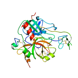
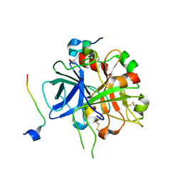
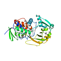
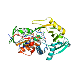
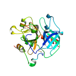
1BXA
 
 | | AMICYANIN REDUCED, PH 4.4, 1.3 ANGSTROMS | | 分子名称: | COPPER (I) ION, PROTEIN (AMICYANIN) | | 著者 | Cunane, L.M, Chen, Z.W, Durley, R.C.E, Mathews, F.S. | | 登録日 | 1998-10-01 | | 公開日 | 1998-10-07 | | 最終更新日 | 2023-08-09 | | 実験手法 | X-RAY DIFFRACTION (1.3 Å) | | 主引用文献 | Molecular basis for interprotein complex-dependent effects on the redox properties of amicyanin.
Biochemistry, 37, 1998
|
|
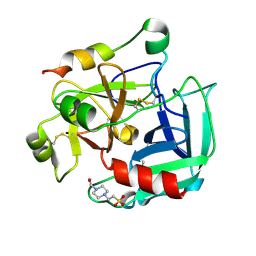
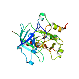
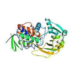
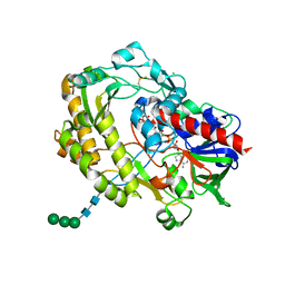
4MLF
 | | Crystal structure for the complex of thrombin mutant D102N and hirudin | | 分子名称: | 2-acetamido-2-deoxy-beta-D-glucopyranose, ACETATE ION, Hirudin variant-1, ... | | 著者 | Vogt, A.D, Pozzi, N, Chen, Z, Di Cera, E. | | 登録日 | 2013-09-06 | | 公開日 | 2013-09-25 | | 最終更新日 | 2023-09-20 | | 実験手法 | X-RAY DIFFRACTION (2.2 Å) | | 主引用文献 | Essential role of conformational selection in ligand binding.
Biophys.Chem., 186C, 2014
|
|
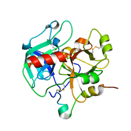
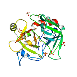
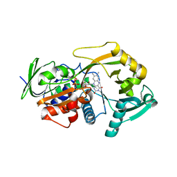
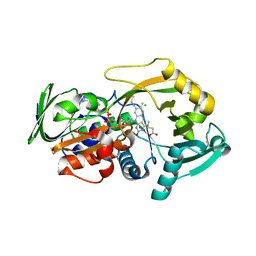
2GP9
 | | Crystal structure of the slow form of thrombin in a self-inhibited conformation | | 分子名称: | 4-(2-HYDROXYETHYL)-1-PIPERAZINE ETHANESULFONIC ACID, Prothrombin | | 著者 | Pineda, A, Chen, Z, Mathews, F.S, Di Cera, E. | | 登録日 | 2006-04-17 | | 公開日 | 2006-09-12 | | 最終更新日 | 2023-08-30 | | 実験手法 | X-RAY DIFFRACTION (1.87 Å) | | 主引用文献 | Crystal structure of thrombin in a self-inhibited conformation.
J.Biol.Chem., 281, 2006
|
|
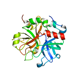
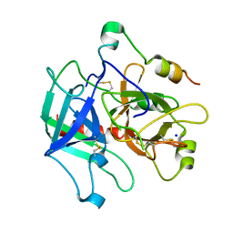
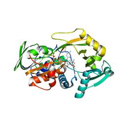
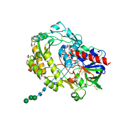
3HK3
 | | Crystal structure of murine thrombin mutant W215A/E217A (one molecule in the asymmetric unit) | | 分子名称: | Thrombin heavy chain, Thrombin light chain | | 著者 | Gandhi, P.S, Page, M.J, Chen, Z, Bush-Pelc, L, Di Cera, E. | | 登録日 | 2009-05-22 | | 公開日 | 2009-07-07 | | 最終更新日 | 2023-09-06 | | 実験手法 | X-RAY DIFFRACTION (1.94 Å) | | 主引用文献 | Mechanism of the Anticoagulant Activity of Thrombin Mutant W215A/E217A.
J.Biol.Chem., 284, 2009
|
|
3HK6
 | | Crystal structure of murine thrombin mutant W215A/E217A (two molecules in the asymmetric unit) | | 分子名称: | Thrombin heavy chain, Thrombin light chain | | 著者 | Gandhi, P.S, Page, M.J, Chen, Z, Bush-Pelc, L, Di Cera, E. | | 登録日 | 2009-05-22 | | 公開日 | 2009-07-07 | | 最終更新日 | 2023-09-06 | | 実験手法 | X-RAY DIFFRACTION (3.2 Å) | | 主引用文献 | Mechanism of the Anticoagulant Activity of Thrombin Mutant W215A/E217A.
J.Biol.Chem., 284, 2009
|
|
3HKI
 | | Crystal structure of murine thrombin mutant W215A/E217A in complex with the extracellular fragment of human PAR1 | | 分子名称: | 2-acetamido-2-deoxy-beta-D-glucopyranose, Proteinase-activated receptor 1, Thrombin heavy chain, ... | | 著者 | Gandhi, P.S, Page, M.J, Chen, Z, Bush-Pelc, L, Di Cera, E. | | 登録日 | 2009-05-23 | | 公開日 | 2009-07-07 | | 最終更新日 | 2023-09-06 | | 実験手法 | X-RAY DIFFRACTION (2.2 Å) | | 主引用文献 | Mechanism of the Anticoagulant Activity of Thrombin Mutant W215A/E217A.
J.Biol.Chem., 284, 2009
|
|
3HKJ
 | | Crystal structure of human thrombin mutant W215A/E217A in complex with the extracellular fragment of human PAR1 | | 分子名称: | 2-acetamido-2-deoxy-beta-D-glucopyranose, Proteinase-activated receptor 1, Thrombin heavy chain, ... | | 著者 | Gandhi, P.S, Page, M.J, Chen, Z, Bush-Pelc, L, Di Cera, E. | | 登録日 | 2009-05-23 | | 公開日 | 2009-07-07 | | 最終更新日 | 2023-09-06 | | 実験手法 | X-RAY DIFFRACTION (2.6 Å) | | 主引用文献 | Mechanism of the Anticoagulant Activity of Thrombin Mutant W215A/E217A.
J.Biol.Chem., 284, 2009
|
|
4H6T
 | | Crystal structure of prethrombin-2 mutant E14eA/D14lA/E18A/S195A | | 分子名称: | PHOSPHATE ION, Prothrombin | | 著者 | Pozzi, N, Chen, Z, Zapata, F, Pelc, L.A, Di Cera, E. | | 登録日 | 2012-09-19 | | 公開日 | 2013-03-13 | | 最終更新日 | 2023-09-20 | | 実験手法 | X-RAY DIFFRACTION (2.4 Å) | | 主引用文献 | Autoactivation of thrombin precursors.
J.Biol.Chem., 288, 2013
|
|
4H6S
 | | Crystal structure of thrombin mutant E14eA/D14lA/E18A/S195A | | 分子名称: | Prothrombin, SODIUM ION | | 著者 | Pozzi, N, Chen, Z, Zapata, F, Pelc, L.A, Di Cera, E. | | 登録日 | 2012-09-19 | | 公開日 | 2013-03-13 | | 最終更新日 | 2023-09-20 | | 実験手法 | X-RAY DIFFRACTION (2.19 Å) | | 主引用文献 | Autoactivation of thrombin precursors.
J.Biol.Chem., 288, 2013
|
|
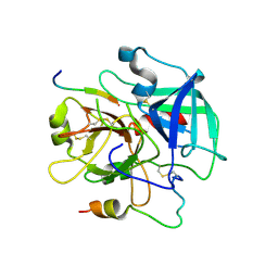
3M12
 | |
3M0O
 | |
3M13
 | | Crystal Structure of the Lys265Arg PEG-crystallized mutant of monomeric sarcosine oxidase | | 分子名称: | CHLORIDE ION, FLAVIN-ADENINE DINUCLEOTIDE, Monomeric sarcosine oxidase, ... | | 著者 | Mathews, F.S, Chen, Z.-W, Jorns, M.S. | | 登録日 | 2010-03-04 | | 公開日 | 2010-04-21 | | 最終更新日 | 2024-02-21 | | 実験手法 | X-RAY DIFFRACTION (2.1 Å) | | 主引用文献 | Structural characterization of mutations at the oxygen activation site in monomeric sarcosine oxidase.
Biochemistry, 49, 2010
|
|
3QSE
 | | Crystal structure for the complex of substrate-reduced msox with sarcosine | | 分子名称: | CHLORIDE ION, FLAVIN-ADENINE DINUCLEOTIDE, Monomeric sarcosine oxidase, ... | | 著者 | Kommoju, P, Chen, Z, Bruckner, R.C, Mathews, F.S, Jorns, M.S. | | 登録日 | 2011-02-21 | | 公開日 | 2011-06-01 | | 最終更新日 | 2023-09-13 | | 実験手法 | X-RAY DIFFRACTION (1.75 Å) | | 主引用文献 | Probing oxygen activation sites in two flavoprotein oxidases using chloride as an oxygen surrogate.
Biochemistry, 50, 2011
|
|
3QSM
 | | Crystal structure for the MSOX.chloride binary complex | | 分子名称: | CHLORIDE ION, FLAVIN-ADENINE DINUCLEOTIDE, Monomeric sarcosine oxidase | | 著者 | Kommoju, P, Chen, Z, Bruckner, R.C, Mathews, F.S, Jorns, M.S. | | 登録日 | 2011-02-21 | | 公開日 | 2011-06-01 | | 最終更新日 | 2023-09-13 | | 実験手法 | X-RAY DIFFRACTION (1.9 Å) | | 主引用文献 | Probing oxygen activation sites in two flavoprotein oxidases using chloride as an oxygen surrogate.
Biochemistry, 50, 2011
|
|
3QVR
 | | Crystal structure of glucose oxidase for space group P3121 at 1.3 A resolution. | | 分子名称: | 2-acetamido-2-deoxy-beta-D-glucopyranose, CHLORIDE ION, FLAVIN-ADENINE DINUCLEOTIDE, ... | | 著者 | Kommoju, P, Chen, Z, Bruckner, R.C, Mathews, F.S, Jorns, M.S. | | 登録日 | 2011-02-25 | | 公開日 | 2011-06-01 | | 最終更新日 | 2023-09-13 | | 実験手法 | X-RAY DIFFRACTION (1.3 Å) | | 主引用文献 | Probing oxygen activation sites in two flavoprotein oxidases using chloride as an oxygen surrogate.
Biochemistry, 50, 2011
|
|
3QSS
 | | Crystal structure for the MSOX.chloride.MTA ternary complex | | 分子名称: | CHLORIDE ION, FLAVIN-ADENINE DINUCLEOTIDE, Monomeric sarcosine oxidase, ... | | 著者 | Kommoju, P, Chen, Z, Bruckner, R.C, Mathews, F.S, Jorns, M.S. | | 登録日 | 2011-02-21 | | 公開日 | 2011-06-01 | | 最終更新日 | 2023-09-13 | | 実験手法 | X-RAY DIFFRACTION (1.85 Å) | | 主引用文献 | Probing oxygen activation sites in two flavoprotein oxidases using chloride as an oxygen surrogate.
Biochemistry, 50, 2011
|
|
3QVP
 | | Crystal structure of glucose oxidase for space group C2221 at 1.2 A resolution | | 分子名称: | 2-acetamido-2-deoxy-beta-D-glucopyranose, CHLORIDE ION, DI(HYDROXYETHYL)ETHER, ... | | 著者 | Kommoju, P, Chen, Z, Bruckner, R.C, Mathews, F.S, Jorns, M.S. | | 登録日 | 2011-02-25 | | 公開日 | 2011-06-01 | | 最終更新日 | 2023-09-13 | | 実験手法 | X-RAY DIFFRACTION (1.2 Å) | | 主引用文献 | Probing oxygen activation sites in two flavoprotein oxidases using chloride as an oxygen surrogate.
Biochemistry, 50, 2011
|
|
3R3G
 | | Structure of human thrombin with residues 145-150 of murine thrombin. | | 分子名称: | 2-acetamido-2-deoxy-beta-D-glucopyranose, SODIUM ION, Thrombin Heavy Chain, ... | | 著者 | Pozzi, N, Chen, R, Chen, Z, Bah, A, Di Cera, E. | | 登録日 | 2011-03-15 | | 公開日 | 2011-05-11 | | 最終更新日 | 2023-09-13 | | 実験手法 | X-RAY DIFFRACTION (1.75 Å) | | 主引用文献 | Rigidification of the autolysis loop enhances Na(+) binding to thrombin.
Biophys.Chem., 159, 2011
|
|
3QDZ
 | | Crystal structure of the human thrombin mutant D102N in complex with the extracellular fragment of human PAR4. | | 分子名称: | Proteinase-activated receptor 4, Thrombin heavy chain, Thrombin light chain | | 著者 | Gandhi, P, Chen, Z, Appelbaum, E, Zapata, F, Di Cera, E. | | 登録日 | 2011-01-19 | | 公開日 | 2011-06-29 | | 最終更新日 | 2023-09-13 | | 実験手法 | X-RAY DIFFRACTION (2.8 Å) | | 主引用文献 | Structural basis of thrombin-protease-receptor interactions
IUBMB LIFE, 63, 2011
|
|


