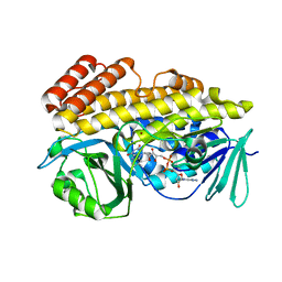
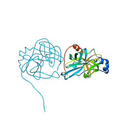
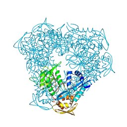
6B3T
 
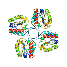
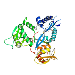 | | Crystal structure of acetyltransferase Eis from Mycobacterium tuberculosis in complex with a 1,2,4-triazino[5,6b]indole-3-thioether inhibitor analogue 39b | | 分子名称: | 8-fluoro-5-methyl-3-{[2-(piperidin-1-yl)ethyl]sulfanyl}-5H-[1,2,4]triazino[5,6-b]indole, COENZYME A, N-acetyltransferase Eis, ... | | 著者 | Gajadeera, C.S, Hou, C, Garneau-Tsodikova, S, Ngo, H.X, Tsodikov, O.V. | | 登録日 | 2017-09-24 | | 公開日 | 2018-04-04 | | 最終更新日 | 2023-10-04 | | 実験手法 | X-RAY DIFFRACTION (2.4 Å) | | 主引用文献 | Potent 1,2,4-Triazino[5,6 b]indole-3-thioether Inhibitors of the Kanamycin Resistance Enzyme Eis from Mycobacterium tuberculosis.
ACS Infect Dis, 4, 2018
|
|
6BZZ
 | |
6BZN
 | |
6BZI
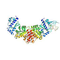
 | | Crystal structure of halogenase PltM in complex with ethyl mercury and mercury | | 分子名称: | CALCIUM ION, ETHYL MERCURY ION, GLYCEROL, ... | | 著者 | Pang, A.H, Garneau-Tsodikova, S, Tsodikov, O.V. | | 登録日 | 2017-12-24 | | 公開日 | 2019-03-20 | | 最終更新日 | 2024-03-13 | | 実験手法 | X-RAY DIFFRACTION (2.4 Å) | | 主引用文献 | Unusual substrate and halide versatility of phenolic halogenase PltM.
Nat Commun, 10, 2019
|
|
6BZQ
 | | Crystal structure of halogenase PltM in complex with FAD | | 分子名称: | BROMIDE ION, CHLORIDE ION, FLAVIN-ADENINE DINUCLEOTIDE, ... | | 著者 | Pang, A.H, Garneau-Tsodikova, S, Tsodikov, O.V. | | 登録日 | 2017-12-25 | | 公開日 | 2019-03-20 | | 最終更新日 | 2023-10-04 | | 実験手法 | X-RAY DIFFRACTION (2.75 Å) | | 主引用文献 | Unusual substrate and halide versatility of phenolic halogenase PltM.
Nat Commun, 10, 2019
|
|
6BZA
 | | Crystal structure of halogenase PltM in complex with phloroglucinol and FAD | | 分子名称: | CHLORIDE ION, FLAVIN-ADENINE DINUCLEOTIDE, Halogenase PltM, ... | | 著者 | Pang, A.H, Garneau-Tsodikova, S, Tsodikov, O.V. | | 登録日 | 2017-12-22 | | 公開日 | 2019-03-20 | | 最終更新日 | 2024-03-13 | | 実験手法 | X-RAY DIFFRACTION (2.6 Å) | | 主引用文献 | Unusual substrate and halide versatility of phenolic halogenase PltM.
Nat Commun, 10, 2019
|
|
6BZT
 | | Crystal structure of halogenase PltM L111Y mutant in complex with FAD | | 分子名称: | BROMIDE ION, CALCIUM ION, CHLORIDE ION, ... | | 著者 | Pang, A.H, Garneau-Tsodikova, S, Tsodikov, O.V. | | 登録日 | 2017-12-26 | | 公開日 | 2019-03-20 | | 最終更新日 | 2023-10-04 | | 実験手法 | X-RAY DIFFRACTION (2.1 Å) | | 主引用文献 | Unusual substrate and halide versatility of phenolic halogenase PltM.
Nat Commun, 10, 2019
|
|
6DXJ
 | |
6DY5
 | |
8D1R
 | | Crystal structure of acetyltransferase Eis from Mycobacterium tuberculosis in complex with inhibitor SGT520 | | 分子名称: | 1-{3-[(4-chlorophenyl)methoxy]phenyl}methanamine, GLYCEROL, N-acetyltransferase Eis | | 著者 | Pang, A.H, Punetha, A, Garneau-Tsodikova, S, Tsodikov, O.V. | | 登録日 | 2022-05-27 | | 公開日 | 2022-09-14 | | 最終更新日 | 2023-10-18 | | 実験手法 | X-RAY DIFFRACTION (2.19 Å) | | 主引用文献 | Discovery of substituted benzyloxy-benzylamine inhibitors of acetyltransferase Eis and their anti-mycobacterial activity.
Eur.J.Med.Chem., 242, 2022
|
|
8D25
 | | Crystal structure of acetyltransferase Eis from Mycobacterium tuberculosis in complex with inhibitor SGT530 | | 分子名称: | 1-{2-[(3-chlorophenyl)methoxy]phenyl}-N-[(pyridin-4-yl)methyl]methanamine, GLYCEROL, N-acetyltransferase Eis | | 著者 | Pang, A.H, Punetha, A, Garneau-Tsodikova, S, Tsodikov, O.V. | | 登録日 | 2022-05-27 | | 公開日 | 2022-09-14 | | 最終更新日 | 2023-10-18 | | 実験手法 | X-RAY DIFFRACTION (2.05 Å) | | 主引用文献 | Discovery of substituted benzyloxy-benzylamine inhibitors of acetyltransferase Eis and their anti-mycobacterial activity.
Eur.J.Med.Chem., 242, 2022
|
|
8D23
 | | Crystal structure of acetyltransferase Eis from Mycobacterium tuberculosis in complex with inhibitor SGT529 | | 分子名称: | 1-{2-[(3-chlorophenyl)methoxy]phenyl}-N-[(pyridin-3-yl)methyl]methanamine, GLYCEROL, N-acetyltransferase Eis | | 著者 | Pang, A.H, Punetha, A, Garneau-Tsodikova, S, Tsodikov, O.V. | | 登録日 | 2022-05-27 | | 公開日 | 2022-09-14 | | 最終更新日 | 2023-10-18 | | 実験手法 | X-RAY DIFFRACTION (2.14 Å) | | 主引用文献 | Discovery of substituted benzyloxy-benzylamine inhibitors of acetyltransferase Eis and their anti-mycobacterial activity.
Eur.J.Med.Chem., 242, 2022
|
|
6DY9
 | |
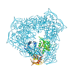
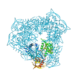
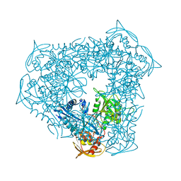
3K6Z
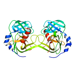 | | Crystal structure of Rv3671c protease, inactive form | | 分子名称: | POSSIBLE MEMBRANE-ASSOCIATED SERINE PROTEASE | | 著者 | Biswas, T, Small, J, Vandal, O, Ehrt, S, Tsodikov, O.V. | | 登録日 | 2009-10-10 | | 公開日 | 2010-10-13 | | 最終更新日 | 2023-09-06 | | 実験手法 | X-RAY DIFFRACTION (1.75 Å) | | 主引用文献 | Structural insight into serine protease Rv3671c that Protects M. tuberculosis from oxidative and acidic stress.
Structure, 18, 2010
|
|
3I6B
 | |
5W34
 | |
3K6Y
 | | Crystal structure of Rv3671c protease from M. tuberculosis, active form | | 分子名称: | POSSIBLE MEMBRANE-ASSOCIATED SERINE PROTEASE | | 著者 | Biswas, T, Small, J, Vandal, O, Ehrt, S, Tsodikov, O.V. | | 登録日 | 2009-10-10 | | 公開日 | 2010-10-13 | | 最終更新日 | 2023-09-06 | | 実験手法 | X-RAY DIFFRACTION (1.3 Å) | | 主引用文献 | Structural insight into serine protease Rv3671c that Protects M. tuberculosis from oxidative and acidic stress.
Structure, 18, 2010
|
|
5W33
 | |
5W35
 | |
5W36
 | |
6P3T
 | | Crystal structure of Eis from Mycobacterium tuberculosis in complex with inhibitor SGT449 | | 分子名称: | AMMONIUM ION, DI(HYDROXYETHYL)ETHER, DIMETHYL SULFOXIDE, ... | | 著者 | Punetha, A, Garneau-Tsodikova, S, Tsodikov, O.V. | | 登録日 | 2019-05-24 | | 公開日 | 2019-09-04 | | 最終更新日 | 2023-10-11 | | 実験手法 | X-RAY DIFFRACTION (2.5 Å) | | 主引用文献 | Probing the Robustness of Inhibitors of Tuberculosis Aminoglycoside Resistance Enzyme Eis by Mutagenesis.
Acs Infect Dis., 5, 2019
|
|
6P3V
 | | Crystal structure of Eis from Mycobacterium tuberculosis in complex with inhibitor SGT416 | | 分子名称: | DIMETHYL SULFOXIDE, N,N-diethyl-2-[(8-fluoro-5-methyl-5H-[1,2,4]triazino[5,6-b]indol-3-yl)sulfanyl]ethan-1-amine, N-acetyltransferase Eis, ... | | 著者 | Punetha, A, Garneau-Tsodikova, S, Tsodikov, O.V. | | 登録日 | 2019-05-24 | | 公開日 | 2019-09-04 | | 最終更新日 | 2023-10-11 | | 実験手法 | X-RAY DIFFRACTION (2.5 Å) | | 主引用文献 | Probing the Robustness of Inhibitors of Tuberculosis Aminoglycoside Resistance Enzyme Eis by Mutagenesis.
Acs Infect Dis., 5, 2019
|
|
6P3U
 | | Crystal structure of Eis from Mycobacterium tuberculosis in complex with inhibitor SGT335 | | 分子名称: | 1-(4-fluorophenyl)-2-[2-(4-fluorophenyl)-2-oxoethyl]pyrrolo[1,2-a]pyrazin-2-ium, DI(HYDROXYETHYL)ETHER, GLYCEROL, ... | | 著者 | Punetha, A, Garneau-Tsodikova, S, Tsodikov, O.V. | | 登録日 | 2019-05-24 | | 公開日 | 2019-09-04 | | 最終更新日 | 2023-10-11 | | 実験手法 | X-RAY DIFFRACTION (2.55 Å) | | 主引用文献 | Probing the Robustness of Inhibitors of Tuberculosis Aminoglycoside Resistance Enzyme Eis by Mutagenesis.
Acs Infect Dis., 5, 2019
|
|
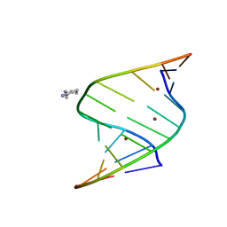
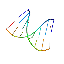
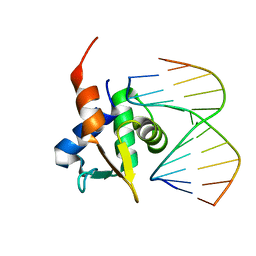
5JW2
 | | Crystal structure of mithramycin analogue MTM SA-Phe in complex with a 10-mer DNA AGGGATCCCT | | 分子名称: | DNA (5'-D(*AP*GP*GP*GP*AP*TP*CP*CP*CP*T)-3'), Plicamycin, mithramycin analogue MTM SA-Phe, ... | | 著者 | Hou, C, Rohr, J, Tsodikov, O.V. | | 登録日 | 2016-05-11 | | 公開日 | 2016-09-14 | | 最終更新日 | 2024-03-06 | | 実験手法 | X-RAY DIFFRACTION (3.1 Å) | | 主引用文献 | Structures of mithramycin analogues bound to DNA and implications for targeting transcription factor FLI1.
Nucleic Acids Res., 44, 2016
|
|
5JVT
 | |


