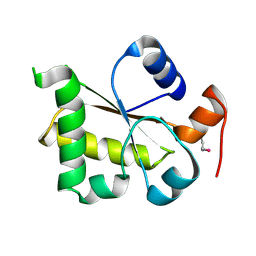
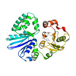
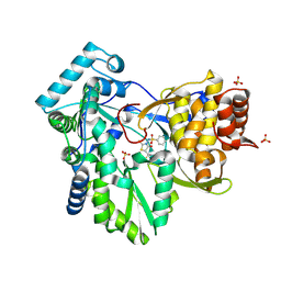
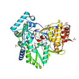
3HXT
 
 | | Structure of human MTHFS | | 分子名称: | 5-formyltetrahydrofolate cyclo-ligase, MAGNESIUM ION, NICKEL (II) ION | | 著者 | Wu, D, Li, Y, Song, G, Cheng, C, Zhang, R, Joachimiak, A, Shaw, N, Liu, Z.-J. | | 登録日 | 2009-06-22 | | 公開日 | 2009-07-14 | | 最終更新日 | 2023-11-01 | | 実験手法 | X-RAY DIFFRACTION (1.9 Å) | | 主引用文献 | Structural basis for the inhibition of human 5,10-methenyltetrahydrofolate synthetase by N10-substituted folate analogues
Cancer Res., 69, 2009
|
|
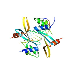
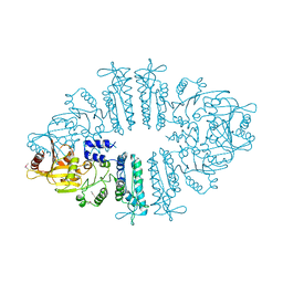
3I2V
 | | Crystal structure of human MOCS3 rhodanese-like domain | | 分子名称: | Adenylyltransferase and sulfurtransferase MOCS3 | | 著者 | Bacik, J.P, Walker, J.R, Lopez, L, Li, Y, Weigelt, J, Bountra, C, Arrowsmith, C.H, Edwards, A.M, Bochkarev, A, Dhe-Paganon, S, Structural Genomics Consortium (SGC) | | 登録日 | 2009-06-29 | | 公開日 | 2009-07-21 | | 最終更新日 | 2011-07-13 | | 実験手法 | X-RAY DIFFRACTION (1.25 Å) | | 主引用文献 | Crystal structure of the human MOCS3 rhodanese-like domain
To be Published
|
|
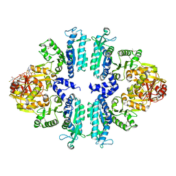
7MWL
 | | The TAM domain of BAZ2A in complex with a 12mer mCG DNA | | 分子名称: | Bromodomain adjacent to zinc finger domain protein 2A, DNA (5'-D(*GP*CP*CP*AP*AP*(5CM)P*GP*TP*TP*GP*GP*C)-3'), GLYCEROL | | 著者 | Liu, K, Dong, A, Li, Y, Loppnau, P, Edwards, A.M, Arrowsmith, C.H, Min, J, Structural Genomics Consortium (SGC) | | 登録日 | 2021-05-17 | | 公開日 | 2021-07-21 | | 最終更新日 | 2023-10-18 | | 実験手法 | X-RAY DIFFRACTION (1.84 Å) | | 主引用文献 | The TAM domain of BAZ2A in complex with a 12mer mCG DNA
To Be Published
|
|
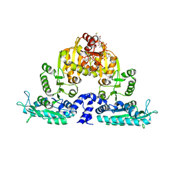
7MIS
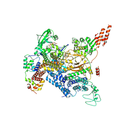 | | Cryo-EM structure of SidJ-SdeC-CaM reaction intermediate complex | | 分子名称: | ADENOSINE MONOPHOSPHATE, ADENOSINE-5'-TRIPHOSPHATE, CALCIUM ION, ... | | 著者 | Osinski, A, Black, M.H, Pawlowski, K, Chen, Z, Li, Y, Tagliabracci, V.S. | | 登録日 | 2021-04-17 | | 公開日 | 2021-08-18 | | 最終更新日 | 2021-11-17 | | 実験手法 | ELECTRON MICROSCOPY (2.8 Å) | | 主引用文献 | Structural and mechanistic basis for protein glutamylation by the kinase fold.
Mol.Cell, 81, 2021
|
|
7MIR
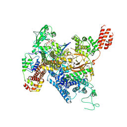 | | Cryo-EM structure of SidJ-SdeA-CaM reaction intermediate complex | | 分子名称: | ADENOSINE MONOPHOSPHATE, ADENOSINE-5'-TRIPHOSPHATE, CALCIUM ION, ... | | 著者 | Osinski, A, Black, M.H, Pawlowski, K, Chen, Z, Li, Y, Tagliabracci, V.S. | | 登録日 | 2021-04-17 | | 公開日 | 2021-08-18 | | 最終更新日 | 2024-05-29 | | 実験手法 | ELECTRON MICROSCOPY (2.5 Å) | | 主引用文献 | Structural and mechanistic basis for protein glutamylation by the kinase fold.
Mol.Cell, 81, 2021
|
|
3GV4
 | | Crystal structure of human HDAC6 zinc finger domain and ubiquitin C-terminal peptide RLRGG | | 分子名称: | CALCIUM ION, Histone deacetylase 6, ZINC ION, ... | | 著者 | Dong, A, Ravichandran, M, Loppnau, P, Li, Y, MacKenzie, F, Kozieradzki, I, Edwards, A.M, Arrowsmith, C.H, Weigelt, J, Bountra, C, Bochkarev, A, Dhe-Paganon, S, Min, J, Ouyang, H, Structural Genomics Consortium (SGC) | | 登録日 | 2009-03-30 | | 公開日 | 2009-04-28 | | 最終更新日 | 2023-09-06 | | 実験手法 | X-RAY DIFFRACTION (1.72 Å) | | 主引用文献 | Crystal structure of human HDAC6 zinc finger domain and ubiquitin C-terminal peptide RLRGG
To be Published
|
|
3GUC
 | | Human Ubiquitin-activating Enzyme 5 in Complex with AMPPNP | | 分子名称: | PHOSPHOAMINOPHOSPHONIC ACID-ADENYLATE ESTER, Ubiquitin-like modifier-activating enzyme 5, ZINC ION | | 著者 | Walker, J.R, Bacik, J.P, Li, Y, Weigelt, J, Bountra, C, Arrowsmith, C.H, Edwards, A.M, Bochkarev, A, Dhe-Paganon, S, Structural Genomics Consortium (SGC) | | 登録日 | 2009-03-29 | | 公開日 | 2009-05-26 | | 最終更新日 | 2023-09-06 | | 実験手法 | X-RAY DIFFRACTION (2.25 Å) | | 主引用文献 | Human Ubiquitin-activating Enzyme 5 in Complex with AMPPNP
To be Published
|
|
3HCX
 | | Crystal structure of E. coli HPPK(N10A) | | 分子名称: | 2-AMINO-2-HYDROXYMETHYL-PROPANE-1,3-DIOL, 2-amino-4-hydroxy-6-hydroxymethyldihydropteridine pyrophosphokinase, CHLORIDE ION | | 著者 | Blaszczyk, J, Li, Y, Yan, H, Ji, X. | | 登録日 | 2009-05-06 | | 公開日 | 2010-05-19 | | 最終更新日 | 2023-09-06 | | 実験手法 | X-RAY DIFFRACTION (1.75 Å) | | 主引用文献 | Role of loop coupling in enzymatic catalysis and conformational dynamics
To be Published
|
|
3HD2
 | | Crystal structure of E. coli HPPK(Q50A) in complex with MgAMPCPP and pterin | | 分子名称: | 2-amino-4-hydroxy-6-hydroxymethyldihydropteridine pyrophosphokinase, ACETATE ION, CHLORIDE ION, ... | | 著者 | Blaszczyk, J, Li, Y, Yan, H, Ji, X. | | 登録日 | 2009-05-06 | | 公開日 | 2010-05-19 | | 最終更新日 | 2023-09-06 | | 実験手法 | X-RAY DIFFRACTION (1.1 Å) | | 主引用文献 | Role of loop coupling in enzymatic catalysis and conformational dynamics
To be Published
|
|
3HD1
 | | Crystal structure of E. coli HPPK(N10A) in complex with MgAMPCPP | | 分子名称: | 2-amino-4-hydroxy-6-hydroxymethyldihydropteridine pyrophosphokinase, ACETATE ION, CHLORIDE ION, ... | | 著者 | Blaszczyk, J, Li, Y, Yan, H, Ji, X. | | 登録日 | 2009-05-06 | | 公開日 | 2010-05-19 | | 最終更新日 | 2023-09-06 | | 実験手法 | X-RAY DIFFRACTION (1.3 Å) | | 主引用文献 | Role of loop coupling in enzymatic catalysis and conformational dynamics
To be Published
|
|
3FCX
 | | Crystal structure of human esterase D | | 分子名称: | CALCIUM ION, MAGNESIUM ION, S-formylglutathione hydrolase | | 著者 | Wu, D, Li, Y, Song, G, Zhang, D, Shaw, N, Liu, Z.J. | | 登録日 | 2008-11-24 | | 公開日 | 2008-12-09 | | 最終更新日 | 2023-11-01 | | 実験手法 | X-RAY DIFFRACTION (1.5 Å) | | 主引用文献 | Crystal structure of human esterase D: a potential genetic marker of retinoblastoma
Faseb J., 23, 2009
|
|
3FL2
 | | Crystal structure of the ring domain of the E3 ubiquitin-protein ligase UHRF1 | | 分子名称: | E3 ubiquitin-protein ligase UHRF1, ZINC ION | | 著者 | Walker, J.R, Avvakumov, G.V, Xue, S, Li, Y, Bountra, C, Weigelt, J, Arrowsmith, C.H, Edwards, A.M, Bochkarev, A, Dhe-Paganon, S, Structural Genomics Consortium (SGC) | | 登録日 | 2008-12-18 | | 公開日 | 2009-01-20 | | 最終更新日 | 2023-09-06 | | 実験手法 | X-RAY DIFFRACTION (1.75 Å) | | 主引用文献 | Structure of the Ring Domain of the E3 Ubiquitin-Protein Ligase Uhrf1
To be Published
|
|
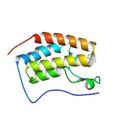
3IV1
 | | Coiled-coil domain of tumor susceptibility gene 101 | | 分子名称: | CHLORIDE ION, SULFATE ION, Tumor susceptibility gene 101 protein | | 著者 | Neculai, D, Avvakumov, G.V, Wernimont, A.K, Xue, S, Walker, J.R, Li, Y, Weigelt, J, Bountra, C, Arrowsmith, C.H, Edwards, A.M, Bochkarev, A, Dhe-Paganon, S, Structural Genomics Consortium (SGC) | | 登録日 | 2009-08-31 | | 公開日 | 2009-11-03 | | 最終更新日 | 2011-07-13 | | 実験手法 | X-RAY DIFFRACTION (2.5 Å) | | 主引用文献 | Coiled-Coil Domain of Human Tsg101
To be Published
|
|
3S44
 | | Crystal Structure of Pasteurella multocida sialyltransferase M144D mutant with CMP bound | | 分子名称: | Alpha-2,3/2,6-sialyltransferase/sialidase, CMP-3F(a)-Neu5Ac | | 著者 | Sugiarto, G, Lau, K, Li, Y, Lim, S, Ames, J.B, Le, D.-T, Fisher, A.J, Chen, X. | | 登録日 | 2011-05-18 | | 公開日 | 2012-08-15 | | 最終更新日 | 2023-09-13 | | 実験手法 | X-RAY DIFFRACTION (1.45 Å) | | 主引用文献 | A Sialyltransferase Mutant with Decreased Donor Hydrolysis and Reduced Sialidase Activities for Directly Sialylating Lewis(x).
Acs Chem.Biol., 7, 2012
|
|
3S6K
 | | Crystal structure of xcNAGS | | 分子名称: | Acetylglutamate kinase | | 著者 | Shi, D, Li, Y, Cabrera-Luque, J, Jin, Z, Yu, X, Allewell, N.M, Tuchman, M. | | 登録日 | 2011-05-25 | | 公開日 | 2012-04-18 | | 実験手法 | X-RAY DIFFRACTION (2.8018 Å) | | 主引用文献 | A Novel N-acetylglutamate synthase architecture revealed by the crystal structure of the bifunctional enzyme from Maricaulis maris.
Plos One, 6, 2011
|
|
3S6G
 | | Crystal structures of Seleno-substituted mutant mmNAGS in space group P212121 | | 分子名称: | 1,2-ETHANEDIOL, COENZYME A, MALONATE ION, ... | | 著者 | Shi, D, Li, Y, Cabrera-Luque, J, Jin, Z, Yu, X, Allewell, N.M, Tuchman, M. | | 登録日 | 2011-05-25 | | 公開日 | 2012-04-18 | | 最終更新日 | 2017-11-08 | | 実験手法 | X-RAY DIFFRACTION (2.6681 Å) | | 主引用文献 | A Novel N-acetylglutamate synthase architecture revealed by the crystal structure of the bifunctional enzyme from Maricaulis maris.
Plos One, 6, 2011
|
|
3S6H
 | | Crystal structure of native mmNAGS/k | | 分子名称: | COENZYME A, GLUTAMIC ACID, N-acetylglutamate kinase / N-acetylglutamate synthase | | 著者 | Shi, D, Li, Y, Cabrera-Luque, J, Jin, Z, Yu, X, Allewell, N.M, Tuchman, M. | | 登録日 | 2011-05-25 | | 公開日 | 2012-04-25 | | 最終更新日 | 2023-09-13 | | 実験手法 | X-RAY DIFFRACTION (3.102 Å) | | 主引用文献 | A Novel N-acetylglutamate synthase architecture revealed by the crystal structure of the bifunctional enzyme from Maricaulis maris.
Plos One, 6, 2011
|
|
2AHV
 | | Crystal Structure of Acyl-CoA transferase from E. coli O157:H7 (YdiF)-thioester complex with CoA- 1 | | 分子名称: | COENZYME A, putative enzyme YdiF | | 著者 | Rangarajan, E.S, Li, Y, Ajamian, E, Iannuzzi, P, Kernaghan, S.D, Fraser, M.E, Cygler, M, Matte, A, Montreal-Kingston Bacterial Structural Genomics Initiative (BSGI) | | 登録日 | 2005-07-28 | | 公開日 | 2005-11-01 | | 最終更新日 | 2023-08-23 | | 実験手法 | X-RAY DIFFRACTION (2 Å) | | 主引用文献 | Crystallographic trapping of the glutamyl-CoA thioester intermediate of family I CoA transferases.
J.Biol.Chem., 280, 2005
|
|
2FD6
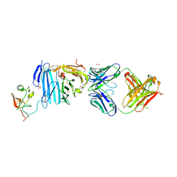 | | Structure of Human Urokinase Plasminogen Activator in Complex with Urokinase Receptor and an anti-upar antibody at 1.9 A | | 分子名称: | 1,2-ETHANEDIOL, 2-ETHOXYETHANOL, 2-acetamido-2-deoxy-alpha-D-glucopyranose, ... | | 著者 | Huang, M, Huai, Q, Li, Y. | | 登録日 | 2005-12-13 | | 公開日 | 2006-02-21 | | 最終更新日 | 2023-08-30 | | 実験手法 | X-RAY DIFFRACTION (1.9 Å) | | 主引用文献 | Structure of human urokinase plasminogen activator in complex with its receptor
Science, 311, 2006
|
|
2AX0
 | | Hepatitis C Virus NS5b RNA Polymerase in complex with a covalent inhibitor (5x) | | 分子名称: | 5R-(2E-METHYL-3-PHENYL-ALLYL)-3-(BENZENESULFONYLAMINO)-4-OXO-2-THIONOTHIAZOLIDINE, Genome polyprotein, SULFATE ION | | 著者 | Powers, J.P, Piper, D.E, Li, Y, Mayorga, V, Anzola, J, Chen, J.M, Jaen, J.C, Lee, G, Liu, J, Peterson, M.G, Tonn, G.R, Ye, Q, Walker, N.P, Wang, Z. | | 登録日 | 2005-09-02 | | 公開日 | 2006-01-24 | | 最終更新日 | 2021-10-20 | | 実験手法 | X-RAY DIFFRACTION (2 Å) | | 主引用文献 | SAR and Mode of Action of Novel Non-Nucleoside Inhibitors of Hepatitis C NS5b RNA Polymerase.
J.Med.Chem., 49, 2006
|
|
2AWZ
 | | Hepatitis C Virus NS5b RNA Polymerase in complex with a covalent inhibitor (5h) | | 分子名称: | 5R-(4-BROMOPHENYLMETHYL)-3-(BENZENESULFONYLAMINO)-4-OXO-2-THIONOTHIAZOLIDINE, Genome polyprotein, SULFATE ION | | 著者 | Powers, J.P, Piper, D.E, Li, Y, Mayorga, V, Anzola, J, Chen, J.M, Jaen, J.C, Lee, G, Liu, J, Peterson, M.G, Tonn, G.R, Ye, Q, Walker, N.P, Wang, Z. | | 登録日 | 2005-09-02 | | 公開日 | 2006-01-24 | | 最終更新日 | 2021-10-20 | | 実験手法 | X-RAY DIFFRACTION (2.15 Å) | | 主引用文献 | SAR and Mode of Action of Novel Non-Nucleoside Inhibitors of Hepatitis C NS5b RNA Polymerase.
J.Med.Chem., 49, 2006
|
|
5YOV
 | | Crystal structure of BRD4-BD1 bound with hjp126 | | 分子名称: | (3~{R})-4-cyclopentyl-~{N}-(2,4-dimethylphenyl)-1,3-dimethyl-2-oxidanylidene-3~{H}-quinoxaline-6-carboxamide, Bromodomain-containing protein 4 | | 著者 | Xiong, B, Hu, J, Li, Y, Cao, D. | | 登録日 | 2017-10-31 | | 公開日 | 2018-11-07 | | 最終更新日 | 2024-03-27 | | 実験手法 | X-RAY DIFFRACTION (1.451 Å) | | 主引用文献 | Structure-based optimization of a series of selective BET inhibitors containing aniline or indoline groups.
Eur.J.Med.Chem., 150, 2018
|
|
3S4Y
 | | Crystal structure of human thiamin pyrophosphokinase 1 | | 分子名称: | CALCIUM ION, SULFATE ION, THIAMINE DIPHOSPHATE, ... | | 著者 | Shen, L, Tempel, W, Tong, Y, Li, Y, Walker, J.R, Arrowsmith, C.H, Edwards, A.M, Bountra, C, Weigelt, J, Park, H, Structural Genomics Consortium (SGC) | | 登録日 | 2011-05-20 | | 公開日 | 2011-06-08 | | 最終更新日 | 2024-02-28 | | 実験手法 | X-RAY DIFFRACTION (1.8 Å) | | 主引用文献 | Crystal structure of human thiamin pyrophosphokinase 1
to be published
|
|
7FI9
 | | Crystal structure of human MICA mutants in complex with natural killer cell receptor NKG2D | | 分子名称: | GLYCEROL, MHC class I polypeptide-related sequence A, NKG2-D type II integral membrane protein | | 著者 | Cai, W, Peng, S, Xu, T, Tian, Y, Li, Y, Liu, J. | | 登録日 | 2021-07-30 | | 公開日 | 2022-08-31 | | 最終更新日 | 2023-11-29 | | 実験手法 | X-RAY DIFFRACTION (2.16 Å) | | 主引用文献 | Crystal structure of human MICA mutants in complex with natural killer cell receptor NKG2D
to be published
|
|
7FI7
 | | Crystal structure of human MICA mutants in complex with natural killer cell receptor NKG2D | | 分子名称: | MHC class I polypeptide-related sequence A, NKG2-D type II integral membrane protein | | 著者 | Cai, W, Peng, S, Xu, T, Tian, Y, Li, Y, Liu, J. | | 登録日 | 2021-07-30 | | 公開日 | 2022-08-31 | | 最終更新日 | 2023-11-29 | | 実験手法 | X-RAY DIFFRACTION (2.78 Å) | | 主引用文献 | Crystal structure of human MICA mutants in complex with natural killer cell receptor NKG2D
to be published
|
|


