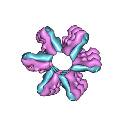
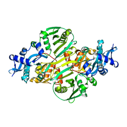
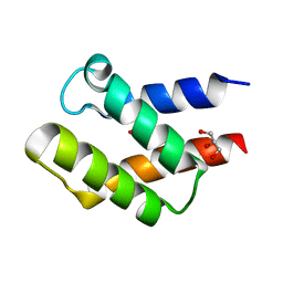
1S5H
 
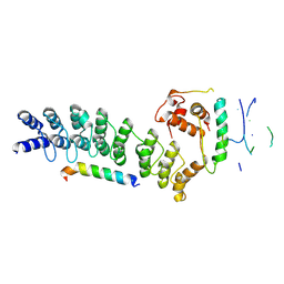
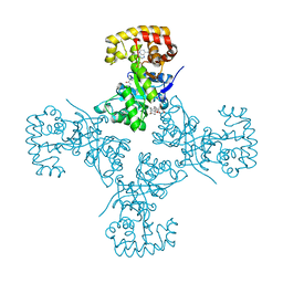
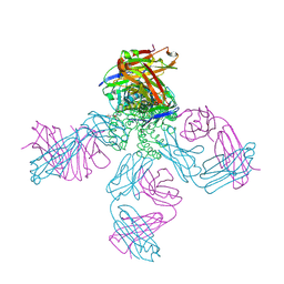 | | Potassium Channel Kcsa-Fab Complex T75C mutant in K+ | | 分子名称: | ANTIBODY FAB FRAGMENT HEAVY CHAIN, ANTIBODY FAB FRAGMENT LIGHT CHAIN, DIACYL GLYCEROL, ... | | 著者 | Mackinnon, R, Zhou, M. | | 登録日 | 2004-01-20 | | 公開日 | 2004-05-18 | | 最終更新日 | 2024-11-20 | | 実験手法 | X-RAY DIFFRACTION (2.2 Å) | | 主引用文献 | A mutant KcsA K(+) channel with altered conduction properties and selectivity filter ion distribution.
J.Mol.Biol., 338, 2004
|
|
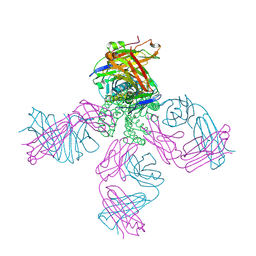
6V4L
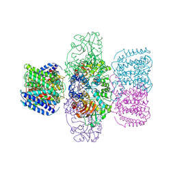 | | Structure of TrkH-TrkA in complex with ATPgammaS | | 分子名称: | PHOSPHOTHIOPHOSPHORIC ACID-ADENYLATE ESTER, Potassium uptake protein TrkA, Trk system potassium uptake protein TrkH | | 著者 | Zhou, M, Zhang, H. | | 登録日 | 2019-11-27 | | 公開日 | 2020-02-12 | | 最終更新日 | 2024-11-06 | | 実験手法 | X-RAY DIFFRACTION (3.8 Å) | | 主引用文献 | TrkA undergoes a tetramer-to-dimer conversion to open TrkH which enables changes in membrane potential.
Nat Commun, 11, 2020
|
|
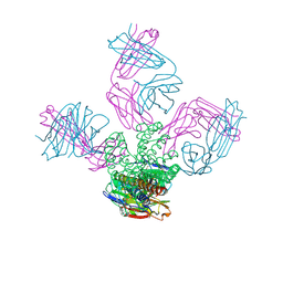
6V4J
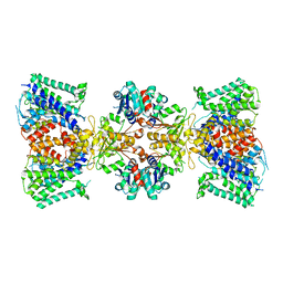 | | Structure of TrkH-TrkA in complex with ATP | | 分子名称: | Potassium uptake protein TrkA, Trk system potassium uptake protein TrkH | | 著者 | Zhou, M, Zhang, H. | | 登録日 | 2019-11-27 | | 公開日 | 2020-02-12 | | 最終更新日 | 2025-05-28 | | 実験手法 | ELECTRON MICROSCOPY (2.97 Å) | | 主引用文献 | TrkA undergoes a tetramer-to-dimer conversion to open TrkH which enables changes in membrane potential.
Nat Commun, 11, 2020
|
|
6V4K
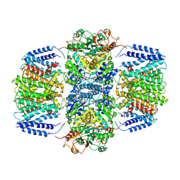 | | Structure of TrkH-TrkA in complex with ADP | | 分子名称: | ADENOSINE-5'-DIPHOSPHATE, Potassium transporter peripheral membrane component, Trk system potassium uptake protein | | 著者 | Zhou, M, Zhang, H. | | 登録日 | 2019-11-27 | | 公開日 | 2020-02-12 | | 最終更新日 | 2023-10-11 | | 実験手法 | X-RAY DIFFRACTION (3.53004146 Å) | | 主引用文献 | TrkA undergoes a tetramer-to-dimer conversion to open TrkH which enables changes in membrane potential.
Nat Commun, 11, 2020
|
|
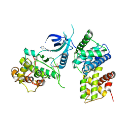
8Y1U
 | | Crystal structure of ASB7-Elongin B/C bound to the LZTS1-degron | | 分子名称: | Ankyrin repeat and SOCS box protein 7, Elongin-B, Elongin-C, ... | | 著者 | Dong, C, Yan, X, Zhou, M. | | 登録日 | 2024-01-25 | | 公開日 | 2024-12-04 | | 実験手法 | X-RAY DIFFRACTION (2.41 Å) | | 主引用文献 | Molecular insights into degron recognition by CRL5 ASB7 ubiquitin ligase.
Nat Commun, 15, 2024
|
|
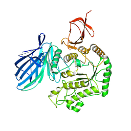
8K5R
 | | CDK9/cyclin T1 in complex with KB-0742 | | 分子名称: | (1S,3S)-N3-(5-pentan-3-ylpyrazolo[1,5-a]pyrimidin-7-yl)cyclopentane-1,3-diamine, Cyclin-T1, Cyclin-dependent kinase 9 | | 著者 | Zhou, M, Li, H, Gao, H, Trotter, B.W, Freeman, D. | | 登録日 | 2023-07-24 | | 公開日 | 2023-12-06 | | 最終更新日 | 2024-11-13 | | 実験手法 | X-RAY DIFFRACTION (3.751 Å) | | 主引用文献 | Discovery of KB-0742, a Potent, Selective, Orally Bioavailable Small Molecule Inhibitor of CDK9 for MYC-Dependent Cancers.
J.Med.Chem., 66, 2023
|
|
8X1B
 | | Cryo-EM structure of FpGalactosaminidase from Flavonifractor plautii in apo state | | 分子名称: | Alpha-galactosidase | | 著者 | Wu, G, Han, P, Su, C, Zhou, M, Luo, K. | | 登録日 | 2023-11-06 | | 公開日 | 2024-11-13 | | 最終更新日 | 2025-07-02 | | 実験手法 | ELECTRON MICROSCOPY (2.59 Å) | | 主引用文献 | Structural basis of FpGalNase and its combination with FpGalNAcDeAc for efficient A-to-O blood group conversion.
Exp Hematol Oncol, 14, 2025
|
|
3CZX
 | |
3J1Z
 | | Inward-Facing Conformation of the Zinc Transporter YiiP revealed by Cryo-electron Microscopy | | 分子名称: | Cation efflux family protein | | 著者 | Coudray, N, Valvo, S, Hu, M, Lasala, R, Kim, C, Vink, M, Zhou, M, Provasi, D, Filizola, M, Tao, J, Fang, J, Penczek, P.A, Ubarretxena-Belandia, I, Stokes, D.L, Transcontinental EM Initiative for Membrane Protein Structure (TEMIMPS) | | 登録日 | 2012-07-24 | | 公開日 | 2012-10-10 | | 最終更新日 | 2024-02-21 | | 実験手法 | ELECTRON MICROSCOPY (13 Å) | | 主引用文献 | Inward-facing conformation of the zinc transporter YiiP revealed by cryoelectron microscopy.
Proc.Natl.Acad.Sci.USA, 110, 2013
|
|
3CRJ
 | |
5JPD
 | | Metal ABC transporter from Listeria monocytogenes with cadmium | | 分子名称: | CADMIUM ION, CHLORIDE ION, Manganese-binding lipoprotein MntA | | 著者 | Osipiuk, J, Zhou, M, Grimshaw, S, Anderson, W.F, Joachimiak, A, Center for Structural Genomics of Infectious Diseases (CSGID) | | 登録日 | 2016-05-03 | | 公開日 | 2016-05-11 | | 最終更新日 | 2024-11-13 | | 実験手法 | X-RAY DIFFRACTION (1.72 Å) | | 主引用文献 | Metal ABC transporter from Listeria monocytogenes with cadmium
to be published
|
|
3S52
 | | Crystal structure of a putative fumarylacetoacetate hydrolase family protein from Yersinia pestis CO92 | | 分子名称: | CHLORIDE ION, Putative fumarylacetoacetate hydrolase family protein, SULFATE ION | | 著者 | Nocek, B, Zhou, M, Grimshaw, S, Anderson, W.F, Joachimiak, A, Center for Structural Genomics of Infectious Diseases (CSGID) | | 登録日 | 2011-05-20 | | 公開日 | 2011-06-29 | | 最終更新日 | 2024-11-06 | | 実験手法 | X-RAY DIFFRACTION (2.012 Å) | | 主引用文献 | Crystal structure of a putative fumarylacetoacetate hydrolase family protein from Yersinia pestis CO92
TO BE PUBLISHED
|
|
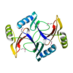
2I00
 | | Crystal structure of acetyltransferase (GNAT family) from Enterococcus faecalis | | 分子名称: | Acetyltransferase, GNAT family | | 著者 | Zhang, R, Zhou, M, Moy, S, Joachimiak, A, Midwest Center for Structural Genomics (MCSG) | | 登録日 | 2006-08-09 | | 公開日 | 2006-09-19 | | 最終更新日 | 2024-02-21 | | 実験手法 | X-RAY DIFFRACTION (2.3 Å) | | 主引用文献 | The crystal structure of the acetyltransferase (GNAT family) from Enterococcus faecalis
To be Published, 2006
|
|
2I7R
 | |
5JQC
 | | Crystal structure putative autolysin from Listeria monocytogenes | | 分子名称: | DI(HYDROXYETHYL)ETHER, GLYCEROL, Lmo1076 protein, ... | | 著者 | Chang, C, Zhou, M, Shatsman, S, Anderson, W.F, Joachimiak, A, Center for Structural Genomics of Infectious Diseases (CSGID) | | 登録日 | 2016-05-04 | | 公開日 | 2016-05-18 | | 最終更新日 | 2024-12-25 | | 実験手法 | X-RAY DIFFRACTION (2.149 Å) | | 主引用文献 | Crystal structure putative autolysin from Listeria monocytogenes
To Be Published
|
|
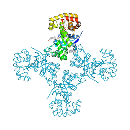
2ITD
 | | Potassium Channel KcsA-Fab complex in Barium Chloride | | 分子名称: | BARIUM ION, Voltage-gated potassium channel, antibody Fab fragment heavy chain, ... | | 著者 | Lockless, S.W, Zhou, M, MacKinnon, R. | | 登録日 | 2006-10-19 | | 公開日 | 2007-05-15 | | 最終更新日 | 2024-10-16 | | 実験手法 | X-RAY DIFFRACTION (2.7 Å) | | 主引用文献 | Structural and Thermodynamic Properties of Selective Ion Binding in a K(+) Channel.
Plos Biol., 5, 2007
|
|
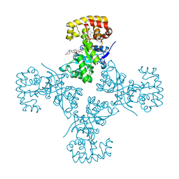
2ITC
 | | Potassium Channel KcsA-Fab complex in Sodium Chloride | | 分子名称: | Antibody Fab fragment heavy chain, Antibody Fab fragment light chain, SODIUM ION, ... | | 著者 | Lockless, S.W, Zhou, M, MacKinnon, R. | | 登録日 | 2006-10-19 | | 公開日 | 2007-05-15 | | 最終更新日 | 2024-11-13 | | 実験手法 | X-RAY DIFFRACTION (3.2 Å) | | 主引用文献 | Structural and Thermodynamic Properties of Selective Ion Binding in a K(+) Channel.
Plos Biol., 5, 2007
|
|
1U84
 | | Crystal Structure of APC36109 from Bacillus stearothermophilus | | 分子名称: | 1,2-ETHANEDIOL, GLYCEROL, Hypothetical protein | | 著者 | Kim, Y, Zhou, M, Collart, F, Joachimiak, A, Midwest Center for Structural Genomics (MCSG) | | 登録日 | 2004-08-04 | | 公開日 | 2004-10-05 | | 最終更新日 | 2024-02-14 | | 実験手法 | X-RAY DIFFRACTION (1.6 Å) | | 主引用文献 | Crystal Structure of APC36109 from Bacillus stearothermophilus
To be Published
|
|
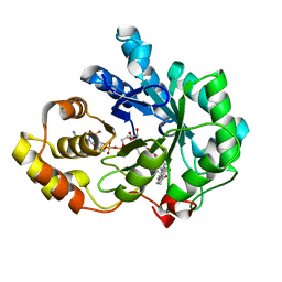
4JAL
 | | Crystal structure of tRNA (Um34/Cm34) methyltransferase TrmL from Escherichia coli with SAH | | 分子名称: | 1,2-ETHANEDIOL, 4-(2-HYDROXYETHYL)-1-PIPERAZINE ETHANESULFONIC ACID, S-ADENOSYL-L-HOMOCYSTEINE, ... | | 著者 | Liu, R.J, Zhou, M, Wang, E.D. | | 登録日 | 2013-02-18 | | 公開日 | 2013-07-31 | | 最終更新日 | 2023-11-08 | | 実験手法 | X-RAY DIFFRACTION (2 Å) | | 主引用文献 | The tRNA recognition mechanism of the minimalist SPOUT methyltransferase, TrmL
Nucleic Acids Res., 41, 2013
|
|
4JAK
 | |
1XF0
 | | Crystal structure of human 17beta-hydroxysteroid dehydrogenase type 5 (AKR1C3) complexed with delta4-androstene-3,17-dione and NADP | | 分子名称: | 4-ANDROSTENE-3-17-DIONE, ACETATE ION, Aldo-keto reductase family 1 member C3, ... | | 著者 | Qiu, W, Zhou, M, Labrie, F, Lin, S.-X. | | 登録日 | 2004-09-13 | | 公開日 | 2004-10-26 | | 最終更新日 | 2023-08-23 | | 実験手法 | X-RAY DIFFRACTION (2 Å) | | 主引用文献 | Crystal structures of the multispecific 17beta-hydroxysteroid dehydrogenase type 5: critical androgen regulation in human peripheral tissues
Mol.Endocrinol., 18, 2004
|
|
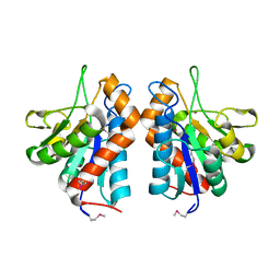
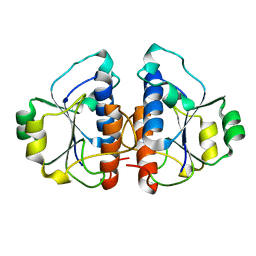
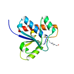
3EB3
 | | Voltage-dependent K+ channel beta subunit (W121A) in complex with cortisone | | 分子名称: | 17,21-DIHYDROXYPREGNA-1,4-DIENE-3,11,20-TRIONE, NADPH DIHYDRO-NICOTINAMIDE-ADENINE-DINUCLEOTIDE PHOSPHATE, Voltage-gated potassium channel subunit beta-2 | | 著者 | Pan, Y, Weng, J, Kabaleeswaran, V, Li, H, Cao, Y, Bhosle, R.C, Zhou, M. | | 登録日 | 2008-08-26 | | 公開日 | 2008-09-23 | | 最終更新日 | 2023-08-30 | | 実験手法 | X-RAY DIFFRACTION (2 Å) | | 主引用文献 | Cortisone dissociates the Shaker family K+ channels from their beta subunits.
Nat.Chem.Biol., 4, 2008
|
|
3EAU
 | | Voltage-dependent K+ channel beta subunit in complex with cortisone | | 分子名称: | 17,21-DIHYDROXYPREGNA-1,4-DIENE-3,11,20-TRIONE, NADPH DIHYDRO-NICOTINAMIDE-ADENINE-DINUCLEOTIDE PHOSPHATE, Voltage-gated potassium channel subunit beta-2 | | 著者 | Pan, Y, Weng, J, Kabaleeswaran, V, Li, H, Cao, Y, Bhosle, R.C, Zhou, M. | | 登録日 | 2008-08-26 | | 公開日 | 2008-09-23 | | 最終更新日 | 2023-08-30 | | 実験手法 | X-RAY DIFFRACTION (1.82 Å) | | 主引用文献 | Cortisone dissociates the Shaker family K+ channels from their beta subunits.
Nat.Chem.Biol., 4, 2008
|
|
3EB4
 | | Voltage-dependent K+ channel beta subunit (I211R) in complex with cortisone | | 分子名称: | 17,21-DIHYDROXYPREGNA-1,4-DIENE-3,11,20-TRIONE, NADPH DIHYDRO-NICOTINAMIDE-ADENINE-DINUCLEOTIDE PHOSPHATE, Voltage-gated potassium channel subunit beta-2 | | 著者 | Pan, Y, Weng, J, Kabaleeswaran, V, Li, H, Cao, Y, Bhosle, R.C, Zhou, M. | | 登録日 | 2008-08-26 | | 公開日 | 2008-09-23 | | 最終更新日 | 2023-08-30 | | 実験手法 | X-RAY DIFFRACTION (2 Å) | | 主引用文献 | Cortisone dissociates the Shaker family K+ channels from their beta subunits.
Nat.Chem.Biol., 4, 2008
|
|
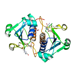
1DG9
 | | CRYSTAL STRUCTURE OF BOVINE LOW MOLECULAR WEIGHT PTPASE COMPLEXED WITH HEPES | | 分子名称: | 4-(2-HYDROXYETHYL)-1-PIPERAZINE ETHANESULFONIC ACID, TYROSINE PHOSPHATASE | | 著者 | Zhang, M, Zhou, M, Van Etten, R.L, Stauffacher, C.V. | | 登録日 | 1999-11-23 | | 公開日 | 1999-12-08 | | 最終更新日 | 2024-02-07 | | 実験手法 | X-RAY DIFFRACTION (1.9 Å) | | 主引用文献 | Crystal structure of bovine low molecular weight phosphotyrosyl phosphatase complexed with the transition state analog vanadate.
Biochemistry, 36, 1997
|
|


