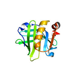
4NK4
 
 | |
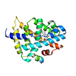
4NK5
 | |
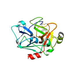
8IN0
 | |
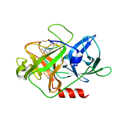
6LC1
 | |
8HBM
 | |
5WXT
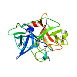
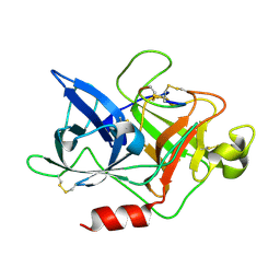
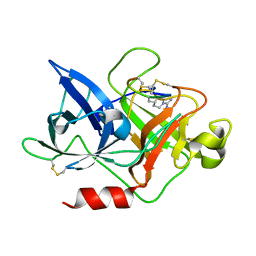
 | | Crystal structure of uPA-S195A in complex with S2444 | | 分子名称: | (2R)-N-[2-[[(2S)-1-[[4-[bis(oxidanyl)amino]phenyl]amino]-5-carbamimidamido-1-oxidanylidene-pentan-2-yl]amino]-2-oxidanylidene-ethyl]-5-oxidanylidene-pyrrolidine-2-carboxamide, Urokinase-type plasminogen activator chain B | | 著者 | Jiang, L, Huang, M. | | 登録日 | 2017-01-08 | | 公開日 | 2018-07-11 | | 最終更新日 | 2023-11-22 | | 実験手法 | X-RAY DIFFRACTION (2.1 Å) | | 主引用文献 | Crystal structure of uPA-S195A in complex with S2444
To Be Published
|
|
5WXQ
 | |
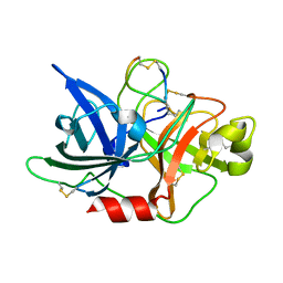
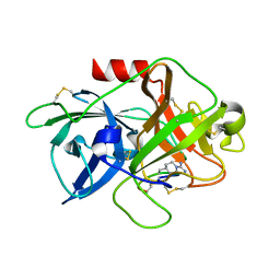
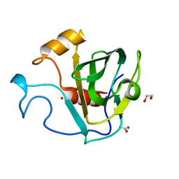
5WXF
 | | Crystal structure of uPA in complex with upain-2-2 | | 分子名称: | SULFATE ION, Urokinase-type plasminogen activator chain B, upain-2-2 peptide | | 著者 | Jiang, L, Huang, M. | | 登録日 | 2017-01-07 | | 公開日 | 2018-07-11 | | 最終更新日 | 2023-11-22 | | 実験手法 | X-RAY DIFFRACTION (1.46 Å) | | 主引用文献 | Cleavage of peptidic inhibitors by target protease is caused by peptide conformational transition.
Biochim. Biophys. Acta, 1862, 2018
|
|
5WXR
 | |
5WXO
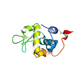
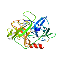
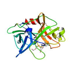
 | | Crystal structure of uPA in complex with upain-2-2-W3A | | 分子名称: | Urokinase-type plasminogen activator chain B, upain-2-2-W3A peptide | | 著者 | Jiang, L, Huang, M. | | 登録日 | 2017-01-08 | | 公開日 | 2018-07-11 | | 最終更新日 | 2023-11-22 | | 実験手法 | X-RAY DIFFRACTION (1.64 Å) | | 主引用文献 | Cleavage of peptidic inhibitors by target protease is caused by peptide conformational transition.
Biochim. Biophys. Acta, 1862, 2018
|
|
5WXS
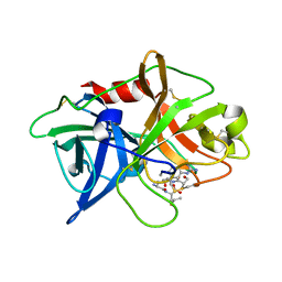
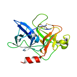
 | | Crystal structure of uPA in complex with S2444 | | 分子名称: | (2R)-N-[2-[[(2S)-5-carbamimidamido-1-oxidanylidene-pentan-2-yl]amino]-2-oxidanylidene-ethyl]-5-oxidanylidene-1,2-dihydropyrrole-2-carboxamide, Urokinase-type plasminogen activator chain B | | 著者 | Jiang, L, Huang, M. | | 登録日 | 2017-01-08 | | 公開日 | 2018-07-11 | | 最終更新日 | 2023-11-22 | | 実験手法 | X-RAY DIFFRACTION (2.3 Å) | | 主引用文献 | Crystal structure of uPA in complex with S2444
To Be Published
|
|
5XUW
 | |
5WXP
 | | Crystal structure of uPA in complex with upain-2-3-W3A | | 分子名称: | ALANINE, CYSTEINE, Urokinase-type plasminogen activator chain B, ... | | 著者 | Jiang, L, Huang, M. | | 登録日 | 2017-01-08 | | 公開日 | 2018-07-11 | | 最終更新日 | 2023-11-22 | | 実験手法 | X-RAY DIFFRACTION (1.75 Å) | | 主引用文献 | Cleavage of peptidic inhibitors by target protease is caused by peptide conformational transition.
Biochim. Biophys. Acta, 1862, 2018
|
|
5Y5C
 | |
7D42
 | |
7CJ1
 | |
4X1N
 | | The crystal structure of mupain-1-16 in complex with murinised human uPA at pH7.4 | | 分子名称: | Urokinase-type plasminogen activator, mupain-1-16, piperidine-1-carboximidamide | | 著者 | Jiang, L, Zhao, B, Xu, P, Andreasen, P, Huang, M. | | 登録日 | 2014-11-25 | | 公開日 | 2015-03-25 | | 最終更新日 | 2023-11-08 | | 実験手法 | X-RAY DIFFRACTION (1.8 Å) | | 主引用文献 | A cyclic peptidic serine protease inhibitor: increasing affinity by increasing peptide flexibility.
Plos One, 9, 2014
|
|
4X1Q
 | | The crystal structure of mupain-1 in complex with murinised human uPA at pH7.4 | | 分子名称: | Urokinase-type plasminogen activator, mupain-1 | | 著者 | Jiang, L, Zhao, B, Xu, P, Andreasen, P, Huang, M. | | 登録日 | 2014-11-25 | | 公開日 | 2015-03-25 | | 最終更新日 | 2015-11-04 | | 実験手法 | X-RAY DIFFRACTION (2.28 Å) | | 主引用文献 | A cyclic peptidic serine protease inhibitor: increasing affinity by increasing peptide flexibility.
Plos One, 9, 2014
|
|
4X1S
 | | The crystal structure of mupain-1-16-D9A in complex with murinised human uPA at pH7.4 | | 分子名称: | Urokinase-type plasminogen activator, mupain-1-16, piperidine-1-carboximidamide | | 著者 | Jiang, L, Zhao, B, Xu, P, Andreasen, P, Huang, M. | | 登録日 | 2014-11-25 | | 公開日 | 2015-10-28 | | 最終更新日 | 2023-11-08 | | 実験手法 | X-RAY DIFFRACTION (1.9 Å) | | 主引用文献 | A cyclic peptidic serine protease inhibitor: increasing affinity by increasing peptide flexibility.
Plos One, 9, 2014
|
|
4X1R
 | | The crystal structure of mupain-1-12 in complex with murinised human uPA at pH7.4 | | 分子名称: | 1-phenylguanidine, Urokinase-type plasminogen activator, mupain-1-12 | | 著者 | Jiang, L, Zhao, B, Xu, P, Andreasen, P, Huang, M. | | 登録日 | 2014-11-25 | | 公開日 | 2015-03-25 | | 最終更新日 | 2023-11-08 | | 実験手法 | X-RAY DIFFRACTION (2.1 Å) | | 主引用文献 | A cyclic peptidic serine protease inhibitor: increasing affinity by increasing peptide flexibility.
Plos One, 9, 2014
|
|
4ZHL
 | |
4ZHM
 | |
5GT1
 | | crystal structure of cbpa from L. salivarius REN | | 分子名称: | 1,2-ETHANEDIOL, ACETATE ION, Choline binding protein A, ... | | 著者 | Jiang, L, Ren, F. | | 登録日 | 2016-08-18 | | 公開日 | 2017-07-19 | | 最終更新日 | 2023-11-08 | | 実験手法 | X-RAY DIFFRACTION (1.85 Å) | | 主引用文献 | The Adhesion of Lactobacillus salivarius REN to a Human Intestinal Epithelial Cell Line Requires S-layer Proteins
Sci Rep, 7, 2017
|
|
5XG4
 | | Crystal structure of uPA in complex with quercetin | | 分子名称: | 1-(2-METHOXY-ETHOXY)-2-{2-[2-(2-METHOXY-ETHOXY]-ETHOXY}-ETHANE, 3,5,7,3',4'-PENTAHYDROXYFLAVONE, Urokinase-type plasminogen activator | | 著者 | Jiang, L, Huang, M. | | 登録日 | 2017-04-11 | | 公開日 | 2017-07-26 | | 最終更新日 | 2023-11-22 | | 実験手法 | X-RAY DIFFRACTION (3 Å) | | 主引用文献 | A structural mechanism of flavonoids in inhibiting serine proteases
Food Funct, 8, 2017
|
|
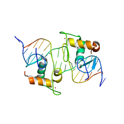
8J54
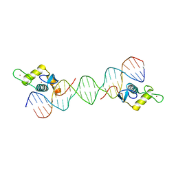
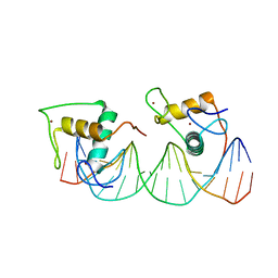 | | Crystal structure of RXR/DR2 complex | | 分子名称: | DNA (5'-D(*CP*AP*TP*GP*AP*CP*CP*TP*AP*CP*TP*GP*AP*CP*CP*TP*AP*G)-3'), DNA (5'-D(*CP*TP*AP*GP*GP*TP*CP*AP*GP*TP*AP*GP*GP*TP*CP*AP*TP*G)-3'), Retinoic acid receptor RXR, ... | | 著者 | Chen, Y, Jiang, L. | | 登録日 | 2023-04-21 | | 公開日 | 2024-01-17 | | 最終更新日 | 2024-04-17 | | 実験手法 | X-RAY DIFFRACTION (2.72 Å) | | 主引用文献 | Structural characterization of the DNA binding mechanism of retinoic acid-related orphan receptor gamma.
Structure, 32, 2024
|
|


