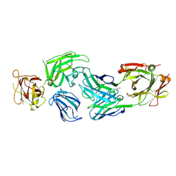
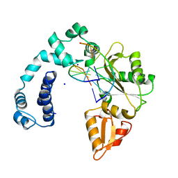
8Z25
 
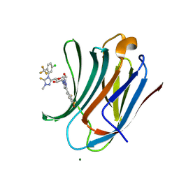 | | Crystal structure of mouse Galectin-3 in complex with small molecule inhibitor | | 分子名称: | 5-[(2~{S},3~{R},4~{R},5~{R},6~{R})-4-[4-[4-bromanyl-2,3-bis(fluoranyl)phenyl]-1,2,3-triazol-1-yl]-6-(hydroxymethyl)-3,5-bis(oxidanyl)oxan-2-yl]-4-[5-chloranyl-2-(trifluoromethyl)phenyl]-2-methyl-1,2,4-triazole-3-thione, Galectin-3, MAGNESIUM ION | | 著者 | Amit, K, Swetha, R, Ghosh, K. | | 登録日 | 2024-04-12 | | 公開日 | 2025-03-05 | | 実験手法 | X-RAY DIFFRACTION (1.72 Å) | | 主引用文献 | Atropisomerism Observed in Galactose-Based Monosaccharide Inhibitors of Galectin-3 Comprising 2-Methyl-4-phenyl-2,4-dihydro-3 H -1,2,4-triazole-3-thione.
J.Med.Chem., 67, 2024
|
|
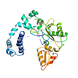
8ZUV
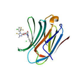 | | Crystal structure of mouse Galectin-3 in complex with small molecule inhibitor | | 分子名称: | 5-[(2~{S},3~{R},4~{R},5~{R},6~{R})-4-[4-[4-bromanyl-2,3-bis(fluoranyl)phenyl]-1,2,3-triazol-1-yl]-6-(hydroxymethyl)-3,5-bis(oxidanyl)oxan-2-yl]-4-[5-chloranyl-2-(trifluoromethyl)phenyl]-2-methyl-1,2,4-triazole-3-thione, CHLORIDE ION, Galectin-3 | | 著者 | Amit, K, Swetha, R, Ghosh, K. | | 登録日 | 2024-06-10 | | 公開日 | 2025-03-05 | | 実験手法 | X-RAY DIFFRACTION (1.45 Å) | | 主引用文献 | Atropisomerism Observed in Galactose-Based Monosaccharide Inhibitors of Galectin-3 Comprising 2-Methyl-4-phenyl-2,4-dihydro-3 H -1,2,4-triazole-3-thione.
J.Med.Chem., 67, 2024
|
|
3DCX
 | |
1NOM
 | |
1KVX
 | |
3DUE
 | |
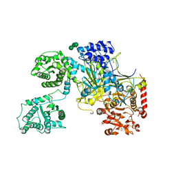
8W2F
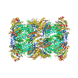 | | Plasmodium falciparum 20S proteasome bound to an inhibitor | | 分子名称: | (3S)-1-[(2-fluoroethoxy)acetyl]-N-{[(4P)-4-(6-methylpyridin-3-yl)-1,3-thiazol-2-yl]methyl}piperidine-3-carboxamide, Proteasome endopeptidase complex, Proteasome subunit alpha type, ... | | 著者 | Han, Y, Deng, X, Ray, S, Chen, Z, Phillips, M. | | 登録日 | 2024-02-20 | | 公開日 | 2024-07-31 | | 最終更新日 | 2025-05-28 | | 実験手法 | ELECTRON MICROSCOPY (3.1 Å) | | 主引用文献 | Identification of potent and reversible piperidine carboxamides that are species-selective orally active proteasome inhibitors to treat malaria.
Cell Chem Biol, 31, 2024
|
|
7M17
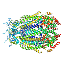 | | SN-407-LRRC8A in MSP1E3D1 lipid nanodiscs (Pose-1) | | 分子名称: | 1,2-dioleoyl-sn-glycero-3-phosphoethanolamine, 7-{[(2S)-2-butyl-6,7-dichloro-2-cyclopentyl-1-oxo-2,3-dihydro-1H-inden-5-yl]oxy}heptanoic acid, Volume-regulated anion channel subunit LRRC8A | | 著者 | Kern, D.M, Gerber, E.E, Brohawn, S.G. | | 登録日 | 2021-03-12 | | 公開日 | 2021-03-24 | | 最終更新日 | 2022-02-23 | | 実験手法 | ELECTRON MICROSCOPY (3.65 Å) | | 主引用文献 | Small molecule SWELL1 complex induction improves glycemic control and nonalcoholic fatty liver disease in murine Type 2 diabetes.
Nat Commun, 13, 2022
|
|
7M19
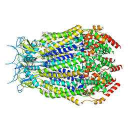 | | SN-407-LRRC8A in MSP1E3D1 lipid nanodiscs (Pose-2) | | 分子名称: | 1,2-dioleoyl-sn-glycero-3-phosphoethanolamine, 7-{[(2S)-2-butyl-6,7-dichloro-2-cyclopentyl-1-oxo-2,3-dihydro-1H-inden-5-yl]oxy}heptanoic acid, Volume-regulated anion channel subunit LRRC8A | | 著者 | Kern, D.M, Gerber, E.E, Brohawn, S.G. | | 登録日 | 2021-03-12 | | 公開日 | 2021-03-24 | | 最終更新日 | 2022-02-23 | | 実験手法 | ELECTRON MICROSCOPY (3.69 Å) | | 主引用文献 | Small molecule SWELL1 complex induction improves glycemic control and nonalcoholic fatty liver disease in murine Type 2 diabetes.
Nat Commun, 13, 2022
|
|
3EQX
 | |
3F1Z
 | |
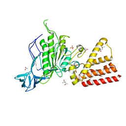
8IVW
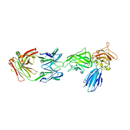 | | Crystal structure of NRP2 in complex with aNRP2-10 Fab fragment | | 分子名称: | Heavy chian of antibody 10V8 Fab fragment, Light chain of antibody 10V8 Fab fragment, Neuropilin-2 | | 著者 | Geng, Y, Zhai, L. | | 登録日 | 2023-03-29 | | 公開日 | 2023-07-12 | | 最終更新日 | 2024-11-06 | | 実験手法 | X-RAY DIFFRACTION (3.205 Å) | | 主引用文献 | Inhibition of VEGF binding to neuropilin-2 enhances chemosensitivity and inhibits metastasis in triple-negative breast cancer.
Sci Transl Med, 15, 2023
|
|
8IVX
 | | Crystal structure of NRP2 in complex with aNRP2-14 Fab fragment | | 分子名称: | 1,2-ETHANEDIOL, Heavy chain of antibody 14V4 Fab fragment, Light chain of antibody 14V4 Fab fragment, ... | | 著者 | Geng, Y, Zhai, L. | | 登録日 | 2023-03-29 | | 公開日 | 2023-07-12 | | 最終更新日 | 2024-11-20 | | 実験手法 | X-RAY DIFFRACTION (1.9 Å) | | 主引用文献 | Inhibition of VEGF binding to neuropilin-2 enhances chemosensitivity and inhibits metastasis in triple-negative breast cancer.
Sci Transl Med, 15, 2023
|
|
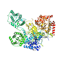
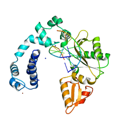
5MZO
 | | UDP-Glucose Glycoprotein Glucosyltransferase from Chaetomium thermophilum (open conformation) | | 分子名称: | 1,2-ETHANEDIOL, 2-acetamido-2-deoxy-beta-D-glucopyranose, 2-acetamido-2-deoxy-beta-D-glucopyranose-(1-4)-2-acetamido-2-deoxy-beta-D-glucopyranose, ... | | 著者 | Roversi, P, Caputo, A.T, Hill, J, Alonzi, D.S, Zitzmann, N. | | 登録日 | 2017-02-01 | | 公開日 | 2017-07-26 | | 最終更新日 | 2024-10-23 | | 実験手法 | X-RAY DIFFRACTION (3.48 Å) | | 主引用文献 | Interdomain conformational flexibility underpins the activity of UGGT, the eukaryotic glycoprotein secretion checkpoint.
Proc. Natl. Acad. Sci. U.S.A., 114, 2017
|
|
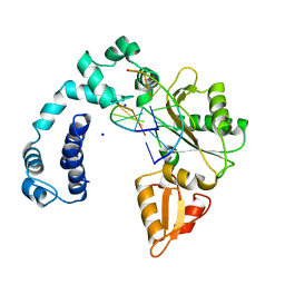
5NV4
 | | UDP-Glucose Glycoprotein Glucosyltransferase from Chaetomium thermophilum double mutant D611C:G1050C | | 分子名称: | 2-acetamido-2-deoxy-beta-D-glucopyranose, FORMIC ACID, UDP-glucose-glycoprotein glucosyltransferase-like protein, ... | | 著者 | Roversi, P, Caputo, A.T, Hill, J, Alonzi, D.S, Zitzmann, N. | | 登録日 | 2017-05-03 | | 公開日 | 2017-07-26 | | 最終更新日 | 2024-10-23 | | 実験手法 | X-RAY DIFFRACTION (2.78 Å) | | 主引用文献 | Interdomain conformational flexibility underpins the activity of UGGT, the eukaryotic glycoprotein secretion checkpoint.
Proc. Natl. Acad. Sci. U.S.A., 114, 2017
|
|
5MU1
 | | UDP-Glucose Glycoprotein Glucosyltransferase from Chaetomium thermophilum soaked with K2PtI6 | | 分子名称: | CALCIUM ION, IODIDE ION, PLATINUM (II) ION, ... | | 著者 | Roversi, P, Caputo, A.T, Hill, J, Alonzi, D.S, Zitzmann, N. | | 登録日 | 2017-01-11 | | 公開日 | 2017-07-26 | | 最終更新日 | 2024-10-16 | | 実験手法 | X-RAY DIFFRACTION (3.48 Å) | | 主引用文献 | Interdomain conformational flexibility underpins the activity of UGGT, the eukaryotic glycoprotein secretion checkpoint.
Proc. Natl. Acad. Sci. U.S.A., 114, 2017
|
|
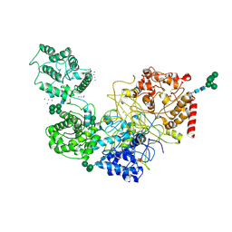
5N2J
 | | UDP-Glucose Glycoprotein Glucosyltransferase from Chaetomium thermophilum (closed form) | | 分子名称: | 1,2-ETHANEDIOL, 2-acetamido-2-deoxy-beta-D-glucopyranose, 2-acetamido-2-deoxy-beta-D-glucopyranose-(1-4)-2-acetamido-2-deoxy-beta-D-glucopyranose, ... | | 著者 | Roversi, P, Caputo, A.T, Hill, J, Alonzi, D.S, Zitzmann, N. | | 登録日 | 2017-02-07 | | 公開日 | 2017-07-26 | | 最終更新日 | 2024-11-06 | | 実験手法 | X-RAY DIFFRACTION (4.4 Å) | | 主引用文献 | Interdomain conformational flexibility underpins the activity of UGGT, the eukaryotic glycoprotein secretion checkpoint.
Proc. Natl. Acad. Sci. U.S.A., 114, 2017
|
|
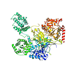
3SE0
 | | Structural characterization of the subunit A mutant F508W of the A-ATP synthase from Pyrococcus horikoshii | | 分子名称: | (4S)-2-METHYL-2,4-PENTANEDIOL, 2-AMINO-2-HYDROXYMETHYL-PROPANE-1,3-DIOL, ACETIC ACID, ... | | 著者 | Tadwal, V.S, Manimekalai, M.S.S, Balakrishna, A.M, Gruber, G. | | 登録日 | 2011-06-09 | | 公開日 | 2012-01-25 | | 最終更新日 | 2023-11-01 | | 実験手法 | X-RAY DIFFRACTION (2.62 Å) | | 主引用文献 | Engineered tryptophan in the adenine-binding pocket of catalytic subunit A of A-ATP synthase demonstrates the importance of aromatic residues in adenine binding, forming a tool for steady-state and time-resolved fluorescence spectroscopy.
Acta Crystallogr.,Sect.F, 67, 2011
|
|
7ICF
 | |
7ICT
 | |
7ICI
 | |
7ICS
 | |
7ICH
 | |
7ICR
 | |
7ICE
 | |


