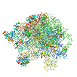
6GAW
 
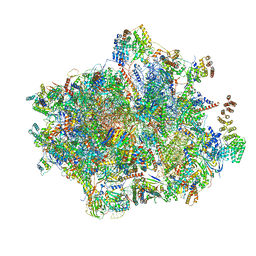 | | Unique features of mammalian mitochondrial translation initiation revealed by cryo-EM. This file contains the complete 55S ribosome. | | 分子名称: | 12S ribosomal RNA, mitochondrial, 16S ribosomal RNA, ... | | 著者 | Kummer, E, Leibundgut, M, Boehringer, D, Ban, N. | | 登録日 | 2018-04-13 | | 公開日 | 2018-08-22 | | 最終更新日 | 2019-12-11 | | 実験手法 | ELECTRON MICROSCOPY (3.2 Å) | | 主引用文献 | Unique features of mammalian mitochondrial translation initiation revealed by cryo-EM.
Nature, 560, 2018
|
|
5OME
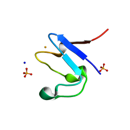 | | The cryofrozen atomic resolution X-ray crystal structure of the reduced form (Fe2+) perdeuterated Pyrococcus furiosus Rubredoxin in D2O (100K, 0.75 Angstrom resolution) | | 分子名称: | FE (III) ION, PHOSPHATE ION, Rubredoxin, ... | | 著者 | Cuypers, M.G, Mason, S.A, Mossou, E, Haertlein, M, Mitchell, E.P, Forsyth, V.T. | | 登録日 | 2017-07-31 | | 公開日 | 2018-09-05 | | 最終更新日 | 2024-06-19 | | 実験手法 | X-RAY DIFFRACTION (0.747 Å) | | 主引用文献 | The cryofrozen atomic resolution X-ray crystal structure of the reduced form (Fe2+) perdeuterated Pyrococcus furiosus Rubredoxin in D2O (100K, 0.75 Angstrom resolution)
To Be Published
|
|
5NW3
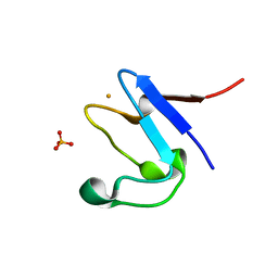 | | The cryofrozen atomic resolution X-ray crystal structure of perdeuterated Pyrococcus furiosus Rubredoxin (100K, 0.59A resolution) | | 分子名称: | FE (III) ION, PHOSPHATE ION, POTASSIUM ION, ... | | 著者 | Cuypers, M.G, Mason, S.A, Mossou, E, Haertlein, M, Forsyth, V.T. | | 登録日 | 2017-05-04 | | 公開日 | 2017-06-07 | | 最終更新日 | 2024-05-08 | | 実験手法 | X-RAY DIFFRACTION (0.59 Å) | | 主引用文献 | The cryofrozen atomic resolution X-ray crystal structure of perdeuterated Pyrococcus furiosus Rubredoxin (100K, 0.59A resolution)
To Be Published
|
|
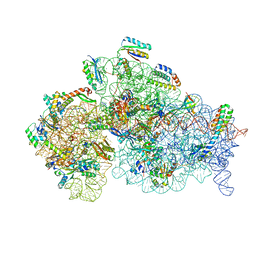
5O61
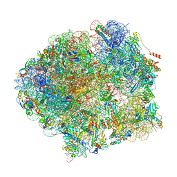 | | The complete structure of the Mycobacterium smegmatis 70S ribosome | | 分子名称: | 16S rRNA, 23S rRNA, 30S ribosomal protein S10, ... | | 著者 | Hentschel, J, Burnside, C, Mignot, I, Leibundgut, M, Boehringer, D, Ban, N. | | 登録日 | 2017-06-03 | | 公開日 | 2017-07-12 | | 最終更新日 | 2024-05-15 | | 実験手法 | ELECTRON MICROSCOPY (3.31 Å) | | 主引用文献 | The Complete Structure of the Mycobacterium smegmatis 70S Ribosome.
Cell Rep, 20, 2017
|
|
5O60
 | | Structure of the 50S large ribosomal subunit from Mycobacterium smegmatis | | 分子名称: | 23S rRNA, 50S ribosomal protein L10, 50S ribosomal protein L11, ... | | 著者 | Hentschel, J, Burnside, C, Mignot, I, Leibundgut, M, Boehringer, D, Ban, N. | | 登録日 | 2017-06-02 | | 公開日 | 2017-07-12 | | 最終更新日 | 2024-05-15 | | 実験手法 | ELECTRON MICROSCOPY (3.18 Å) | | 主引用文献 | The Complete Structure of the Mycobacterium smegmatis 70S Ribosome.
Cell Rep, 20, 2017
|
|
5O5J
 | | Structure of the 30S small ribosomal subunit from Mycobacterium smegmatis | | 分子名称: | 16S rRNA, 30S ribosomal protein S10, 30S ribosomal protein S11, ... | | 著者 | Hentschel, J, Burnside, C, Mignot, I, Leibundgut, M, Boehringer, D, Ban, N. | | 登録日 | 2017-06-02 | | 公開日 | 2017-07-12 | | 最終更新日 | 2024-05-15 | | 実験手法 | ELECTRON MICROSCOPY (3.451 Å) | | 主引用文献 | The Complete Structure of the Mycobacterium smegmatis 70S Ribosome.
Cell Rep, 20, 2017
|
|
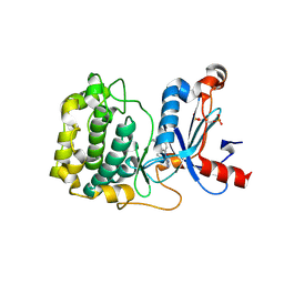
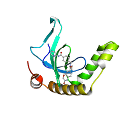
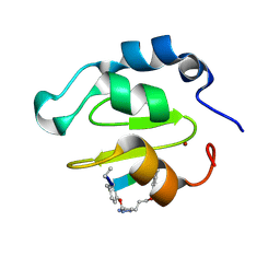
6BPH
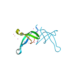 | | Crystal structure of the chromodomain of RBBP1 | | 分子名称: | AT-rich interactive domain-containing protein 4A, UNKNOWN ATOM OR ION | | 著者 | Liu, Y, Tempel, W, Bountra, C, Arrowsmith, C.H, Edwards, A.M, Min, J, Structural Genomics Consortium (SGC) | | 登録日 | 2017-11-23 | | 公開日 | 2017-12-20 | | 最終更新日 | 2023-10-04 | | 実験手法 | X-RAY DIFFRACTION (1.85 Å) | | 主引用文献 | Crystal structure of chromo barrel domain of RBBP1.
Biochem. Biophys. Res. Commun., 496, 2018
|
|
6C1Y
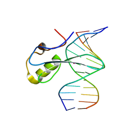 | | mbd of human mecp2 in complex with methylated DNA | | 分子名称: | 12-mer DNA, Methyl-CpG-binding protein 2, UNKNOWN ATOM OR ION | | 著者 | Liu, K, Bian, C, Tempel, W, Wernimont, A.K, Arrowsmith, C.H, Bountra, C, Edwards, A.M, Min, J, Structural Genomics Consortium (SGC) | | 登録日 | 2018-01-05 | | 公開日 | 2018-02-14 | | 最終更新日 | 2023-10-04 | | 実験手法 | X-RAY DIFFRACTION (2.3 Å) | | 主引用文献 | Plasticity at the DNA recognition site of the MeCP2 mCG-binding domain.
Biochim Biophys Acta Gene Regul Mech, 1862, 2019
|
|
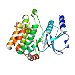
5F9E
 | | Structure of Protein Kinase C theta with compound 10: 2,2-dimethyl-7-(2-oxidanylidene-3~{H}-imidazo[4,5-b]pyridin-1-yl)-1-(phenylmethyl)-3~{H}-quinazolin-4-one | | 分子名称: | 2,2-dimethyl-7-(2-oxidanylidene-3~{H}-imidazo[4,5-b]pyridin-1-yl)-1-(phenylmethyl)-3~{H}-quinazolin-4-one, Protein kinase C theta type | | 著者 | Klein, M. | | 登録日 | 2015-12-09 | | 公開日 | 2016-05-11 | | 実験手法 | X-RAY DIFFRACTION (2 Å) | | 主引用文献 | Discovery and optimization of 1,7-disubstituted-2,2-dimethyl-2,3-dihydroquinazolin-4(1H)-ones as potent and selective PKC theta inhibitors.
Bioorg.Med.Chem., 24, 2016
|
|
5VFC
 | | WDR5 bound to inhibitor MM-589 | | 分子名称: | 1,2-ETHANEDIOL, N-{(3R,6S,9S,12R)-6-ethyl-12-methyl-9-[3-(N'-methylcarbamimidamido)propyl]-2,5,8,11-tetraoxo-3-phenyl-1,4,7,10-tetraazacyclotetradecan-12-yl}-2-methylpropanamide, SULFATE ION, ... | | 著者 | Stuckey, J.A. | | 登録日 | 2017-04-07 | | 公開日 | 2017-06-28 | | 最終更新日 | 2024-03-13 | | 実験手法 | X-RAY DIFFRACTION (1.64 Å) | | 主引用文献 | Discovery of a Highly Potent, Cell-Permeable Macrocyclic Peptidomimetic (MM-589) Targeting the WD Repeat Domain 5 Protein (WDR5)-Mixed Lineage Leukemia (MLL) Protein-Protein Interaction.
J. Med. Chem., 60, 2017
|
|
6KIU
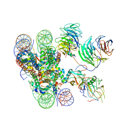 | | Cryo-EM structure of human MLL1-ubNCP complex (3.2 angstrom) | | 分子名称: | DNA (145-MER), GLUTAMINE, Histone H2A, ... | | 著者 | Huang, J, Xue, H, Yao, T. | | 登録日 | 2019-07-20 | | 公開日 | 2019-09-11 | | 最終更新日 | 2024-03-27 | | 実験手法 | ELECTRON MICROSCOPY (3.2 Å) | | 主引用文献 | Structural basis of nucleosome recognition and modification by MLL methyltransferases.
Nature, 573, 2019
|
|
6KIW
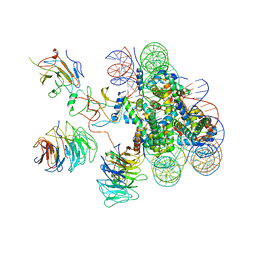 | | Cryo-EM structure of human MLL3-ubNCP complex (4.0 angstrom) | | 分子名称: | DNA (144-MER), DNA (145-MER), Histone H2A, ... | | 著者 | Huang, J, Xue, H, Yao, T. | | 登録日 | 2019-07-20 | | 公開日 | 2019-09-11 | | 最終更新日 | 2024-03-27 | | 実験手法 | ELECTRON MICROSCOPY (4 Å) | | 主引用文献 | Structural basis of nucleosome recognition and modification by MLL methyltransferases.
Nature, 573, 2019
|
|
6XCG
 | | Histone-lysine N-methyltransferase NSD2-PWWP1 with compound UNC6934 | | 分子名称: | Histone-lysine N-methyltransferase NSD2, N-cyclopropyl-3-oxo-N-({4-[(pyrimidin-4-yl)carbamoyl]phenyl}methyl)-3,4-dihydro-2H-1,4-benzoxazine-7-carboxamide, UNKNOWN ATOM OR ION | | 著者 | Zhou, M.Q, Dong, A, Ingerman, L.A, Hanley, R.P, Bountra, C, Arrowsmith, C.H, Edwards, A.M, Min, J, Structural Genomics Consortium (SGC) | | 登録日 | 2020-06-08 | | 公開日 | 2020-07-22 | | 最終更新日 | 2023-10-18 | | 実験手法 | X-RAY DIFFRACTION (1.64 Å) | | 主引用文献 | A chemical probe targeting the PWWP domain alters NSD2 nucleolar localization.
Nat.Chem.Biol., 18, 2022
|
|
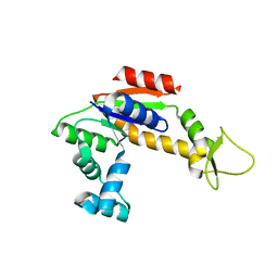
2RGX
 | | Crystal Structure of Adenylate Kinase from Aquifex Aeolicus in complex with Ap5A | | 分子名称: | Adenylate kinase, BIS(ADENOSINE)-5'-PENTAPHOSPHATE, ZINC ION | | 著者 | Thai, V, Wolf-Watz, M, Fenn, T, Pozharski, E, Wilson, M.A, Petsko, G.A, Kern, D. | | 登録日 | 2007-10-05 | | 公開日 | 2007-12-18 | | 最終更新日 | 2023-08-30 | | 実験手法 | X-RAY DIFFRACTION (1.9 Å) | | 主引用文献 | Intrinsic motions along an enzymatic reaction trajectory.
Nature, 450, 2007
|
|
2RH5
 | | Structure of Apo Adenylate Kinase from Aquifex Aeolicus | | 分子名称: | Adenylate kinase | | 著者 | Thai, V, Wolf-Watz, M, Fenn, T, Pozharski, E, Wilson, M.A, Petsko, G.A, Kern, D. | | 登録日 | 2007-10-05 | | 公開日 | 2007-12-18 | | 最終更新日 | 2024-02-21 | | 実験手法 | X-RAY DIFFRACTION (2.48 Å) | | 主引用文献 | Intrinsic motions along an enzymatic reaction trajectory.
Nature, 450, 2007
|
|
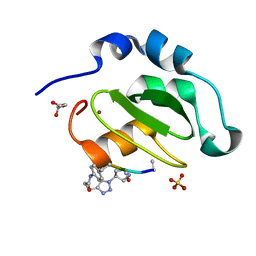
4Z3C
 | | Zinc finger region of human TET3 in complex with CpG DNA | | 分子名称: | DNA (5'-D(*GP*CP*CP*AP*AP*CP*GP*TP*TP*GP*GP*C)-3'), Methylcytosine dioxygenase, UNKNOWN ATOM OR ION, ... | | 著者 | Liu, K, Xu, C, Tempel, W, Dong, A, Arrowsmith, C.H, Bountra, C, Edwards, A.M, Min, J, Structural Genomics Consortium (SGC) | | 登録日 | 2015-03-31 | | 公開日 | 2015-04-29 | | 最終更新日 | 2023-09-27 | | 実験手法 | X-RAY DIFFRACTION (1.57 Å) | | 主引用文献 | DNA Sequence Recognition of Human CXXC Domains and Their Structural Determinants.
Structure, 2017
|
|
2HY8
 | | PAK1 complex with ST2001 | | 分子名称: | (5S,6R,7R,9R)-12-HYDROXY-6-METHOXY-5-METHYL-7-(METHYLAMINO)-6,7,8,9-TETRAHYDRO-5H,14H-5,9-EPOXY-4B,9A,15-TRIAZADIBENZO[ B,H]CYCLONONA[1,2,3,4-JKL]CYCLOPENTA[E]-AS-INDACEN-14-ONE, Serine/threonine-protein kinase PAK 1 | | 著者 | Schulze-Gahmen, U, Lu, H. | | 登録日 | 2006-08-04 | | 公開日 | 2006-11-07 | | 最終更新日 | 2024-02-14 | | 実験手法 | X-RAY DIFFRACTION (2 Å) | | 主引用文献 | Crystal Structure of the Complex Between Human Pak1-kinase and 3-Hydroxystaurosporine
To be Published
|
|
4WVT
 | | Crystal structure of XIAP-BIR2 domain complexed with ligand bound | | 分子名称: | 3,11-DIFLUORO-6,8,13-TRIMETHYL-8H-QUINO[4,3,2-KL]ACRIDIN-13-IUM, E3 ubiquitin-protein ligase XIAP, ZINC ION | | 著者 | Pokross, M.E. | | 登録日 | 2014-11-07 | | 公開日 | 2015-03-04 | | 最終更新日 | 2015-04-08 | | 実験手法 | X-RAY DIFFRACTION (1.96 Å) | | 主引用文献 | The Discovery of Macrocyclic XIAP Antagonists from a DNA-Programmed Chemistry Library, and Their Optimization To Give Lead Compounds with in Vivo Antitumor Activity.
J.Med.Chem., 58, 2015
|
|
4WVU
 | | CRYSTAL STRUCTURE OF XIAP-BIR2 DOMAIN COMPLEXED WITH LIGAND BOUND | | 分子名称: | 3,11-DIFLUORO-6,8,13-TRIMETHYL-8H-QUINO[4,3,2-KL]ACRIDIN-13-IUM, E3 ubiquitin-protein ligase XIAP, GLYCEROL, ... | | 著者 | Pokross, M.E. | | 登録日 | 2014-11-07 | | 公開日 | 2015-03-04 | | 最終更新日 | 2023-11-15 | | 実験手法 | X-RAY DIFFRACTION (2.02 Å) | | 主引用文献 | The Discovery of Macrocyclic XIAP Antagonists from a DNA-Programmed Chemistry Library, and Their Optimization To Give Lead Compounds with in Vivo Antitumor Activity.
J.Med.Chem., 58, 2015
|
|
4WVS
 | | Crystal structure of XIAP-BIR2 domain complexed with (S)-3-(4-methoxyphenyl)-2-((S)-2-((S)-1-((S)-2-((S)-2-(methylamino)propanamido)pent-4-ynoyl)pyrrolidine-2-carboxamido)-3-phenylpropanamido)propanoic acid | | 分子名称: | 3,11-DIFLUORO-6,8,13-TRIMETHYL-8H-QUINO[4,3,2-KL]ACRIDIN-13-IUM, E3 ubiquitin-protein ligase XIAP, GLYCEROL, ... | | 著者 | Pokross, M.E. | | 登録日 | 2014-11-07 | | 公開日 | 2015-05-06 | | 最終更新日 | 2023-11-15 | | 実験手法 | X-RAY DIFFRACTION (2.09 Å) | | 主引用文献 | The Discovery of Macrocyclic XIAP Antagonists from a DNA-Programmed Chemistry Library, and Their Optimization To Give Lead Compounds with in Vivo Antitumor Activity.
J.Med.Chem., 58, 2015
|
|
6LI1
 | | Crystal structure of GPR52 ligand free form with flavodoxin fusion | | 分子名称: | (2R)-2,3-dihydroxypropyl (9Z)-octadec-9-enoate, Chimera of G-protein coupled receptor 52 and Flavodoxin, DI(HYDROXYETHYL)ETHER, ... | | 著者 | Luo, Z.P, Lin, X, Xu, F, Han, G.W. | | 登録日 | 2019-12-10 | | 公開日 | 2020-02-26 | | 最終更新日 | 2024-04-03 | | 実験手法 | X-RAY DIFFRACTION (2.9 Å) | | 主引用文献 | Structural basis of ligand recognition and self-activation of orphan GPR52.
Nature, 579, 2020
|
|
6LI0
 | | Crystal structure of GPR52 in complex with agonist c17 | | 分子名称: | (2R)-2,3-dihydroxypropyl (9Z)-octadec-9-enoate, CITRATE ANION, Chimera of G-protein coupled receptor 52 and Flavodoxin, ... | | 著者 | Luo, Z.P, Lin, X, Xu, F, Han, G.W. | | 登録日 | 2019-12-10 | | 公開日 | 2020-02-26 | | 最終更新日 | 2024-04-03 | | 実験手法 | X-RAY DIFFRACTION (2.2 Å) | | 主引用文献 | Structural basis of ligand recognition and self-activation of orphan GPR52.
Nature, 579, 2020
|
|
6LI2
 | | Crystal structure of GPR52 ligand free form with rubredoxin fusion | | 分子名称: | (2R)-2,3-dihydroxypropyl (9Z)-octadec-9-enoate, Chimera of G-protein coupled receptor 52 and Rubredoxin, DI(HYDROXYETHYL)ETHER, ... | | 著者 | Luo, Z.P, Lin, X, Xu, F, Han, G.W. | | 登録日 | 2019-12-10 | | 公開日 | 2020-02-26 | | 最終更新日 | 2024-04-03 | | 実験手法 | X-RAY DIFFRACTION (2.8 Å) | | 主引用文献 | Structural basis of ligand recognition and self-activation of orphan GPR52.
Nature, 579, 2020
|
|
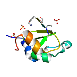
4PZI
 | | Zinc finger region of MLL2 in complex with CpG DNA | | 分子名称: | DNA (5'-D(*GP*CP*CP*AP*CP*CP*GP*GP*TP*GP*GP*C)-3'), Histone-lysine N-methyltransferase 2B, UNKNOWN ATOM OR ION, ... | | 著者 | Chao, X, Tempel, W, Liu, K, Dong, A, Bountra, C, Weigelt, J, Arrowsmith, C.H, Edwards, A.M, Min, J, Structural Genomics Consortium (SGC) | | 登録日 | 2014-03-31 | | 公開日 | 2014-06-04 | | 最終更新日 | 2024-04-03 | | 実験手法 | X-RAY DIFFRACTION (2.15 Å) | | 主引用文献 | DNA Sequence Recognition of Human CXXC Domains and Their Structural Determinants.
Structure, 26, 2018
|
|
4QQG
 | | Crystal structure of an N-terminal HTATIP fragment | | 分子名称: | Histone acetyltransferase KAT5, UNKNOWN ATOM OR ION | | 著者 | Liu, Y, Tempel, W, Wernimont, A.K, Dobrovetsky, E, Bountra, C, Arrowsmith, C.H, Edwards, A.M, Min, J, Structural Genomics Consortium (SGC) | | 登録日 | 2014-06-27 | | 公開日 | 2014-07-09 | | 最終更新日 | 2023-09-20 | | 実験手法 | X-RAY DIFFRACTION (2.8 Å) | | 主引用文献 | Structural and histone binding studies of the chromo barrel domain of TIP60.
FEBS Lett., 592, 2018
|
|


