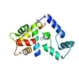
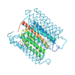
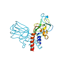
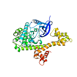
5J1A
 
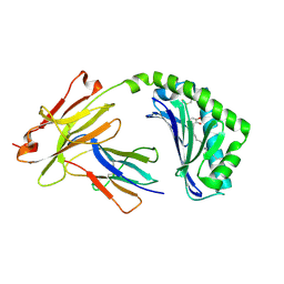 | | Antigen presenting molecule | | 分子名称: | 3-[(8Z,11Z)-pentadeca-8,11-dien-1-yl]benzene-1,2-diol, Beta-2-microglobulin, T-cell surface glycoprotein CD1a | | 著者 | Yongqing, T, Rossjohn, J, Le Nours, J, Marquez, E, Winau, F. | | 登録日 | 2016-03-29 | | 公開日 | 2016-08-03 | | 最終更新日 | 2018-04-04 | | 実験手法 | X-RAY DIFFRACTION (1.86 Å) | | 主引用文献 | CD1a on Langerhans cells controls inflammatory skin disease.
Nat. Immunol., 17, 2016
|
|
8D76
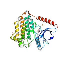 | | Crystal Structure of EGFR LRTM with compound 24 | | 分子名称: | (3S,4R)-3-fluoro-1-(4-{[8-{3-[(methanesulfonyl)methyl]azetidin-1-yl}-5-(propan-2-yl)-2,7-naphthyridin-3-yl]amino}pyrimidin-2-yl)-3-methylpiperidin-4-ol, Epidermal growth factor receptor, GLYCEROL | | 著者 | Kim, J.L. | | 登録日 | 2022-06-07 | | 公開日 | 2022-07-27 | | 最終更新日 | 2023-10-18 | | 実験手法 | X-RAY DIFFRACTION (2.4 Å) | | 主引用文献 | Discovery of BLU-945, a Reversible, Potent, and Wild-Type-Sparing Next-Generation EGFR Mutant Inhibitor for Treatment-Resistant Non-Small-Cell Lung Cancer.
J.Med.Chem., 65, 2022
|
|
8D73
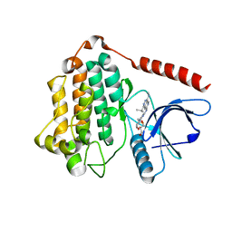 | | Crystal Structure of EGFR LRTM with compound 7 | | 分子名称: | (3S,4R)-3-fluoro-1-(4-{[4-(methylamino)-1-(propan-2-yl)pyrido[3,4-d]pyridazin-7-yl]amino}pyrimidin-2-yl)piperidin-4-ol, Epidermal growth factor receptor, GLYCEROL | | 著者 | Kim, J.L. | | 登録日 | 2022-06-07 | | 公開日 | 2022-07-27 | | 最終更新日 | 2023-10-18 | | 実験手法 | X-RAY DIFFRACTION (2.17 Å) | | 主引用文献 | Discovery of BLU-945, a Reversible, Potent, and Wild-Type-Sparing Next-Generation EGFR Mutant Inhibitor for Treatment-Resistant Non-Small-Cell Lung Cancer.
J.Med.Chem., 65, 2022
|
|
8HTG
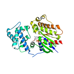 | |
5K8Q
 | | Crystal Structure of Calcium-loaded Calmodulin in complex with STRA6 CaMBP2-site peptide. | | 分子名称: | CALCIUM ION, Calmodulin, IMIDAZOLE, ... | | 著者 | Stowe, S.D, Clarke, O.B, Cavalier, M.C, Godoy-Ruiz, R, Mancia, F, Weber, D.J. | | 登録日 | 2016-05-30 | | 公開日 | 2016-08-24 | | 最終更新日 | 2024-04-03 | | 実験手法 | X-RAY DIFFRACTION (1.739 Å) | | 主引用文献 | Structure of the STRA6 receptor for retinol uptake.
Science, 353, 2016
|
|
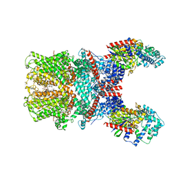
8GVW
 | | Cryo-EM structure of the human TRPC5 ion channel in lipid nanodiscs, class2 | | 分子名称: | (2S)-2-(hexadecanoyloxy)-3-hydroxypropyl (9Z)-octadec-9-enoate, (2S)-3-(hexadecanoyloxy)-2-[(9Z)-octadec-9-enoyloxy]propyl 2-(trimethylammonio)ethyl phosphate, CALCIUM ION, ... | | 著者 | Won, J, Jeong, H, Lee, H.H. | | 登録日 | 2022-09-16 | | 公開日 | 2023-05-24 | | 実験手法 | ELECTRON MICROSCOPY (3.59 Å) | | 主引用文献 | Molecular architecture of the G alpha i -bound TRPC5 ion channel.
Nat Commun, 14, 2023
|
|
8GVX
 | | Cryo-EM structure of the human TRPC5 ion channel in complex with G alpha i3 subunits, class2 | | 分子名称: | (2S)-2-(hexadecanoyloxy)-3-hydroxypropyl (9Z)-octadec-9-enoate, CALCIUM ION, CHOLESTEROL HEMISUCCINATE, ... | | 著者 | Won, J, Jeong, H, Lee, H.H. | | 登録日 | 2022-09-16 | | 公開日 | 2023-05-24 | | 実験手法 | ELECTRON MICROSCOPY (3.91 Å) | | 主引用文献 | Molecular architecture of the G alpha i -bound TRPC5 ion channel.
Nat Commun, 14, 2023
|
|
6A6A
 | | VanYB in complex with D-Alanine | | 分子名称: | ACETATE ION, D-ALANINE, D-alanyl-D-alanine carboxypeptidase, ... | | 著者 | Kim, H.S, Hahn, H. | | 登録日 | 2018-06-27 | | 公開日 | 2018-09-05 | | 最終更新日 | 2023-11-22 | | 実験手法 | X-RAY DIFFRACTION (2.26 Å) | | 主引用文献 | Structural basis for the substrate recognition of peptidoglycan pentapeptides by Enterococcus faecalis VanYB.
Int. J. Biol. Macromol., 119, 2018
|
|
8IU0
 | | Cryo-EM structure of the potassium-selective channelrhodopsin HcKCR1 H225F mutant in lipid nanodisc | | 分子名称: | (7R,17E,20E)-4-HYDROXY-N,N,N-TRIMETHYL-9-OXO-7-[(PALMITOYLOXY)METHYL]-3,5,8-TRIOXA-4-PHOSPHAHEXACOSA-17,20-DIEN-1-AMINIUM 4-OXIDE, HcKCR1, PALMITIC ACID, ... | | 著者 | Tajima, S, Kim, Y, Nakamura, S, Yamashita, K, Fukuda, M, Deisseroth, K, Kato, H.E. | | 登録日 | 2023-03-23 | | 公開日 | 2023-09-06 | | 最終更新日 | 2024-05-01 | | 実験手法 | ELECTRON MICROSCOPY (2.66 Å) | | 主引用文献 | Structural basis for ion selectivity in potassium-selective channelrhodopsins.
Cell, 186, 2023
|
|
4UAB
 | | Crystal structure of a TRAP periplasmic solute binding protein from Chromohalobacter salexigens DSM 3043 (Csal_0678), Target EFI-501078, with bound ethanolamine | | 分子名称: | CHLORIDE ION, ETHANOLAMINE, Twin-arginine translocation pathway signal | | 著者 | Vetting, M.W, Al Obaidi, N.F, Morisco, L.L, Wasserman, S.R, Sojitra, S, Stead, M, Attonito, J.D, Scott Glenn, A, Chowdhury, S, Evans, B, Hillerich, B, Love, J, Seidel, R.D, Imker, H.J, Gerlt, J.A, Almo, S.C, Enzyme Function Initiative (EFI) | | 登録日 | 2014-08-08 | | 公開日 | 2014-09-03 | | 最終更新日 | 2023-11-15 | | 実験手法 | X-RAY DIFFRACTION (1.4 Å) | | 主引用文献 | Experimental strategies for functional annotation and metabolism discovery: targeted screening of solute binding proteins and unbiased panning of metabolomes.
Biochemistry, 54, 2015
|
|
7X6I
 | | Cryo-EM structure of the human TRPC5 ion channel in complex with G alpha i3 subunits, class1 | | 分子名称: | (2S)-2-(hexadecanoyloxy)-3-hydroxypropyl (9Z)-octadec-9-enoate, CALCIUM ION, CHOLESTEROL HEMISUCCINATE, ... | | 著者 | Won, J, Jeong, H, Lee, H.H. | | 登録日 | 2022-03-07 | | 公開日 | 2023-04-26 | | 最終更新日 | 2023-05-24 | | 実験手法 | ELECTRON MICROSCOPY (3.93 Å) | | 主引用文献 | Molecular architecture of the G alpha i -bound TRPC5 ion channel.
Nat Commun, 14, 2023
|
|
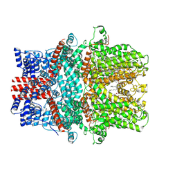
5J1M
 | | Crystal structure of Csd1-Csd2 dimer II | | 分子名称: | ToxR-activated gene (TagE), ZINC ION | | 著者 | An, D.R, Suh, S.W. | | 登録日 | 2016-03-29 | | 公開日 | 2016-10-19 | | 最終更新日 | 2024-03-20 | | 実験手法 | X-RAY DIFFRACTION (2.35 Å) | | 主引用文献 | Structural Basis of the Heterodimer Formation between Cell Shape-Determining Proteins Csd1 and Csd2 from Helicobacter pylori
Plos One, 11, 2016
|
|
7X6C
 | | Cryo-EM structure of the human TRPC5 ion channel in lipid nanodiscs, class1 | | 分子名称: | (2S)-2-(hexadecanoyloxy)-3-hydroxypropyl (9Z)-octadec-9-enoate, (2S)-3-(hexadecanoyloxy)-2-[(9Z)-octadec-9-enoyloxy]propyl 2-(trimethylammonio)ethyl phosphate, CALCIUM ION, ... | | 著者 | Won, J, Jeong, H, Lee, H.H. | | 登録日 | 2022-03-07 | | 公開日 | 2023-05-24 | | 実験手法 | ELECTRON MICROSCOPY (3.15 Å) | | 主引用文献 | Molecular architecture of the G alpha i -bound TRPC5 ion channel.
Nat Commun, 14, 2023
|
|
5J1K
 | | Crystal structure of Csd2-Csd2 dimer | | 分子名称: | GLYCEROL, ToxR-activated gene (TagE) | | 著者 | An, D.R, Suh, S.W. | | 登録日 | 2016-03-29 | | 公開日 | 2016-10-19 | | 最終更新日 | 2024-03-20 | | 実験手法 | X-RAY DIFFRACTION (1.81 Å) | | 主引用文献 | Structural Basis of the Heterodimer Formation between Cell Shape-Determining Proteins Csd1 and Csd2 from Helicobacter pylori
Plos One, 11, 2016
|
|
5J1L
 | | Crystal structure of Csd1-Csd2 dimer I | | 分子名称: | ToxR-activated gene (TagE), ZINC ION | | 著者 | An, D.R, Suh, S.W. | | 登録日 | 2016-03-29 | | 公開日 | 2016-10-19 | | 最終更新日 | 2024-03-20 | | 実験手法 | X-RAY DIFFRACTION (2.27 Å) | | 主引用文献 | Structural Basis of the Heterodimer Formation between Cell Shape-Determining Proteins Csd1 and Csd2 from Helicobacter pylori
Plos One, 11, 2016
|
|
3UY5
 | |
4WG5
 | |
4WG3
 | |
4WG4
 | |
7BYP
 | |
7BYO
 | |
7D04
 | |
7D05
 | |
7D01
 | |
7D02
 | |


