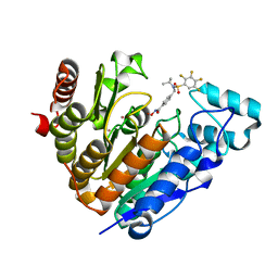
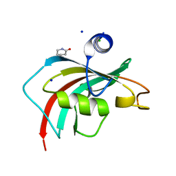
Loading
PDBj
MenuPDBj@FacebookPDBj@TwitterPDBj@YouTubewwPDB FoundationwwPDB
RCSB PDBPDBeBMRBAdv. SearchSearch help
Search by PDB author
6XTV
DownloadVisualize
BU of 6xtv by Molmil
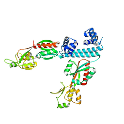
FULL-LENGTH LTTR LYSG FROM CORYNEBACTERIUM GLUTAMICUM WITH BOUND EFFECTOR ARG
Descriptor: ARGININE, Lysine export transcriptional regulatory protein LysG
Authors:Hofmann, E, Syberg, F, Schlicker, C, Eggeling, L, Schendzielorz, G.
Deposit date:2020-01-16
Release date:2020-10-07
Last modified:2024-01-24
Method:X-RAY DIFFRACTION (3.3 Å)
Cite:Engineering and application of a biosensor with focused ligand specificity.
Nat Commun, 11, 2020
6YLV
DownloadVisualize
BU of 6ylv by Molmil
Translation initiation factor 4E in complex with 4-Cl-Bn7GpppG mRNA 5' cap analog
Descriptor: 4-Cl-Bn7GpppG mRNA 5' cap analog, Eukaryotic translation initiation factor 4E, GLYCEROL
Authors:Kubacka, D, Wojcik, R, Baranowski, M.R, Kowalska, J, Jemielity, J.
Deposit date:2020-04-07
Release date:2020-06-10
Last modified:2024-01-24
Method:X-RAY DIFFRACTION (2.66005659 Å)
Cite:Novel N7-Arylmethyl Substituted Dinucleotide mRNA 5' cap Analogs: Synthesis and Evaluation as Modulators of Translation.
Pharmaceutics, 13, 2021
6DHO
DownloadVisualize
BU of 6dho by Molmil
RT XFEL structure of the two-flash state of Photosystem II (2F, S3-rich) at 2.07 Angstrom resolution
Descriptor: 1,2-DI-O-ACYL-3-O-[6-DEOXY-6-SULFO-ALPHA-D-GLUCOPYRANOSYL]-SN-GLYCEROL, 1,2-DIPALMITOYL-PHOSPHATIDYL-GLYCEROLE, 1,2-DISTEAROYL-MONOGALACTOSYL-DIGLYCERIDE, ...
Authors:Kern, J, Chatterjee, R, Young, I.D, Fuller, F.D, Lassalle, L, Ibrahim, M, Gul, S, Fransson, T, Brewster, A.S, Alonso-Mori, R, Hussein, R, Zhang, M, Douthit, L, de Lichtenberg, C, Cheah, M.H, Shevela, D, Wersig, J, Seufert, I, Sokaras, D, Pastor, E, Weninger, C, Kroll, T, Sierra, R.G, Aller, P, Butryn, A, Orville, A.M, Liang, M, Batyuk, A, Koglin, J.E, Carbajo, S, Boutet, S, Moriarty, N.W, Holton, J.M, Dobbek, H, Adams, P.D, Bergmann, U, Sauter, N.K, Zouni, A, Messinger, J, Yano, J, Yachandra, V.K.
Deposit date:2018-05-20
Release date:2018-11-21
Last modified:2024-03-13
Method:X-RAY DIFFRACTION (2.07 Å)
Cite:Structures of the intermediates of Kok's photosynthetic water oxidation clock.
Nature, 563, 2018
5EBJ
DownloadVisualize
BU of 5ebj by Molmil
Joint X-ray/neutron structure of reversibly photoswitching chromogenic protein, Dathail
Descriptor: photoswitching chromogenic protein
Authors:Kovalevsky, A.Y, Langan, P.S, Bradbury, A.R.M.
Deposit date:2015-10-19
Release date:2016-04-06
Last modified:2023-11-15
Method:NEUTRON DIFFRACTION (2.5 Å), X-RAY DIFFRACTION
Cite:Evolution and characterization of a new reversibly photoswitching chromogenic protein, Dathail.
J.Mol.Biol., 428, 2016
3KBW
DownloadVisualize
BU of 3kbw by Molmil
Room temperature X-ray mixed-metal structure of D-Xylose Isomerase in complex with Ni(2+) and Mg(2+) co-factors
Descriptor: MAGNESIUM ION, NICKEL (II) ION, Xylose isomerase
Authors:Kovalevsky, A.Y, Hanson, L, Langan, P.
Deposit date:2009-10-20
Release date:2010-06-16
Last modified:2024-02-21
Method:X-RAY DIFFRACTION (1.6 Å)
Cite:Metal ion roles and the movement of hydrogen during reaction catalyzed by D-xylose isomerase: a joint x-ray and neutron diffraction study.
Structure, 18, 2010
3KBM
DownloadVisualize
BU of 3kbm by Molmil
Room Temperature X-ray structure of D-Xylose Isomerase complexed with 2Cd(2+) co-factors and d12-D-alpha-glucose in the cyclic form
Descriptor: CADMIUM ION, Xylose isomerase, alpha-D-glucopyranose
Authors:Kovalevsky, A.Y, Hanson, L, Langan, P.
Deposit date:2009-10-20
Release date:2010-06-16
Last modified:2023-09-06
Method:X-RAY DIFFRACTION (2 Å)
Cite:Metal ion roles and the movement of hydrogen during reaction catalyzed by D-xylose isomerase: a joint x-ray and neutron diffraction study.
Structure, 18, 2010
3KCJ
DownloadVisualize
BU of 3kcj by Molmil
Room temperature neutron structure of apo-D-Xylose Isomerase (refined jointly with X-ray structure 3KBJ)
Descriptor: Xylose isomerase
Authors:Kovalevsky, A.Y, Langan, P.
Deposit date:2009-10-21
Release date:2010-09-29
Last modified:2024-04-03
Method:NEUTRON DIFFRACTION (1.8 Å), X-RAY DIFFRACTION
Cite:Identification of the Elusive Hydronium Ion Exchanging Roles with a Proton in an Enzyme at Lower pH Values
Angew.Chem.Int.Ed.Engl., 50, 2011
3KCL
DownloadVisualize
BU of 3kcl by Molmil
Room temperature neutron structure of D-Xylose Isomerase in complex with two Cd2+ cations and d12-D-alpha-glucose in the ring form (refined jointly with X-ray structure 3KBM)
Descriptor: CADMIUM ION, Xylose isomerase, alpha-D-glucopyranose
Authors:Kovalevsky, A.Y, Langan, P.
Deposit date:2009-10-21
Release date:2010-06-16
Last modified:2024-04-03
Method:NEUTRON DIFFRACTION (2 Å), X-RAY DIFFRACTION
Cite:Metal ion roles and the movement of hydrogen during reaction catalyzed by D-xylose isomerase: a joint x-ray and neutron diffraction study.
Structure, 18, 2010
3KBJ
DownloadVisualize
BU of 3kbj by Molmil
Room temperature X-ray structure of apo-D-Xylose Isomerase
Descriptor: Xylose isomerase
Authors:Kovalevsky, A.Y, Hanson, L, Langan, P.
Deposit date:2009-10-20
Release date:2010-09-29
Last modified:2024-02-21
Method:X-RAY DIFFRACTION (2 Å)
Cite:Identification of the Elusive Hydronium Ion Exchanging Roles with a Proton in an Enzyme at Lower pH Values.
Angew.Chem.Int.Ed.Engl., 50, 2011
3KCO
DownloadVisualize
BU of 3kco by Molmil
Room temperature neutron structure of D-Xylose Isomerase in complex with two Ni2+ cations and d12-D-glucose in the linear form (refined jointly with X-ray structure 3KBN)
Descriptor: D-glucose, NICKEL (II) ION, Xylose isomerase
Authors:Kovalevsky, A.Y, Langan, P.
Deposit date:2009-10-21
Release date:2010-06-16
Last modified:2024-04-03
Method:NEUTRON DIFFRACTION (1.8 Å), X-RAY DIFFRACTION
Cite:Metal ion roles and the movement of hydrogen during reaction catalyzed by D-xylose isomerase: a joint x-ray and neutron diffraction study.
Structure, 18, 2010
3KBN
DownloadVisualize
BU of 3kbn by Molmil
Room temperature structure of D-Xylose Isomerase in complex with 2Ni(2+) co-factors and d12-D-glucose in the linear form
Descriptor: D-glucose, NICKEL (II) ION, Xylose isomerase
Authors:Kovalevsky, A.Y, Hanson, L, Langan, P.
Deposit date:2009-10-20
Release date:2010-06-16
Last modified:2023-09-06
Method:X-RAY DIFFRACTION (1.53 Å)
Cite:Metal ion roles and the movement of hydrogen during reaction catalyzed by D-xylose isomerase: a joint x-ray and neutron diffraction study.
Structure, 18, 2010
3KBS
DownloadVisualize
BU of 3kbs by Molmil
Room Temperature X-ray structure of D-Xylose Isomerase in complex with 2Cd(2+) co-factors
Descriptor: CADMIUM ION, Xylose isomerase
Authors:Kovalevsky, A.Y, Hanson, L, Langan, P.
Deposit date:2009-10-20
Release date:2010-06-16
Last modified:2024-02-21
Method:X-RAY DIFFRACTION (1.8 Å)
Cite:Metal ion roles and the movement of hydrogen during reaction catalyzed by D-xylose isomerase: a joint x-ray and neutron diffraction study.
Structure, 18, 2010
3KBV
DownloadVisualize
BU of 3kbv by Molmil
Room temperature structure of D-Xylose Isomerase in complex with 2Ni(2+) co-factors
Descriptor: NICKEL (II) ION, Xylose isomerase
Authors:Kovalevsky, A.Y, Hanson, L, Langan, P.
Deposit date:2009-10-20
Release date:2010-06-16
Last modified:2024-02-21
Method:X-RAY DIFFRACTION (1.8 Å)
Cite:Metal ion roles and the movement of hydrogen during reaction catalyzed by D-xylose isomerase: a joint x-ray and neutron diffraction study.
Structure, 18, 2010
3KKX
DownloadVisualize
BU of 3kkx by Molmil
Neutron structure of human carbonic anhydrase II
Descriptor: Carbonic anhydrase 2, ZINC ION
Authors:Fisher, S.Z, Langan, P.A.
Deposit date:2009-11-06
Release date:2010-01-12
Last modified:2023-09-06
Method:NEUTRON DIFFRACTION (2 Å)
Cite:Neutron structure of human carbonic anhydrase II: implications for proton transfer.
Biochemistry, 49, 2010
3KMF
DownloadVisualize
BU of 3kmf by Molmil
Room Temperature Time-of-Flight Neutron Diffraction Study of Deoxy Human Normal Adult Hemoglobin
Descriptor: Hemoglobin subunit alpha, Hemoglobin subunit beta, PROTOPORPHYRIN IX CONTAINING FE
Authors:Kovalevsky, A.Y, Morimoto, Y, Chatake, T.
Deposit date:2009-11-10
Release date:2010-04-21
Last modified:2023-11-01
Method:NEUTRON DIFFRACTION (2 Å)
Cite:Direct Determination of Protonation States of Histidine Residues in a 2 A Neutron Structure of Deoxy-Human Normal Adult Hemoglobin and Implications for the Bohr Effect.
J.Mol.Biol., 398, 2010
8G6C
DownloadVisualize
BU of 8g6c by Molmil
GTP Cyclohydrolase-IB with manganese
Descriptor: 1,2-ETHANEDIOL, GTP cyclohydrolase FolE2, MANGANESE (II) ION
Authors:McWhorter, K.L, Amaya Lopez, C.Y, Davis, K.M.
Deposit date:2023-02-14
Release date:2024-02-21
Method:X-RAY DIFFRACTION (2.82 Å)
Cite:Combatting melioidosis with chemical synthetic lethality
To Be Published
7UK2
DownloadVisualize
BU of 7uk2 by Molmil
Crystal structure of Danio rerio histone deacetylase 6 catalytic domain 2 complexed with NN-390
Descriptor: Hdac6 protein, N-hydroxy-4-{[(propan-2-yl)(2,3,4,5-tetrafluorobenzene-1-sulfonyl)amino]methyl}benzamide, POTASSIUM ION, ...
Authors:Erdogan, F, Seo, H.-S, Dhe-Paganon, S.
Deposit date:2022-03-31
Release date:2022-11-02
Last modified:2023-10-25
Method:X-RAY DIFFRACTION (1.6 Å)
Cite:High Efficacy and Drug Synergy of HDAC6-Selective Inhibitor NN-429 in Natural Killer (NK)/T-Cell Lymphoma.
Pharmaceuticals, 15, 2022
7AYG
DownloadVisualize
BU of 7ayg by Molmil
oxalyl-CoA decarboxylase from Methylorubrum extorquens with bound TPP and ADP
Descriptor: ADENOSINE-5'-DIPHOSPHATE, MAGNESIUM ION, Putative oxalyl-CoA decarboxylase (Oxc, ...
Authors:Pfister, P, Burgener, S, Nattermann, M, Zarzycki, J, Erb, T.J.
Deposit date:2020-11-12
Release date:2021-04-28
Last modified:2024-01-31
Method:X-RAY DIFFRACTION (1.9 Å)
Cite:Engineering a Highly Efficient Carboligase for Synthetic One-Carbon Metabolism.
Acs Catalysis, 11, 2021
7B2E
DownloadVisualize
BU of 7b2e by Molmil
quadruple mutant of oxalyl-CoA decarboxylase from Methylorubrum extorquens with bound TPP and ADP
Descriptor: ADENOSINE-5'-DIPHOSPHATE, MAGNESIUM ION, Putative oxalyl-CoA decarboxylase (Oxc, ...
Authors:Pfister, P, Burgener, S, Nattermann, M, Zarzycki, J, Erb, T.J.
Deposit date:2020-11-26
Release date:2021-04-28
Last modified:2024-01-31
Method:X-RAY DIFFRACTION (2.8 Å)
Cite:Engineering a Highly Efficient Carboligase for Synthetic One-Carbon Metabolism.
Acs Catalysis, 11, 2021
6TX4
DownloadVisualize
BU of 6tx4 by Molmil
CRYSTAL STRUCTURE OF HUMAN FKBP51 FK1 DOMAIN A19T MUTANT IN COMPLEX WITH 2-PYRIDONE
Descriptor: 1~{H}-pyridin-2-one, CHLORIDE ION, Peptidyl-prolyl cis-trans isomerase FKBP5, ...
Authors:Fiegen, D, Draxler, S.W.
Deposit date:2020-01-13
Release date:2020-05-27
Last modified:2024-01-24
Method:X-RAY DIFFRACTION (1.06 Å)
Cite:Hybrid Screening Approach for Very Small Fragments: X-ray and Computational Screening on FKBP51.
J.Med.Chem., 63, 2020
6TX7
DownloadVisualize
BU of 6tx7 by Molmil
CRYSTAL STRUCTURE OF HUMAN FKBP51 FK1 DOMAIN A19T MUTANT IN COMPLEX WITH 2-PIPERIDONE
Descriptor: CHLORIDE ION, Peptidyl-prolyl cis-trans isomerase FKBP5, SODIUM ION, ...
Authors:Fiegen, D, Draxler, S.W.
Deposit date:2020-01-13
Release date:2020-05-27
Last modified:2024-01-24
Method:X-RAY DIFFRACTION (1.13 Å)
Cite:Hybrid Screening Approach for Very Small Fragments: X-ray and Computational Screening on FKBP51.
J.Med.Chem., 63, 2020
5H31
DownloadVisualize
BU of 5h31 by Molmil
Structural basis for dimerization of the death effector domains of Caspase-8
Descriptor: Caspase-8
Authors:Shen, C, Pei, J, Guo, X, Quan, J.
Deposit date:2016-10-19
Release date:2017-10-25
Last modified:2024-03-20
Method:X-RAY DIFFRACTION (3.16953516 Å)
Cite:Structural basis for dimerization of the death effector domains of Caspase-8
To Be Published
6TX5
DownloadVisualize
BU of 6tx5 by Molmil
CRYSTAL STRUCTURE OF HUMAN FKBP51 FK1 DOMAIN A19T MUTANT IN COMPLEX WITH 4-METHYLIMIDAZOLE
Descriptor: 4-METHYLIMIDAZOLE, Peptidyl-prolyl cis-trans isomerase FKBP5, SODIUM ION
Authors:Fiegen, D, Draxler, S.W.
Deposit date:2020-01-13
Release date:2020-05-27
Last modified:2024-01-24
Method:X-RAY DIFFRACTION (1.08 Å)
Cite:Hybrid Screening Approach for Very Small Fragments: X-ray and Computational Screening on FKBP51.
J.Med.Chem., 63, 2020
5T8H
DownloadVisualize
BU of 5t8h by Molmil
Joint X-ray/neutron structure of HIV-1 protease triple mutant (V32I,I47V,V82I) with amprenavir at pH 6.0
Descriptor: Protease, {3-[(4-AMINO-BENZENESULFONYL)-ISOBUTYL-AMINO]-1-BENZYL-2-HYDROXY-PROPYL}-CARBAMIC ACID TETRAHYDRO-FURAN-3-YL ESTER
Authors:Kovalevsky, A.Y, Gerlits, O.O.
Deposit date:2016-09-07
Release date:2017-03-01
Last modified:2024-04-03
Method:NEUTRON DIFFRACTION (1.85 Å), X-RAY DIFFRACTION
Cite:Room Temperature Neutron Crystallography of Drug Resistant HIV-1 Protease Uncovers Limitations of X-ray Structural Analysis at 100 K.
J. Med. Chem., 60, 2017
7ABB
DownloadVisualize
BU of 7abb by Molmil
The truncated structure of the Bottromycin biosynthetic protein SalCYP
Descriptor: HEME C, SalCYP truncation
Authors:Adam, S, Koehnke, J.
Deposit date:2020-09-07
Release date:2020-12-02
Last modified:2024-01-31
Method:X-RAY DIFFRACTION (1.50004339 Å)
Cite:Characterization of the Stereoselective P450 Enzyme BotCYP Enables the In Vitro Biosynthesis of the Bottromycin Core Scaffold.
J.Am.Chem.Soc., 142, 2020

224931

PDB entries from 2024-09-11

PDB statisticsPDBj update infoContact PDBjnumon