Loading
PDBj
MenuPDBj@FacebookPDBj@TwitterPDBj@YouTubewwPDB FoundationwwPDB
RCSB PDBPDBeBMRBAdv. SearchSearch help
Search by PDB author
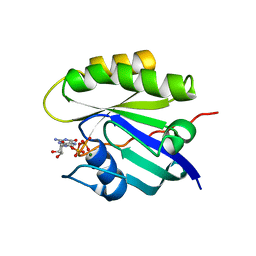
6Q21
DownloadVisualize
BU of 6q21 by Molmil
MOLECULAR SWITCH FOR SIGNAL TRANSDUCTION: STRUCTURAL DIFFERENCES BETWEEN ACTIVE AND INACTIVE FORMS OF PROTOONCOGENIC RAS PROTEINS
Descriptor: C-H-RAS P21 PROTEIN CATALYTIC DOMAIN, MAGNESIUM ION, PHOSPHOMETHYLPHOSPHONIC ACID GUANYLATE ESTER
Authors:Kim, S.-H.
Deposit date:1992-07-27
Release date:1992-07-28
Last modified:2024-03-13
Method:X-RAY DIFFRACTION (1.95 Å)
Cite:Molecular switch for signal transduction: structural differences between active and inactive forms of protooncogenic ras proteins.
Science, 247, 1990
4Q21
DownloadVisualize
BU of 4q21 by Molmil
MOLECULAR SWITCH FOR SIGNAL TRANSDUCTION: STRUCTURAL DIFFERENCES BETWEEN ACTIVE AND INACTIVE FORMS OF PROTOONCOGENIC RAS PROTEINS
Descriptor: C-H-RAS P21 PROTEIN CATALYTIC DOMAIN, GUANOSINE-5'-DIPHOSPHATE, MAGNESIUM ION
Authors:Kim, S.-H.
Deposit date:1991-09-25
Release date:1992-07-15
Last modified:2024-02-28
Method:X-RAY DIFFRACTION (2 Å)
Cite:Molecular switch for signal transduction: structural differences between active and inactive forms of protooncogenic ras proteins.
Science, 247, 1990
1U0L
DownloadVisualize
BU of 1u0l by Molmil
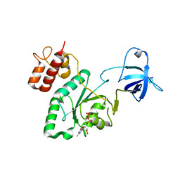
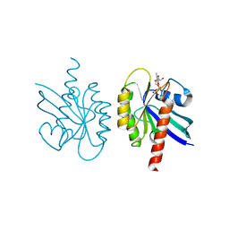
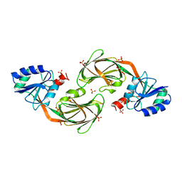
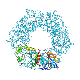
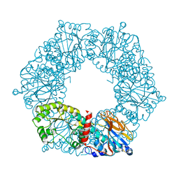
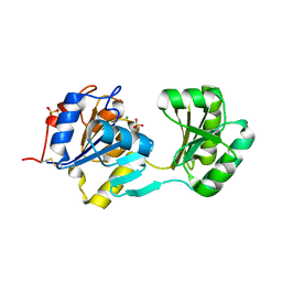
Crystal structure of YjeQ from Thermotoga maritima
Descriptor: GUANOSINE-5'-DIPHOSPHATE, Probable GTPase engC, ZINC ION
Authors:Shin, D.H, Lou, Y, Jaru, J, Kim, R, Yokota, H, Kim, S.H, Berkeley Structural Genomics Center (BSGC)
Deposit date:2004-07-13
Release date:2004-09-07
Last modified:2024-02-14
Method:X-RAY DIFFRACTION (2.8 Å)
Cite:Crystal structure of YjeQ from Thermotoga maritima contains a circularly permuted GTPase domain
Proc.Natl.Acad.Sci.Usa, 101, 2004
2P3U
DownloadVisualize
BU of 2p3u by Molmil
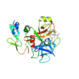
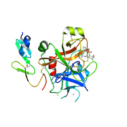
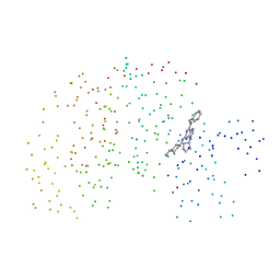
Crystal structure of human factor XA complexed with 3-chloro-N-(4-chloro-2-{[(5-chloropyridin-2-yl)amino]carbonyl}-6-methoxyphenyl)-4-[(1-methyl-1H-imidazol-2-yl)methyl]thiophene-2-carboxamide {Pfizer 320663}
Descriptor: 3-CHLORO-N-(4-CHLORO-2-{[(5-CHLOROPYRIDIN-2-YL)AMINO]CARBONYL}-6-METHOXYPHENYL)-4-[(1-METHYL-1H-IMIDAZOL-2-YL)METHYL]THIOPHENE-2-CARBOXAMIDE, CALCIUM ION, Coagulation factor X
Authors:Adler, M, Whitlow, M.
Deposit date:2007-03-09
Release date:2007-09-11
Last modified:2023-08-30
Method:X-RAY DIFFRACTION (1.62 Å)
Cite:

2LIG
DownloadVisualize
BU of 2lig by Molmil
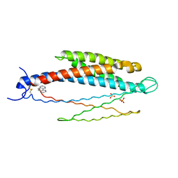
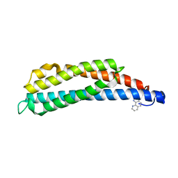
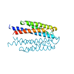
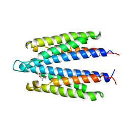
THREE-DIMENSIONAL STRUCTURES OF THE LIGAND-BINDING DOMAIN OF THE BACTERIAL ASPARTATE RECEPTOR WITH AND WITHOUT A LIGAND
Descriptor: 1,10-PHENANTHROLINE, ASPARTATE RECEPTOR, ASPARTIC ACID, ...
Authors:Kim, S.-H, Yeh, J.I, Prive, G.G, Milburn, M, Scott, W, Koshland Junior, D.E.
Deposit date:1995-04-18
Release date:1995-09-15
Last modified:2024-10-23
Method:X-RAY DIFFRACTION (2 Å)
Cite:Three-dimensional structures of the ligand-binding domain of the bacterial aspartate receptor with and without a ligand.
Science, 254, 1991
1CKP
DownloadVisualize
BU of 1ckp by Molmil
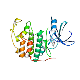
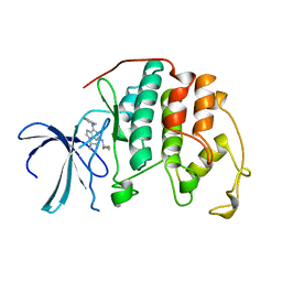
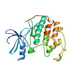
HUMAN CYCLIN DEPENDENT KINASE 2 COMPLEXED WITH THE INHIBITOR PURVALANOL B
Descriptor: 1,2-ETHANEDIOL, PROTEIN (CYCLIN-DEPENDENT PROTEIN KINASE 2), PURVALANOL B
Authors:Gray, N.S, Thunnissen, A.M.W.H, Schultz, P.G, Kim, S.H.
Deposit date:1998-07-14
Release date:1999-01-13
Last modified:2023-08-09
Method:X-RAY DIFFRACTION (2.05 Å)
Cite:Exploiting chemical libraries, structure, and genomics in the search for kinase inhibitors.
Science, 281, 1998
2Q21
DownloadVisualize
BU of 2q21 by Molmil
CRYSTAL STRUCTURES AT 2.2 ANGSTROMS RESOLUTION OF THE CATALYTIC DOMAINS OF NORMAL RAS PROTEIN AND AN ONCOGENIC MUTANT COMPLEXED WITH GSP
Descriptor: C-H-RAS P21 PROTEIN CATALYTIC DOMAIN, GUANOSINE-5'-DIPHOSPHATE, MAGNESIUM ION
Authors:Kim, S.-H.
Deposit date:1991-09-25
Release date:1992-07-15
Last modified:2024-02-21
Method:X-RAY DIFFRACTION (2.2 Å)
Cite:Crystal structures at 2.2 A resolution of the catalytic domains of normal ras protein and an oncogenic mutant complexed with GDP.
J.Mol.Biol., 217, 1991
2P3T
DownloadVisualize
BU of 2p3t by Molmil
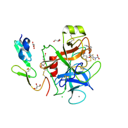
Crystal structure of human factor XA complexed with 3-Chloro-4-(2-methylamino-imidazol-1-ylmethyl)-thiophene-2-carboxylic acid [4-chloro-2-(5-chloro-pyridin-2-ylcarbamoyl)-6-methoxy-phenyl]-amide
Descriptor: 3-CHLORO-4-(2-METHYLAMINO-IMIDAZOL-1-YLMETHYL)-THIOPHENE-2-CARBOXYLIC ACID [4-CHLORO-2-(5-CHLORO-PYRIDIN-2-YLCARBAMOYL)-6-METHOXY-PHENYL]-AMIDE, CALCIUM ION, CHLORIDE ION, ...
Authors:Adler, M, Whitlow, M.
Deposit date:2007-03-09
Release date:2008-01-22
Last modified:2024-11-06
Method:X-RAY DIFFRACTION (1.92 Å)
Cite:Thiophene-anthranilamides as highly potent and orally available factor xa inhibitors.
J.Med.Chem., 50, 2007
1FJS
DownloadVisualize
BU of 1fjs by Molmil
CRYSTAL STRUCTURE OF THE INHIBITOR ZK-807834 (CI-1031) COMPLEXED WITH FACTOR XA
Descriptor: CALCIUM ION, CHLORIDE ION, COAGULATION FACTOR XA, ...
Authors:Adler, M, Whitlow, M.
Deposit date:2000-08-08
Release date:2000-10-04
Last modified:2024-03-13
Method:X-RAY DIFFRACTION (1.92 Å)
Cite:Preparation, characterization, and the crystal structure of the inhibitor ZK-807834 (CI-1031) complexed with factor Xa.
Biochemistry, 39, 2000
2FVO
DownloadVisualize
BU of 2fvo by Molmil
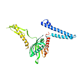
Docking of the modified RF1 X-ray structure into the Low Resolution Cryo-EM map of E.coli 70S Ribosome bound with RF1
Descriptor: Peptide chain release factor 1
Authors:Rawat, U, Gao, H, Zavialov, A, Gursky, R, Ehrenberg, M, Frank, J.
Deposit date:2006-01-31
Release date:2006-04-04
Last modified:2024-02-14
Method:ELECTRON MICROSCOPY (12.8 Å)
Cite:Interactions of the Release Factor RF1 with the Ribosome as Revealed by Cryo-EM.
J.Mol.Biol., 357, 2006
1AQ1
DownloadVisualize
BU of 1aq1 by Molmil
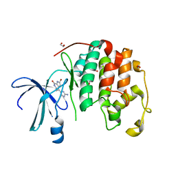
HUMAN CYCLIN DEPENDENT KINASE 2 COMPLEXED WITH THE INHIBITOR STAUROSPORINE
Descriptor: CYCLIN-DEPENDENT PROTEIN KINASE 2, STAUROSPORINE
Authors:Endicott, J.A, Noble, M.E.M, Johnson, L.N, Lawrie, A, Tunnah, P, Brown, N.R.
Deposit date:1997-08-05
Release date:1997-11-12
Last modified:2024-05-22
Method:X-RAY DIFFRACTION (2 Å)
Cite:Protein kinase inhibition by staurosporine revealed in details of the molecular interaction with CDK2.
Nat.Struct.Biol., 4, 1997
1YT5
DownloadVisualize
BU of 1yt5 by Molmil
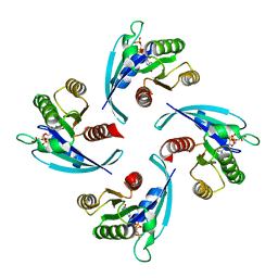
Crystal structure of NAD kinase from Thermotoga maritima
Descriptor: SULFATE ION, inorganic polyphosphate/ATP-NAD kinase
Authors:Berkeley Structural Genomics Center (BSGC)
Deposit date:2005-02-09
Release date:2005-04-05
Last modified:2024-02-14
Method:X-RAY DIFFRACTION (2.3 Å)
Cite:Structure of a NAD kinase from Thermotoga maritima at 2.3 A resolution.
Acta Crystallogr.,Sect.F, 61, 2005
1YTK
DownloadVisualize
BU of 1ytk by Molmil
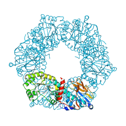
Crystal structure of a nicotinate phosphoribosyltransferase from Thermoplasma acidophilum with nicotinate mononucleotide
Descriptor: 2-AMINO-2-HYDROXYMETHYL-PROPANE-1,3-DIOL, NICOTINATE MONONUCLEOTIDE, nicotinate phosphoribosyltransferase from Thermoplasma acidophilum
Authors:Shin, D.H, Berkeley Structural Genomics Center (BSGC)
Deposit date:2005-02-10
Release date:2005-03-08
Last modified:2011-07-13
Method:X-RAY DIFFRACTION (2.65 Å)
Cite:Crystal structure of a nicotinate phosphoribosyltransferase from Thermoplasma acidophilum.
J.Biol.Chem., 280, 2005
1DM2
DownloadVisualize
BU of 1dm2 by Molmil
HUMAN CYCLIN-DEPENDENT KINASE 2 COMPLEXED WITH THE INHIBITOR HYMENIALDISINE
Descriptor: 1,2-ETHANEDIOL, 4-(5-AMINO-4-OXO-4H-PYRAZOL-3-YL)-2-BROMO-4,5,6,7-TETRAHYDRO-3AH-PYRROLO[2,3-C]AZEPIN-8-ONE, CYCLIN-DEPENDENT KINASE 2
Authors:Thunnissen, A.M, Kim, S.-H.
Deposit date:1999-12-13
Release date:2000-05-31
Last modified:2024-02-07
Method:X-RAY DIFFRACTION (2.1 Å)
Cite:Inhibition of cyclin-dependent kinases, GSK-3beta and CK1 by hymenialdisine, a marine sponge constituent.
Chem.Biol., 7, 2000
1YTE
DownloadVisualize
BU of 1yte by Molmil
Crystal structure of a nicotinate phosphoribosyltransferase from Thermoplasma acidophilum, phosphoribosylpyrophosphate bound structure
Descriptor: 1-ALPHA-PYROPHOSPHORYL-2-ALPHA,3-ALPHA-DIHYDROXY-4-BETA-CYCLOPENTANE-METHANOL-5-PHOSPHATE, nicotinate phosphoribosyltransferase from Thermoplasma acidophilum
Authors:Shin, D.H, Berkeley Structural Genomics Center (BSGC)
Deposit date:2005-02-10
Release date:2005-03-08
Last modified:2024-10-16
Method:X-RAY DIFFRACTION (2.75 Å)
Cite:Crystal structure of a nicotinate phosphoribosyltransferase from Thermoplasma acidophilum.
J.Biol.Chem., 280, 2005
1YTD
DownloadVisualize
BU of 1ytd by Molmil
Crystal structure of a nicotinate phosphoribosyltransferase from Thermoplasma acidophilum, Native Structure
Descriptor: nicotinate phosphoribosyltransferase from Thermoplasma acidophilum
Authors:Shin, D.H, Berkeley Structural Genomics Center (BSGC)
Deposit date:2005-02-10
Release date:2005-03-08
Last modified:2024-10-30
Method:X-RAY DIFFRACTION (2.8 Å)
Cite:Crystal structure of a nicotinate phosphoribosyltransferase from Thermoplasma acidophilum.
J.Biol.Chem., 280, 2005
1LIH
DownloadVisualize
BU of 1lih by Molmil
THREE-DIMENSIONAL STRUCTURES OF THE LIGAND-BINDING DOMAIN OF THE BACTERIAL ASPARTATE RECEPTOR WITH AND WITHOUT A LIGAND
Descriptor: 1,10-PHENANTHROLINE, ASPARTATE RECEPTOR
Authors:Kim, S.-H, Scott, W, Yeh, J.I, Prive, G.G, Milburn, M.
Deposit date:1995-04-18
Release date:1995-09-15
Last modified:2024-02-14
Method:X-RAY DIFFRACTION (2.2 Å)
Cite:Three-dimensional structures of the ligand-binding domain of the bacterial aspartate receptor with and without a ligand.
Science, 254, 1991
1VLS
DownloadVisualize
BU of 1vls by Molmil
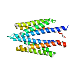
LIGAND BINDING DOMAIN OF THE WILD-TYPE ASPARTATE RECEPTOR
Descriptor: ASPARTATE RECEPTOR
Authors:Kim, S.-H, Yeh, J.I, Biemann, H.-P, Prive, G, Pandit, J, Koshland Junior, D.E.
Deposit date:1996-09-17
Release date:1997-04-21
Last modified:2024-02-14
Method:X-RAY DIFFRACTION (1.85 Å)
Cite:High-resolution structures of the ligand binding domain of the wild-type bacterial aspartate receptor.
J.Mol.Biol., 262, 1996
1VLT
DownloadVisualize
BU of 1vlt by Molmil
LIGAND BINDING DOMAIN OF THE WILD-TYPE ASPARTATE RECEPTOR WITH ASPARTATE
Descriptor: ASPARTATE RECEPTOR, ASPARTIC ACID
Authors:Kim, S.-H, Yeh, J.I, Biemann, H.-P, Prive, G, Pandit, J, Koshland Junior, D.E.
Deposit date:1996-09-17
Release date:1997-05-15
Last modified:2024-02-14
Method:X-RAY DIFFRACTION (2.2 Å)
Cite:High-resolution structures of the ligand binding domain of the wild-type bacterial aspartate receptor.
J.Mol.Biol., 262, 1996
2A4L
DownloadVisualize
BU of 2a4l by Molmil
Human cyclin-dependent kinase 2 in complex with roscovitine
Descriptor: Homo sapiens cyclin-dependent kinase 2, R-ROSCOVITINE
Authors:De Azevedo Jr, W.F, Kim, S.H.
Deposit date:2005-06-29
Release date:2006-10-03
Last modified:2024-02-14
Method:X-RAY DIFFRACTION (2.4 Å)
Cite:Inhibition of cyclin-dependent kinases by purine analogues: crystal structure of human cdk2 complexed with roscovitine.
Eur.J.Biochem., 243, 1997
1WAT
DownloadVisualize
BU of 1wat by Molmil
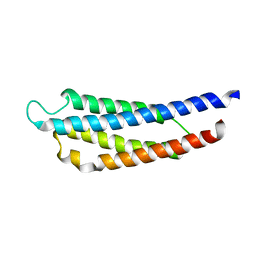
THE THREE-DIMENSIONAL STRUCTURE OF THE LIGAND-BINDING DOMAIN OF A WILD-TYPE BACTERIAL CHEMOTAXIS RECEPTOR
Descriptor: ASPARTATE RECEPTOR, ASPARTIC ACID
Authors:Kim, S.-H.
Deposit date:1993-03-09
Release date:1994-12-20
Last modified:2024-02-14
Method:X-RAY DIFFRACTION (3 Å)
Cite:The three-dimensional structure of the ligand-binding domain of a wild-type bacterial chemotaxis receptor. Structural comparison to the cross-linked mutant forms and conformational changes upon ligand binding.
J.Biol.Chem., 268, 1993
1WAS
DownloadVisualize
BU of 1was by Molmil
THE THREE-DIMENSIONAL STRUCTURE OF THE LIGAND-BINDING DOMAIN OF A WILD-TYPE BACTERIAL CHEMOTAXIS RECEPTOR
Descriptor: BACTERIAL ASPARTATE RECEPTOR
Authors:Kim, S.-H.
Deposit date:1993-03-09
Release date:1994-12-20
Last modified:2024-02-14
Method:X-RAY DIFFRACTION (2.7 Å)
Cite:The three-dimensional structure of the ligand-binding domain of a wild-type bacterial chemotaxis receptor. Structural comparison to the cross-linked mutant forms and conformational changes upon ligand binding.
J.Biol.Chem., 268, 1993
1NF2
DownloadVisualize
BU of 1nf2 by Molmil
X-ray crystal structure of TM0651 from Thermotoga maritima
Descriptor: MAGNESIUM ION, SULFATE ION, phosphatase
Authors:Shin, D.H, Berkeley Structural Genomics Center (BSGC)
Deposit date:2002-12-12
Release date:2003-09-16
Last modified:2011-07-13
Method:X-RAY DIFFRACTION (2.2 Å)
Cite:Crystal structure of a phosphatase with a unique substrate binding domain from Thermotoga maritima
Protein Sci., 12, 2003
1G5S
DownloadVisualize
BU of 1g5s by Molmil
CRYSTAL STRUCTURE OF HUMAN CYCLIN DEPENDENT KINASE 2 (CDK2) IN COMPLEX WITH THE INHIBITOR H717
Descriptor: 2-[TRANS-(4-AMINOCYCLOHEXYL)AMINO]-6-(BENZYL-AMINO)-9-CYCLOPENTYLPURINE, CELL DIVISION PROTEIN KINASE 2
Authors:Dreyer, M.K, Borcherding, D.R, Dumont, J.A, Peet, N.P, Tsay, J.T, Wright, P.S, Bitonti, A.J, Shen, J, Kim, S.-H.
Deposit date:2000-11-02
Release date:2001-11-02
Last modified:2023-08-09
Method:X-RAY DIFFRACTION (2.61 Å)
Cite:Crystal structure of human cyclin-dependent kinase 2 in complex with the adenine-derived inhibitor H717.
J.Med.Chem., 44, 2001
1HCL
DownloadVisualize
BU of 1hcl by Molmil
HUMAN CYCLIN-DEPENDENT KINASE 2
Descriptor: HUMAN CYCLIN-DEPENDENT KINASE 2
Authors:Schulze-Gahmen, U, De Bondt, H.L, Kim, S.-H.
Deposit date:1996-06-03
Release date:1996-12-07
Last modified:2024-02-07
Method:X-RAY DIFFRACTION (1.8 Å)
Cite:High-resolution crystal structures of human cyclin-dependent kinase 2 with and without ATP: bound waters and natural ligand as guides for inhibitor design.
J.Med.Chem., 39, 1996

227111

PDB entries from 2024-11-06

PDB statisticsPDBj update infoContact PDBjnumon