Loading
PDBj
MenuPDBj@FacebookPDBj@TwitterPDBj@YouTubewwPDB FoundationwwPDB
RCSB PDBPDBeBMRBAdv. SearchSearch help
Search by PDB author
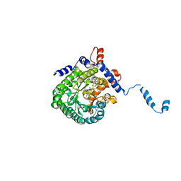
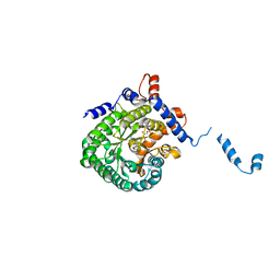
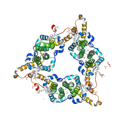
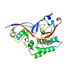
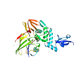
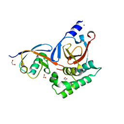
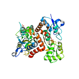
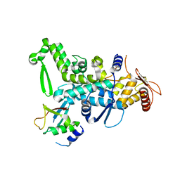
5C7J
DownloadVisualize
BU of 5c7j by Molmil
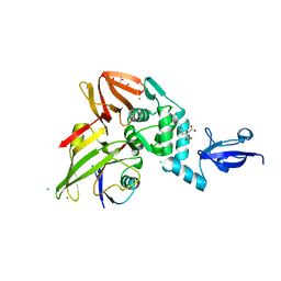
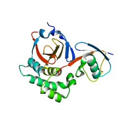
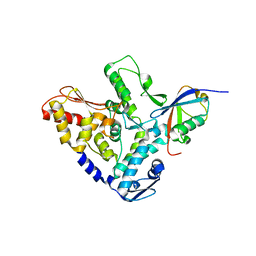
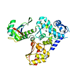
CRYSTAL STRUCTURE OF NEDD4 WITH A UB VARIANT
Descriptor: E3 ubiquitin-protein ligase NEDD4, Polyubiquitin-C
Authors:Walker, J.R, Hu, J, Dong, A, Bountra, C, Edwards, A.M, Arrowsmith, C.H, Tong, Y, Structural Genomics Consortium (SGC)
Deposit date:2015-06-24
Release date:2016-03-16
Last modified:2023-09-27
Method:X-RAY DIFFRACTION (3 Å)
Cite:System-Wide Modulation of HECT E3 Ligases with Selective Ubiquitin Variant Probes.
Mol.Cell, 62, 2016
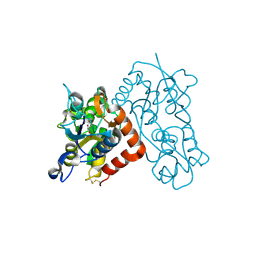
2FZM
DownloadVisualize
BU of 2fzm by Molmil
Structure of the E. coli PutA proline dehydrogenase domain reduced by dithionite and complexed with SO2
Descriptor: Bifunctional protein putA, Proline dehydrogenase (EC 1.5.99.8) (Proline oxidase), FLAVIN-ADENINE DINUCLEOTIDE, ...
Authors:Tanner, J.J.
Deposit date:2006-02-09
Release date:2007-04-03
Last modified:2023-08-30
Method:X-RAY DIFFRACTION (2.3 Å)
Cite:Redox-induced changes in flavin structure and roles of flavin N(5) and the ribityl 2'-OH group in regulating PutA--membrane binding.
Biochemistry, 46, 2007
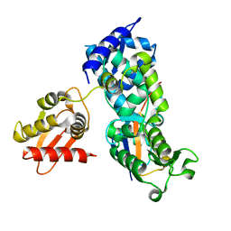
2FZN
DownloadVisualize
BU of 2fzn by Molmil
Structure of the E. coli PutA proline dehydrogenase domain reduced by dithionite and complexed with proline
Descriptor: Bifunctional protein putA, Proline dehydrogenase (EC 1.5.99.8) (Proline oxidase), FLAVIN-ADENINE DINUCLEOTIDE, ...
Authors:Tanner, J.J.
Deposit date:2006-02-09
Release date:2006-12-26
Last modified:2023-08-30
Method:X-RAY DIFFRACTION (2 Å)
Cite:Hydrogen Bonding Interactions of the 2-OH Ribityl Group and the N(5) Position of the FAD Cofactor Regulate PutA-membrane Associations in Escherichia coli
To be Published
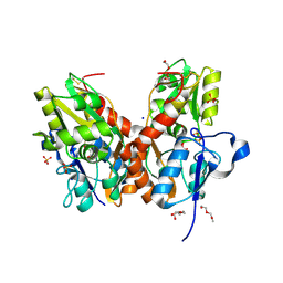
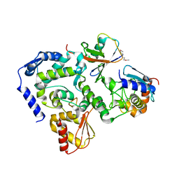
5NS9
DownloadVisualize
BU of 5ns9 by Molmil
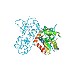
Crystal structure of the GluA2 LBD (L483Y-N754S-L758V) in complex with glutamate
Descriptor: 2-(2-METHOXYETHOXY)ETHANOL, GLUTAMIC ACID, Glutamate receptor 2,Glutamate receptor 2, ...
Authors:Eibl, C, Plested, A.J.R.
Deposit date:2017-04-25
Release date:2017-09-13
Last modified:2024-11-06
Method:X-RAY DIFFRACTION (1.44 Å)
Cite:Unitary Properties of AMPA Receptors with Reduced Desensitization.
Biophys. J., 113, 2017
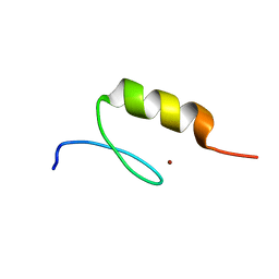
3IUF
DownloadVisualize
BU of 3iuf by Molmil
Crystal structure of the C2H2-type zinc finger domain of human ubi-d4
Descriptor: ZINC ION, Zinc finger protein ubi-d4
Authors:Tempel, W, Xu, C, Bian, C, Adams-Cioaba, M, Eryilmaz, J, Bountra, C, Weigelt, J, Arrowsmith, C.H, Edwards, A.M, Bochkarev, A, Min, J, Structural Genomics Consortium (SGC)
Deposit date:2009-08-31
Release date:2009-11-03
Last modified:2024-02-21
Method:X-RAY DIFFRACTION (1.8 Å)
Cite:Crystal structure of the Cys2His2-type zinc finger domain of human DPF2.
Biochem.Biophys.Res.Commun., 413, 2011
8DEA
DownloadVisualize
BU of 8dea by Molmil
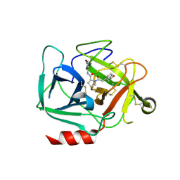
Scaffold Hopping via Ring Opening Enables Identification of Acyclic Compounds as New Complement Factor D Inhibitors
Descriptor: 1-[(3-acetylphenyl)acetyl]-N-(6-bromopyridin-2-yl)-L-prolinamide, Complement factor D, GLYCEROL
Authors:Raman, K, Babu, Y.S.
Deposit date:2022-06-20
Release date:2022-11-02
Last modified:2024-10-23
Method:X-RAY DIFFRACTION (2.214 Å)
Cite:Scaffold hopping via ring opening enables identification of acyclic compounds as new complement Factor D inhibitors.
Bioorg.Med.Chem., 74, 2022
8DG6
DownloadVisualize
BU of 8dg6 by Molmil
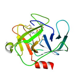
Scaffold Hopping via Ring Opening Enables Identification of Acyclic Compounds as New Complement Factor D Inhibitors
Descriptor: 1-{2-[(2S)-2-{[(3-chloro-2-fluorophenyl)methyl]carbamoyl}pyrrolidin-1-yl]-2-oxoethyl}-1H-indazole-3-carboxamide, Complement factor D
Authors:Raman, K, Babu, Y.S.
Deposit date:2022-06-23
Release date:2022-11-02
Last modified:2024-10-16
Method:X-RAY DIFFRACTION (1.986 Å)
Cite:Scaffold hopping via ring opening enables identification of acyclic compounds as new complement Factor D inhibitors.
Bioorg.Med.Chem., 74, 2022
8D95
DownloadVisualize
BU of 8d95 by Molmil
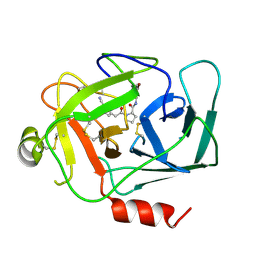
Scaffold Hopping via Ring Opening Enables Identification of Acyclic Compounds as New Complement Factor D Inhibitors
Descriptor: Complement factor D, N-(6-bromopyridin-2-yl)-1-[(3-cyanophenyl)acetyl]-L-prolinamide
Authors:Raman, K, Babu, Y.S.
Deposit date:2022-06-09
Release date:2022-11-02
Last modified:2023-10-18
Method:X-RAY DIFFRACTION (2.166 Å)
Cite:Scaffold hopping via ring opening enables identification of acyclic compounds as new complement Factor D inhibitors.
Bioorg.Med.Chem., 74, 2022
5V6A
DownloadVisualize
BU of 5v6a by Molmil
Crystal structure of the Middle East respiratory syndrome coronavirus papain-like protease bound to ubiquitin variant ME.2
Descriptor: CHLORIDE ION, CITRATE ANION, ME.2, ...
Authors:Bailey-Elkin, B.A, Mark, B.L.
Deposit date:2017-03-16
Release date:2017-05-10
Last modified:2024-03-06
Method:X-RAY DIFFRACTION (2.7 Å)
Cite:Potent and selective inhibition of pathogenic viruses by engineered ubiquitin variants.
PLoS Pathog., 13, 2017
8I71
DownloadVisualize
BU of 8i71 by Molmil
Hepatitis B virus core protein Y132A mutant in complex with Linvencorvir (RG7907), a Hepatitis B Virus (HBV) Core Protein Allosteric Modulator (CpAM)
Descriptor: 3-[(8~{a}~{S})-7-[[5-ethoxycarbonyl-4-(3-fluoranyl-2-methyl-phenyl)-2-(1,3-thiazol-2-yl)-1,4-dihydropyrimidin-6-yl]methyl]-3-oxidanylidene-5,6,8,8~{a}-tetrahydro-1~{H}-imidazo[1,5-a]pyrazin-2-yl]-2,2-dimethyl-propanoic acid, CHLORIDE ION, Capsid protein, ...
Authors:Zhou, Z, Xu, Z.H.
Deposit date:2023-01-30
Release date:2023-03-22
Last modified:2024-10-23
Method:X-RAY DIFFRACTION (1.6 Å)
Cite:Discovery of Linvencorvir (RG7907), a Hepatitis B Virus Core Protein Allosteric Modulator, for the Treatment of Chronic HBV Infection.
J.Med.Chem., 66, 2023
5V5H
DownloadVisualize
BU of 5v5h by Molmil
OTU protease of Crimean Congo Hemorrhagic Fever Virus bound to ubiquitin variant CC.2
Descriptor: 4-(2-HYDROXYETHYL)-1-PIPERAZINE ETHANESULFONIC ACID, RNA-directed RNA polymerase L, SODIUM ION, ...
Authors:Khare, B, Mark, B.L.
Deposit date:2017-03-14
Release date:2017-05-10
Last modified:2023-10-04
Method:X-RAY DIFFRACTION (1.5 Å)
Cite:Potent and selective inhibition of pathogenic viruses by engineered ubiquitin variants.
PLoS Pathog., 13, 2017
5V69
DownloadVisualize
BU of 5v69 by Molmil
Crystal structure of the Middle East respiratory syndrome coronavirus papain-like protease bound to ubiquitin variant ME.4
Descriptor: CHLORIDE ION, ME.4, MERS-CoV PLpro, ...
Authors:Bailey-Elkin, B.A, Mark, B.L.
Deposit date:2017-03-16
Release date:2017-05-10
Last modified:2024-03-06
Method:X-RAY DIFFRACTION (2.55 Å)
Cite:Potent and selective inhibition of pathogenic viruses by engineered ubiquitin variants.
PLoS Pathog., 13, 2017
5V5G
DownloadVisualize
BU of 5v5g by Molmil
OTU protease of Crimean Congo Hemorrhagic Fever Virus bound to ubiquitin variant CC.4
Descriptor: 1,2-ETHANEDIOL, RNA-directed RNA polymerase L, SODIUM ION, ...
Authors:Khare, B, Mark, B.L.
Deposit date:2017-03-14
Release date:2017-05-10
Last modified:2023-10-04
Method:X-RAY DIFFRACTION (2.1 Å)
Cite:Potent and selective inhibition of pathogenic viruses by engineered ubiquitin variants.
PLoS Pathog., 13, 2017
5V5I
DownloadVisualize
BU of 5v5i by Molmil
OTU protease of Crimean Congo Hemorrhagic Fever Virus bound to ubiquitin variant CC.1
Descriptor: RNA-directed RNA polymerase L, Ubiquitin variant CC.1
Authors:Khare, B, Mark, B.L.
Deposit date:2017-03-14
Release date:2017-05-10
Last modified:2020-01-08
Method:X-RAY DIFFRACTION (2.201 Å)
Cite:OTU protease of Crimean Congo Hemorrhagic Fever Virus bound to ubiquitin variant CC.1
To be published
8SRO
DownloadVisualize
BU of 8sro by Molmil
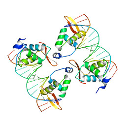
FoxP3 tetramer on TTTG repeats
Descriptor: DNA 72-mer, Forkhead box protein P3
Authors:Leng, F, Hur, S.
Deposit date:2023-05-05
Release date:2023-10-18
Last modified:2023-12-27
Method:ELECTRON MICROSCOPY (3.3 Å)
Cite:FOXP3 recognizes microsatellites and bridges DNA through multimerization.
Nature, 624, 2023
8SRP
DownloadVisualize
BU of 8srp by Molmil
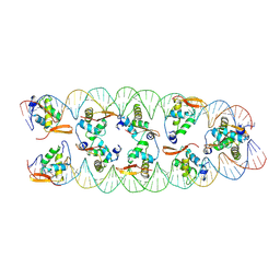
FoxP3 forms Ladder-like multimer to bridge TTTG repeats
Descriptor: DNA 72-mer, Forkhead box protein P3
Authors:Leng, F, Hur, S.
Deposit date:2023-05-05
Release date:2023-10-18
Last modified:2023-12-27
Method:ELECTRON MICROSCOPY (3.7 Å)
Cite:FOXP3 recognizes microsatellites and bridges DNA through multimerization.
Nature, 624, 2023
3B6Q
DownloadVisualize
BU of 3b6q by Molmil
Crystal Structure of the GLUR2 Ligand Binding Core (S1S2J) Mutant T686A in Complex with Glutamate at 2.0 Resolution
Descriptor: GLUTAMIC ACID, Glutamate receptor 2, SULFATE ION
Authors:Cho, Y, Lolis, E, Howe, J.R.
Deposit date:2007-10-29
Release date:2008-02-05
Last modified:2021-10-20
Method:X-RAY DIFFRACTION (2 Å)
Cite:Structural and single-channel results indicate that the rates of ligand binding domain closing and opening directly impact AMPA receptor gating.
J.Neurosci., 28, 2008
3B6W
DownloadVisualize
BU of 3b6w by Molmil
Crystal Structure of the GLUR2 Ligand Binding Core (S1S2J) T686S Mutant in Complex with Glutamate at 1.7 Resolution
Descriptor: GLUTAMIC ACID, Glutamate receptor 2, SULFATE ION
Authors:Cho, Y, Lolis, E, Howe, J.R.
Deposit date:2007-10-29
Release date:2008-02-05
Last modified:2024-10-30
Method:X-RAY DIFFRACTION (1.7 Å)
Cite:Structural and single-channel results indicate that the rates of ligand binding domain closing and opening directly impact AMPA receptor gating.
J.Neurosci., 28, 2008
3B6T
DownloadVisualize
BU of 3b6t by Molmil
Crystal Structure of the GLUR2 Ligand Binding Core (S1S2J) T686A Mutant in Complex with Quisqualate at 2.1 Resolution
Descriptor: (S)-2-AMINO-3-(3,5-DIOXO-[1,2,4]OXADIAZOLIDIN-2-YL)-PROPIONIC ACID, Glutamate receptor 2, SULFATE ION
Authors:Cho, Y, Lolis, E, Howe, J.R.
Deposit date:2007-10-29
Release date:2008-02-05
Last modified:2024-11-06
Method:X-RAY DIFFRACTION (2.1 Å)
Cite:Structural and single-channel results indicate that the rates of ligand binding domain closing and opening directly impact AMPA receptor gating.
J.Neurosci., 28, 2008
5HPK
DownloadVisualize
BU of 5hpk by Molmil
System-wide modulation of HECT E3 ligases with selective ubiquitin variant probes: NEDD4L and UbV NL.1
Descriptor: E3 ubiquitin-protein ligase NEDD4-like, Ubiquitin variant NL.1
Authors:Wu, K.-P, Mukherjee, M, Mercredi, P.Y, Schulman, B.A.
Deposit date:2016-01-20
Release date:2016-03-16
Last modified:2023-09-27
Method:X-RAY DIFFRACTION (2.431 Å)
Cite:System-Wide Modulation of HECT E3 Ligases with Selective Ubiquitin Variant Probes.
Mol.Cell, 62, 2016
5HPT
DownloadVisualize
BU of 5hpt by Molmil
System-wide modulation of HECT E3 ligases with selective ubiquitin variant probes: WWP1, Ubv P2.3 and UBCH7
Descriptor: NEDD4-like E3 ubiquitin-protein ligase WWP1, Ubiquitin variant P2.3, Ubiquitin-conjugating enzyme E2 L3
Authors:Wu, K.-P, Schulman, B.A.
Deposit date:2016-01-20
Release date:2016-03-16
Last modified:2024-11-06
Method:X-RAY DIFFRACTION (2.84 Å)
Cite:System-Wide Modulation of HECT E3 Ligases with Selective Ubiquitin Variant Probes.
Mol.Cell, 62, 2016
5HPL
DownloadVisualize
BU of 5hpl by Molmil
System-wide modulation of HECT E3 ligases with selective ubiquitin variant probes: Rsp5 and UbV R5.4
Descriptor: Rsp5, Ubiquitin variant R5.4
Authors:Wu, K.-P, Kamadurai, H.B, Schulman, B.A.
Deposit date:2016-01-20
Release date:2016-03-16
Last modified:2023-09-27
Method:X-RAY DIFFRACTION (2.31 Å)
Cite:System-Wide Modulation of HECT E3 Ligases with Selective Ubiquitin Variant Probes.
Mol.Cell, 62, 2016
5HPS
DownloadVisualize
BU of 5hps by Molmil
System-wide modulation of HECT E3 ligases with selective ubiquitin variant probes: WWP1 and UbV P1.1
Descriptor: Ubiquitin variant P1.1, WWP1 HECT
Authors:Wu, K.-P, Mercredi, P.Y, Schulman, B.A.
Deposit date:2016-01-20
Release date:2016-03-16
Last modified:2023-09-27
Method:X-RAY DIFFRACTION (2.05 Å)
Cite:System-Wide Modulation of HECT E3 Ligases with Selective Ubiquitin Variant Probes.
Mol.Cell, 62, 2016
7EQ2
DownloadVisualize
BU of 7eq2 by Molmil
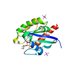
Crystal structure of GDP-bound Rab1a-T75D
Descriptor: (4S)-2-METHYL-2,4-PENTANEDIOL, CACODYLATE ION, COBALT HEXAMMINE(III), ...
Authors:Cao, Y.L, Gu, D.D, Gao, S.
Deposit date:2021-04-28
Release date:2022-11-02
Last modified:2024-01-10
Method:X-RAY DIFFRACTION (1.55090284 Å)
Cite:Aurora kinase A-mediated phosphorylation triggers structural alteration of Rab1A to enhance ER complexity during mitosis
Nat.Struct.Mol.Biol., 2024
8JNJ
DownloadVisualize
BU of 8jnj by Molmil
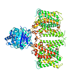
Structure of R932A/K1147A/H1148A mutant AE2
Descriptor: Anion exchange protein 2
Authors:Yin, Y.X, Ding, D.
Deposit date:2023-06-06
Release date:2024-02-07
Method:ELECTRON MICROSCOPY (3.3 Å)
Cite:Structural and functional insights into the lipid regulation of human anion exchanger 2.
Nat Commun, 15, 2024

227111

PDB entries from 2024-11-06

PDB statisticsPDBj update infoContact PDBjnumon