Loading
PDBj
MenuPDBj@FacebookPDBj@X(formerly Twitter)PDBj@BlueSkyPDBj@YouTubewwPDB FoundationwwPDB
RCSB PDBPDBeBMRBAdv. SearchSearch help
Search by PDB author
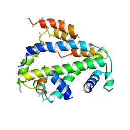
6XWS
DownloadVisualize
BU of 6xws by Molmil
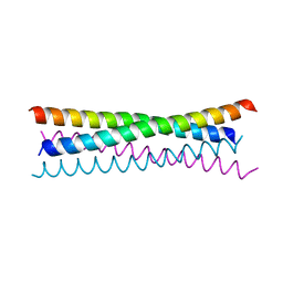
Crystal Structure of Drosophila CAL1 1-160 bound to CENP-A/H4
Descriptor: Chromosome alignment defect 1,Chromosome alignment defect 1, Histone H3-like centromeric protein cid, Histone H4
Authors:Jeyaprakash, A.A, Medina-Pritchard, B, Lazou, V, Zou, J, Byron, O, Abad, M.A, Rappsilber, J, Heun, P.
Deposit date:2020-01-24
Release date:2020-04-15
Last modified:2024-01-24
Method:X-RAY DIFFRACTION (4.36 Å)
Cite:Structural basis for centromere maintenance by Drosophila CENP-A chaperone CAL1.
Embo J., 39, 2020
6YLV
DownloadVisualize
BU of 6ylv by Molmil
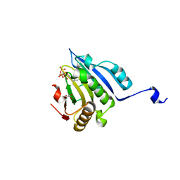
Translation initiation factor 4E in complex with 4-Cl-Bn7GpppG mRNA 5' cap analog
Descriptor: 4-Cl-Bn7GpppG mRNA 5' cap analog, Eukaryotic translation initiation factor 4E, GLYCEROL
Authors:Kubacka, D, Wojcik, R, Baranowski, M.R, Kowalska, J, Jemielity, J.
Deposit date:2020-04-07
Release date:2020-06-10
Last modified:2024-01-24
Method:X-RAY DIFFRACTION (2.66005659 Å)
Cite:Novel N7-Arylmethyl Substituted Dinucleotide mRNA 5' cap Analogs: Synthesis and Evaluation as Modulators of Translation.
Pharmaceutics, 13, 2021
6WYG
DownloadVisualize
BU of 6wyg by Molmil
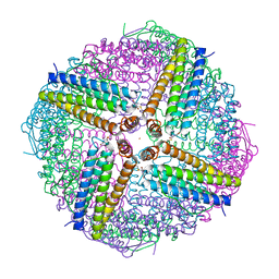
Crystal structure of Human H-chain Ferritin variant 157C Delta C-star Modified with a RAFT agent
Descriptor: CALCIUM ION, FE (III) ION, Ferritin heavy chain, ...
Authors:Bailey, J.B, Zhang, L.
Deposit date:2020-05-12
Release date:2020-11-11
Last modified:2023-10-18
Method:X-RAY DIFFRACTION (2.27 Å)
Cite:Anisotropic Dynamics and Mechanics of Macromolecular Crystals Containing Lattice-Patterned Polymer Networks.
J.Am.Chem.Soc., 142, 2020
6D54
DownloadVisualize
BU of 6d54 by Molmil
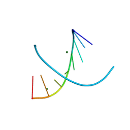
Low Temperature joint X-ray/neutron structure of DNA oligonucleotide d(GTGGCCAC)2 with 2'-SeCH3 modification on Cyt5
Descriptor: DNA (5'-D(*GP*TP*GP*GP*(CSL)P*CP*AP*C)-3'), MAGNESIUM ION
Authors:Kovalevsky, A, Huang, Z, Vandavasi, V.G.
Deposit date:2018-04-19
Release date:2018-10-17
Last modified:2023-10-04
Method:NEUTRON DIFFRACTION (1.65 Å), X-RAY DIFFRACTION
Cite:Temperature-Induced Replacement of Phosphate Proton with Metal Ion Captured in Neutron Structures of A-DNA.
Structure, 26, 2018
6GER
DownloadVisualize
BU of 6ger by Molmil
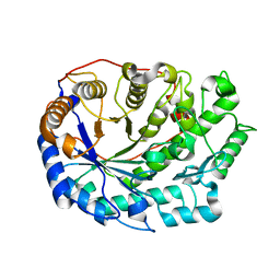
Wheat b-amylase, a clinically relevant food allergen
Descriptor: Beta-amylase
Authors:Hofer, G, Keller, W.
Deposit date:2018-04-27
Release date:2018-12-19
Last modified:2024-05-15
Method:X-RAY DIFFRACTION (2.00004673 Å)
Cite:Three-dimensional structure of the wheat beta-amylase Tri a 17, a clinically relevant food allergen.
Allergy, 74, 2019
7LB7
DownloadVisualize
BU of 7lb7 by Molmil
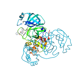
Joint X-ray/neutron structure of SARS-CoV-2 main protease (3CL Mpro) in complex with Telaprevir
Descriptor: (1S,3aR,6aS)-2-[(2S)-2-({(2S)-2-cyclohexyl-2-[(pyrazin-2-ylcarbonyl)amino]acetyl}amino)-3,3-dimethylbutanoyl]-N-[(2R,3S)-1-(cyclopropylamino)-2-hydroxy-1-oxohexan-3-yl]octahydrocyclopenta[c]pyrrole-1-carboxamide, 3C-like proteinase
Authors:Kovalevsky, A.Y, Kneller, D.W, Coates, L.
Deposit date:2021-01-07
Release date:2021-01-20
Last modified:2024-11-13
Method:NEUTRON DIFFRACTION (2 Å), X-RAY DIFFRACTION
Cite:Direct Observation of Protonation State Modulation in SARS-CoV-2 Main Protease upon Inhibitor Binding with Neutron Crystallography.
J.Med.Chem., 64, 2021
6OPJ
DownloadVisualize
BU of 6opj by Molmil
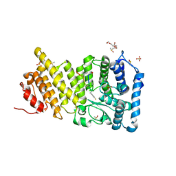
Menin in complex with peptide inhibitor 25
Descriptor: DIMETHYL SULFOXIDE, Menin, Peptide inhibitor 25, ...
Authors:Linhares, B.M, Fortuna, P, Cierpicki, T, Grembecka, J, Berlicki, L.
Deposit date:2019-04-25
Release date:2020-09-02
Last modified:2023-11-15
Method:X-RAY DIFFRACTION (1.5006572 Å)
Cite:Covalent and noncovalent constraints yield a figure eight-like conformation of a peptide inhibiting the menin-MLL interaction.
Eur.J.Med.Chem., 207, 2020
6O9M
DownloadVisualize
BU of 6o9m by Molmil
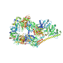
Structure of the human apo TFIIH
Descriptor: CDK-activating kinase assembly factor MAT1, General transcription factor IIH subunit 1, General transcription factor IIH subunit 2, ...
Authors:Yan, C.L, Dodd, T, He, Y, Tainer, J.A, Tsutakawa, S.E, Ivanov, I.
Deposit date:2019-03-14
Release date:2019-05-29
Last modified:2024-03-13
Method:ELECTRON MICROSCOPY (4.4 Å)
Cite:Transcription preinitiation complex structure and dynamics provide insight into genetic diseases.
Nat.Struct.Mol.Biol., 26, 2019
6P0J
DownloadVisualize
BU of 6p0j by Molmil
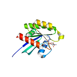
Crystal structure of GDP-bound human RalA
Descriptor: CALCIUM ION, GUANOSINE-5'-DIPHOSPHATE, Ras-related protein Ral-A
Authors:Bum-Erdene, K, Gonzalez-Gutierrez, G, Liu, D, Meroueh, S.O.
Deposit date:2019-05-17
Release date:2020-03-04
Last modified:2023-10-11
Method:X-RAY DIFFRACTION (1.31 Å)
Cite:Small-molecule covalent bond formation at tyrosine creates a binding site and inhibits activation of Ral GTPases.
Proc.Natl.Acad.Sci.USA, 117, 2020
6PHJ
DownloadVisualize
BU of 6phj by Molmil
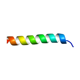
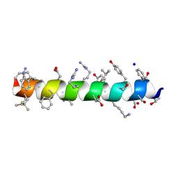
Crystal structure of native glucagon in space group P213 at 1.99 A resolution
Descriptor: Glucagon
Authors:Mroz, P.A, Gonzalez-Gutierrez, G, DiMarchi, R.D.
Deposit date:2019-06-25
Release date:2020-07-01
Last modified:2023-10-11
Method:X-RAY DIFFRACTION (1.99 Å)
Cite:High resolution X-ray structure of glucagon and selected stereo-inversed analogs in novel crystallographic packing arrangement.
To Be Published
6PHO
DownloadVisualize
BU of 6pho by Molmil
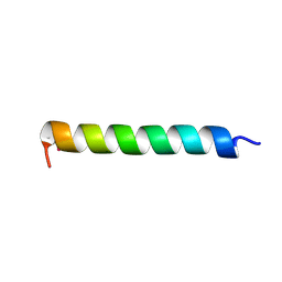
Crystal structure of glucagon analog with selenomethionine substitutions at position 1 and 27 in space group I41 at 1.42 A resolution
Descriptor: Glucagon
Authors:Mroz, P.A, Gonzalez-Gutierrez, G, DiMarchi, R.D.
Deposit date:2019-06-25
Release date:2020-07-01
Last modified:2024-10-09
Method:X-RAY DIFFRACTION (1.42 Å)
Cite:High resolution X-ray structure of glucagon and selected stereo-inversed analogs in novel crystallographic packing arrangement.
To Be Published
6PHK
DownloadVisualize
BU of 6phk by Molmil
Crystal structure of glucagon analog with mono-stereoinversion at position 21 (D-Asp21) in space group I41 at 1.18 A resolution
Descriptor: Glucagon
Authors:Mroz, P.A, Gonzalez-Gutierrez, G, DiMarchi, R.D.
Deposit date:2019-06-25
Release date:2020-07-01
Last modified:2024-10-16
Method:X-RAY DIFFRACTION (1.18 Å)
Cite:High resolution X-ray structure of glucagon and selected stereo-inversed analogs in novel crystallographic packing arrangement.
To Be Published
6PHP
DownloadVisualize
BU of 6php by Molmil
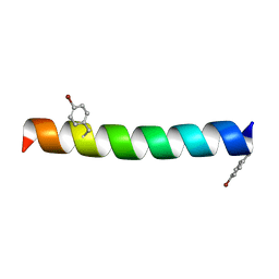
Crystal structure of glucagon analog with 4-bromo-phenylalanine substitutions at position 6 and 22 in space group I41 at 1.65 A resolution
Descriptor: Glucagon
Authors:Mroz, P.A, Gonzalez-Gutierrez, G, DiMarchi, R.D.
Deposit date:2019-06-25
Release date:2020-07-01
Last modified:2023-11-15
Method:X-RAY DIFFRACTION (1.65 Å)
Cite:High resolution X-ray structure of glucagon and selected stereo-inversed analogs in novel crystallographic packing arrangement.
To Be Published
6PQ7
DownloadVisualize
BU of 6pq7 by Molmil
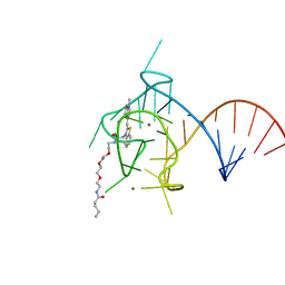
Structure of the iMango-III fluorescent aptamer at room temperature.
Descriptor: MAGNESIUM ION, POTASSIUM ION, RNA (37-MER), ...
Authors:Trachman III, R.J, Ferre-D'Amare, A.R.
Deposit date:2019-07-08
Release date:2019-07-31
Last modified:2023-10-11
Method:X-RAY DIFFRACTION (3 Å)
Cite:Co-crystal structure of the iMango-III fluorescent RNA aptamer using an X-ray free-electron laser.
Acta Crystallogr.,Sect.F, 75, 2019
6PHN
DownloadVisualize
BU of 6phn by Molmil
Crystal structure of glucagon analog composed of D-amino acids with mono-stereoinversion at position 23 (L-Val23) in space group I41 at 1.33 A resolution
Descriptor: D-glucagon L-Val23, SODIUM ION
Authors:Mroz, P.A, Gonzalez-Gutierrez, G, DiMarchi, R.D.
Deposit date:2019-06-25
Release date:2020-07-01
Last modified:2024-10-30
Method:X-RAY DIFFRACTION (1.33 Å)
Cite:High resolution X-ray structure of glucagon and selected stereo-inversed analogs in novel crystallographic packing arrangement.
To Be Published
6PHI
DownloadVisualize
BU of 6phi by Molmil
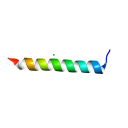
Crystal structure of native glucagon in space group I41 at 1.1 A resolution
Descriptor: CHLORIDE ION, Glucagon
Authors:Mroz, P.A, Gonzalez-Gutierrez, G, DiMarchi, R.D.
Deposit date:2019-06-25
Release date:2020-07-01
Last modified:2023-10-11
Method:X-RAY DIFFRACTION (1.1 Å)
Cite:High resolution X-ray structure of glucagon and selected stereo-inversed analogs in novel crystallographic packing arrangement.
To Be Published
6PHM
DownloadVisualize
BU of 6phm by Molmil
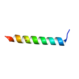
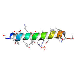
Crystal structure of glucagon analog fully composed of D-amino acids in space group I41 at 1.1 A resolution
Descriptor: D-glucagon, SULFATE ION
Authors:Mroz, P.A, Gonzalez-Gutierrez, G, DiMarchi, R.D.
Deposit date:2019-06-25
Release date:2020-07-01
Last modified:2024-10-23
Method:X-RAY DIFFRACTION (1.1 Å)
Cite:High resolution X-ray structure of glucagon and selected stereo-inversed analogs in novel crystallographic packing arrangement.
To Be Published
6PHL
DownloadVisualize
BU of 6phl by Molmil
Crystal structure of glucagon analog with mono-stereoinversion at position 23 (D-Val23) in space group I41 at 1.44 A resolution
Descriptor: Glucagon
Authors:Mroz, P.A, Gonzalez-Gutierrez, G, DiMarchi, R.D.
Deposit date:2019-06-25
Release date:2020-07-01
Last modified:2024-10-16
Method:X-RAY DIFFRACTION (1.443 Å)
Cite:High resolution X-ray structure of glucagon and selected stereo-inversed analogs in novel crystallographic packing arrangement.
To Be Published
6PHQ
DownloadVisualize
BU of 6phq by Molmil
Crystal structure of glucagon analog fully composed of D-amino acids with 4-bromo-D-phenylalanine substitutions at position 6 and 22 in space group I41 at 1.1 A resolution
Descriptor: D-glucagon D-BrPhe 6,22
Authors:Mroz, P.A, Gonzalez-Gutierrez, G, DiMarchi, R.D.
Deposit date:2019-06-25
Release date:2020-07-01
Last modified:2023-11-15
Method:X-RAY DIFFRACTION (1.32 Å)
Cite:High resolution X-ray structure of glucagon and selected stereo-inversed analogs in novel crystallographic packing arrangement.
To Be Published
6P7M
DownloadVisualize
BU of 6p7m by Molmil
Cryo-EM structure of LbCas12a-crRNA: AcrVA4 (1:2 complex)
Descriptor: Cas12a, MAGNESIUM ION, anti-CRISPR VA4, ...
Authors:Knott, G.J, Liu, J.J, Doudna, J.A.
Deposit date:2019-06-06
Release date:2019-08-21
Last modified:2024-03-20
Method:ELECTRON MICROSCOPY (3 Å)
Cite:Structural basis for AcrVA4 inhibition of specific CRISPR-Cas12a.
Elife, 8, 2019
6P7N
DownloadVisualize
BU of 6p7n by Molmil
Cryo-EM structure of LbCas12a-crRNA: AcrVA4 (2:2 complex)
Descriptor: Cas12a, MAGNESIUM ION, anti-CRISPR VA4, ...
Authors:Knott, G.J, Liu, J.J, Doudna, J.A.
Deposit date:2019-06-06
Release date:2019-08-21
Last modified:2023-08-16
Method:ELECTRON MICROSCOPY (4.9 Å)
Cite:Structural basis for AcrVA4 inhibition of specific CRISPR-Cas12a.
Elife, 8, 2019
6TOC
DownloadVisualize
BU of 6toc by Molmil
Crystal structure of the oligomerisation domain of the transcription factor PHOSPHATE STARVATION RESPONSE 1 from Arabidopsis (crystal form 3).
Descriptor: Protein PHOSPHATE STARVATION RESPONSE 1
Authors:Hothorn, M.
Deposit date:2019-12-11
Release date:2021-01-13
Last modified:2024-01-24
Method:X-RAY DIFFRACTION (1.853 Å)
Cite:Inositol pyrophosphates promote the interaction of SPX domains with the coiled-coil motif of PHR transcription factors to regulate plant phosphate homeostasis.
Nat Commun, 12, 2021
6T6H
DownloadVisualize
BU of 6t6h by Molmil
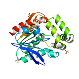
Apo structure of the Bottromycin epimerase BotH
Descriptor: BotH, SODIUM ION, SULFATE ION
Authors:Koehnke, J, Sikandar, A.
Deposit date:2019-10-18
Release date:2020-07-15
Last modified:2024-05-15
Method:X-RAY DIFFRACTION (1.18 Å)
Cite:The bottromycin epimerase BotH defines a group of atypical alpha / beta-hydrolase-fold enzymes.
Nat.Chem.Biol., 16, 2020
6T6Z
DownloadVisualize
BU of 6t6z by Molmil
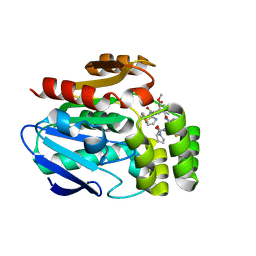
Structure of the Bottromycin epimerase BotH in complex with a bottromycin A2 derivative
Descriptor: BotH, Bottromycin A2 derivative
Authors:Koehnke, J, Sikandar, A.
Deposit date:2019-10-20
Release date:2020-07-15
Last modified:2024-01-24
Method:X-RAY DIFFRACTION (1.7 Å)
Cite:The bottromycin epimerase BotH defines a group of atypical alpha / beta-hydrolase-fold enzymes.
Nat.Chem.Biol., 16, 2020
6T6Y
DownloadVisualize
BU of 6t6y by Molmil
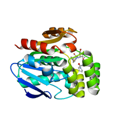
Structure of the Bottromycin epimerase BotH in complex with Bottromycin A2
Descriptor: BotH, Bottromycin A2
Authors:Koehnke, J, Sikandar, A.
Deposit date:2019-10-20
Release date:2020-07-15
Last modified:2024-01-24
Method:X-RAY DIFFRACTION (1.4 Å)
Cite:The bottromycin epimerase BotH defines a group of atypical alpha / beta-hydrolase-fold enzymes.
Nat.Chem.Biol., 16, 2020

227344

PDB entries from 2024-11-13

PDB statisticsPDBj update infoContact PDBjnumon