 Movie
Movie Controller

[English] 日本語
 Yorodumi
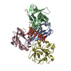
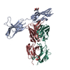
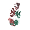
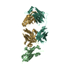
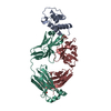
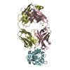
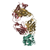
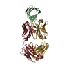
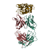
- PDB-4wv1: Crystal structure of the FGFR2 D2 domain in complex with Fab 2B.1.3 -
+ Open data

- Basic information
| Entry | Database: PDB / ID: 4wv1 | ||||||
|---|---|---|---|---|---|---|---|
| Title | Crystal structure of the FGFR2 D2 domain in complex with Fab 2B.1.3 | ||||||
 Components Components |
| ||||||
 Keywords Keywords | TRANSFERASE/IMMUNE SYSTEM /  FGFR2 / Fab / Complex / Antibody / TRANSFERASE-IMMUNE SYSTEM complex FGFR2 / Fab / Complex / Antibody / TRANSFERASE-IMMUNE SYSTEM complex | ||||||
| Function / homology |  Function and homology information Function and homology informationSignaling by FGFR2 amplification mutants / Signaling by FGFR2 fusions / fibroblast growth factor receptor signaling pathway involved in negative regulation of apoptotic process in bone marrow cell / fibroblast growth factor receptor signaling pathway involved in hemopoiesis / fibroblast growth factor receptor signaling pathway involved in positive regulation of cell proliferation in bone marrow / fibroblast growth factor receptor signaling pathway involved in mammary gland specification / lateral sprouting from an epithelium / mammary gland bud formation / branch elongation involved in salivary gland morphogenesis / mesenchymal cell differentiation involved in lung development ...Signaling by FGFR2 amplification mutants / Signaling by FGFR2 fusions / fibroblast growth factor receptor signaling pathway involved in negative regulation of apoptotic process in bone marrow cell / fibroblast growth factor receptor signaling pathway involved in hemopoiesis / fibroblast growth factor receptor signaling pathway involved in positive regulation of cell proliferation in bone marrow / fibroblast growth factor receptor signaling pathway involved in mammary gland specification / lateral sprouting from an epithelium / mammary gland bud formation / branch elongation involved in salivary gland morphogenesis / mesenchymal cell differentiation involved in lung development / lacrimal gland development / otic vesicle formation / prostate gland morphogenesis / regulation of smooth muscle cell differentiation / orbitofrontal cortex development / regulation of morphogenesis of a branching structure / squamous basal epithelial stem cell differentiation involved in prostate gland acinus development / branching morphogenesis of a nerve / embryonic organ morphogenesis / endochondral bone growth / morphogenesis of embryonic epithelium / epidermis morphogenesis / bud elongation involved in lung branching / positive regulation of epithelial cell proliferation involved in lung morphogenesis / pyramidal neuron development / membranous septum morphogenesis / reproductive structure development / limb bud formation / lung lobe morphogenesis / gland morphogenesis / fibroblast growth factor receptor signaling pathway involved in orbitofrontal cortex development / ventricular zone neuroblast division / embryonic digestive tract morphogenesis / epithelial cell proliferation involved in salivary gland morphogenesis / mesenchymal cell differentiation / branching involved in prostate gland morphogenesis / mesenchymal cell proliferation involved in lung development / branching involved in labyrinthine layer morphogenesis / FGFR2b ligand binding and activation / FGFR2c ligand binding and activation / Activated point mutants of FGFR2 / regulation of osteoblast proliferation / Phospholipase C-mediated cascade; FGFR2 / fibroblast growth factor receptor activity / branching involved in salivary gland morphogenesis / embryonic pattern specification / outflow tract septum morphogenesis / positive regulation of phospholipase activity / lung-associated mesenchyme development / mesodermal cell differentiation / digestive tract development / regulation of smoothened signaling pathway / embryonic cranial skeleton morphogenesis / bone morphogenesis / skeletal system morphogenesis / odontogenesis / ureteric bud development / positive regulation of mesenchymal cell proliferation / regulation of osteoblast differentiation / ventricular cardiac muscle tissue morphogenesis / inner ear morphogenesis / Signaling by FGFR2 IIIa TM / organ growth / midbrain development / hair follicle morphogenesis / lung alveolus development / fibroblast growth factor binding / prostate epithelial cord arborization involved in prostate glandular acinus morphogenesis / PI-3K cascade:FGFR2 / bone mineralization / prostate epithelial cord elongation / positive regulation of cell division / excitatory synapse / positive regulation of Wnt signaling pathway / PI3K Cascade / epithelial to mesenchymal transition / negative regulation of keratinocyte proliferation / fibroblast growth factor receptor signaling pathway / embryonic organ development / cell fate commitment / positive regulation of cell cycle / SHC-mediated cascade:FGFR2 / cellular response to retinoic acid / FRS-mediated FGFR2 signaling / positive regulation of cardiac muscle cell proliferation / positive regulation of vascular associated smooth muscle cell proliferation / cellular response to transforming growth factor beta stimulus / Signaling by FGFR2 in disease / epithelial cell differentiation / axonogenesis / post-embryonic development / regulation of ERK1 and ERK2 cascade / positive regulation of epithelial cell proliferation / Negative regulation of FGFR2 signaling / animal organ morphogenesis / lung development / bone development / receptor protein-tyrosine kinase / peptidyl-tyrosine phosphorylation / positive regulation of canonical Wnt signaling pathwaySimilarity search - Function | ||||||
| Biological species |  Homo sapiens (human) Homo sapiens (human) | ||||||
| Method | X-RAY DIFFRACTION / SYNCHROTRON / MOLECULAR REPLACEMENT / Resolution: 2.362 Å | ||||||
 Authors Authors | Yin, Y. / Carter, P.J. | ||||||
 Citation Citation | Journal: Mol.Cancer Ther. / Year: 2015 Title: Redesigning a Monospecific Anti-FGFR3 Antibody to Add Selectivity for FGFR2 and Expand Antitumor Activity. Authors: Yin, Y. / Djakovic, S. / Marsters, S. / Tien, J. / Peng, J. / Tremayne, J. / Lee, G. / Neve, R.M. / Wu, Y. / Merchant, M. / Ashkenazi, A. / Carter, P.J. | ||||||
| History |
|
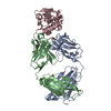
- Structure visualization
| Structure viewer | Molecule: MolmilJmol/JSmol |
|---|
- Downloads & links
-Download
| PDBx/mmCIF format | 4wv1.cif.gz | 215.5 KB | Display | PDBx/mmCIF format |
|---|---|---|---|---|
| PDB format | pdb4wv1.ent.gz | 172.1 KB | Display | PDB format |
| PDBx/mmJSON format | 4wv1.json.gz | Tree view | PDBx/mmJSON format | |
| Others |  Other downloads Other downloads |
-Validation report
| Arichive directory | https://data.pdbj.org/pub/pdb/validation_reports/wv/4wv1ftp://data.pdbj.org/pub/pdb/validation_reports/wv/4wv1 | HTTPS FTP |
|---|
-Related structure data


-Links
 PDBj
PDBj


- Assembly
| Deposited unit | 
| ||||||||
|---|---|---|---|---|---|---|---|---|---|
| 1 | 
| ||||||||
| 2 | 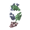
| ||||||||
| Unit cell |
|
-Components
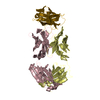
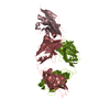
| #1: Protein | / FGFR-2 / K-sam / KGFR / Keratinocyte growth factor receptor Mass: 11494.089 Da / Num. of mol.: 2 / Fragment: UNP residues 153-251 Source method: isolated from a genetically manipulated source Source: (gene. exp.) Homo sapiens (human) / Gene: FGFR2, BEK, KGFR, KSAM / Production host:  Escherichia coli (E. coli) Escherichia coli (E. coli)References: UniProt: P21802, receptor protein-tyrosine kinase#2: Antibody | Fragment antigen-bindingMass: 24344.121 Da / Num. of mol.: 2 Source method: isolated from a genetically manipulated source Source: (gene. exp.) Homo sapiens (human) / Production host: Escherichia coli (E. coli)#3: Antibody | Fragment antigen-bindingMass: 23308.816 Da / Num. of mol.: 2 Source method: isolated from a genetically manipulated source Source: (gene. exp.) Homo sapiens (human) / Production host: Escherichia coli (E. coli)#4: Water | ChemComp-HOH / | Water Mass: 18.015 Da / Num. of mol.: 149 / Source method: isolated from a natural source / Formula: H2O Mass: 18.015 Da / Num. of mol.: 149 / Source method: isolated from a natural source / Formula: H2O |
|---|
-Experimental details
-Experiment
| Experiment | Method: X-RAY DIFFRACTION / Number of used crystals: 1 |
|---|
- Sample preparation
| Crystal | Density Matthews: 2.52 Å3/Da / Density % sol: 51.18 % |
|---|---|
| Crystal grow | Temperature: 292 K / Method: vapor diffusion, sitting drop / Details: PEG 3350, potassium nitrate |
-Data collection
| Diffraction | Mean temperature: 100 K |
|---|---|
| Diffraction source | Source: SYNCHROTRON / Site: ALS  / Beamline: 5.0.2 / Wavelength: 1 Å / Beamline: 5.0.2 / Wavelength: 1 Å |
| Detector | Type: ADSC QUANTUM 315r / Detector: CCD / Date: Mar 26, 2014 |
| Radiation | Protocol: SINGLE WAVELENGTH / Monochromatic (M) / Laue (L): M / Scattering type: x-ray |
| Radiation wavelength | Wavelength: 1 Å / Relative weight: 1 |
| Reflection | Resolution: 2.36→45.64 Å / Num. obs: 46583 / % possible obs: 98 % / Redundancy: 2.5 % / Rsym value: 0.094 / Net I/σ(I): 14.4 |
| Reflection shell | Resolution: 2.36→2.47 Å / Redundancy: 2.5 % / Rmerge(I) obs: 0.489 / Mean I/σ(I) obs: 1.9 / % possible all: 99.3 |
- Processing
| Software |
| ||||||||||||||||||||||||||||||||||||||||||||||||||||||||||||||||||||||||||||||||||||||||||||||||||||||||||||||||||||||||||||||
|---|---|---|---|---|---|---|---|---|---|---|---|---|---|---|---|---|---|---|---|---|---|---|---|---|---|---|---|---|---|---|---|---|---|---|---|---|---|---|---|---|---|---|---|---|---|---|---|---|---|---|---|---|---|---|---|---|---|---|---|---|---|---|---|---|---|---|---|---|---|---|---|---|---|---|---|---|---|---|---|---|---|---|---|---|---|---|---|---|---|---|---|---|---|---|---|---|---|---|---|---|---|---|---|---|---|---|---|---|---|---|---|---|---|---|---|---|---|---|---|---|---|---|---|---|---|---|---|
| Refinement | Method to determine structure: MOLECULAR REPLACEMENT Starting model: PDB entries 3GRW and 3CU1 Resolution: 2.362→45.64 Å / SU ML: 0.32 / Cross valid method: FREE R-VALUE / σ(F): 1.36 / Phase error: 26.73 / Stereochemistry target values: ML
| ||||||||||||||||||||||||||||||||||||||||||||||||||||||||||||||||||||||||||||||||||||||||||||||||||||||||||||||||||||||||||||||
| Solvent computation | Shrinkage radii: 0.9 Å / VDW probe radii: 1.11 Å / Solvent model: FLAT BULK SOLVENT MODEL | ||||||||||||||||||||||||||||||||||||||||||||||||||||||||||||||||||||||||||||||||||||||||||||||||||||||||||||||||||||||||||||||
| Refinement step | Cycle: LAST / Resolution: 2.362→45.64 Å
| ||||||||||||||||||||||||||||||||||||||||||||||||||||||||||||||||||||||||||||||||||||||||||||||||||||||||||||||||||||||||||||||
| Refine LS restraints |
| ||||||||||||||||||||||||||||||||||||||||||||||||||||||||||||||||||||||||||||||||||||||||||||||||||||||||||||||||||||||||||||||
| LS refinement shell |
|
