 Movie
Movie Controller

[English] 日本語
 Yorodumi
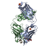
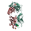
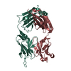
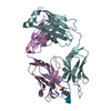
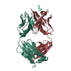
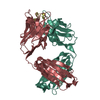
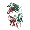
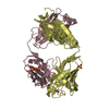
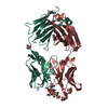
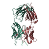
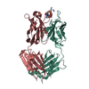
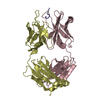
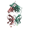
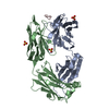
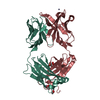
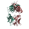
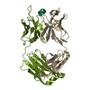
- PDB-3f58: IGG1 FAB FRAGMENT (58.2) COMPLEX WITH 12-RESIDUE CYCLIC PEPTIDE (... -
+ Open data

- Basic information
| Entry | Database: PDB / ID: 3f58 | ||||||
|---|---|---|---|---|---|---|---|
| Title | IGG1 FAB FRAGMENT (58.2) COMPLEX WITH 12-RESIDUE CYCLIC PEPTIDE (INCLUDING RESIDUES 315-324 OF HIV-1 GP120 (MN ISOLATE); H315S MUTATION | ||||||
 Components Components |
| ||||||
 Keywords Keywords | IMMUNE SYSTEM / IMMUNOGLOBULIN / FAB / HIV-1 / GP120 / V3 | ||||||
| Function / homology | Immunoglobulins / Immunoglobulin-like / Sandwich / Mainly Beta Function and homology information Function and homology information | ||||||
| Biological species |  | ||||||
| Method |  X-RAY DIFFRACTION / MOLECULAR REPLACEMENT / Resolution: 2.8 Å X-RAY DIFFRACTION / MOLECULAR REPLACEMENT / Resolution: 2.8 Å | ||||||
 Authors Authors | Stanfield, R.L. / Cabezas, E. / Satterthwait, A.C. / Stura, E.A. / Profy, A.T. / Wilson, I.A. | ||||||
 Citation Citation | Journal: Structure Fold.Des. / Year: 1999 Title: Dual conformations for the HIV-1 gp120 V3 loop in complexes with different neutralizing fabs. Authors: Stanfield, R. / Cabezas, E. / Satterthwait, A. / Stura, E. / Profy, A. / Wilson, I. | ||||||
| History |
|
- Structure visualization
| Structure viewer | Molecule: MolmilJmol/JSmol |
|---|
- Downloads & links
-Download
| PDBx/mmCIF format | 3f58.cif.gz | 91.4 KB | Display | PDBx/mmCIF format |
|---|---|---|---|---|
| PDB format | pdb3f58.ent.gz | 70.4 KB | Display | PDB format |
| PDBx/mmJSON format | 3f58.json.gz | Tree view | PDBx/mmJSON format | |
| Others |  Other downloads Other downloads |
-Validation report
| Arichive directory | https://data.pdbj.org/pub/pdb/validation_reports/f5/3f58ftp://data.pdbj.org/pub/pdb/validation_reports/f5/3f58 | HTTPS FTP |
|---|
-Related structure data
| Related structure data | 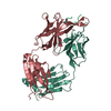 1f58C 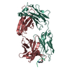 2f58C 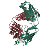 1acyS S: Starting model for refinement C: citing same article ( |
|---|---|
| Similar structure data |


-Links
 PDBj
PDBj


- Assembly
| Deposited unit | 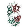
| ||||||||
|---|---|---|---|---|---|---|---|---|---|
| 1 |
| ||||||||
| Unit cell |
|
-Components
| #1: Antibody | Mass: 23501.934 Da / Num. of mol.: 1 / Fragment: LIGHT CHAIN OF FAB / Source method: isolated from a natural source / Source: (natural) |
|---|---|
| #2: Antibody | Mass: 24864.744 Da / Num. of mol.: 1 / Fragment: HEAVY CHAIN OF FAB / Source method: isolated from a natural source / Source: (natural) |
| #3: Protein/peptide | Mass: 976.070 Da / Num. of mol.: 1 / Fragment: RESIDUES 315-324 FROM HIV-1 GP120 / Mutation: H315S / Source method: obtained synthetically Details: 12 RESIDUE HYDRAZONE-LINKED CYCLIC PEPTIDE WAS CHEMICALLY SYNTHESIZED FROM RESIDUES 316-324 OF HIV-1 GP120 STRAIN MN. |
| #4: Water | ChemComp-HOH /  Mass: 18.015 Da / Num. of mol.: 1 / Source method: isolated from a natural source / Formula: H2O Mass: 18.015 Da / Num. of mol.: 1 / Source method: isolated from a natural source / Formula: H2O |
| Has protein modification | Y |
| Sequence details | THE FAB FRAGMENT IS NUMBERED BY THE CONVENTION OF E. KABAT (E.A. KABAT, T.T. WU, M. REID-MILLER, H. ...THE FAB FRAGMENT IS NUMBERED BY THE CONVENTION |
-Experimental details
-Experiment
| Experiment | Method: X-RAY DIFFRACTION / Number of used crystals: 1 |
|---|
- Sample preparation
| Crystal | Density Matthews: 2.73 Å3/Da / Density % sol: 55 % | ||||||||||||||||||||||||||||||
|---|---|---|---|---|---|---|---|---|---|---|---|---|---|---|---|---|---|---|---|---|---|---|---|---|---|---|---|---|---|---|---|
| Crystal grow | pH: 5 / Details: pH 5.0 | ||||||||||||||||||||||||||||||
| Crystal | *PLUS | ||||||||||||||||||||||||||||||
| Crystal grow | *PLUS Temperature: 22.5 ℃ / Method: vapor diffusion, sitting drop | ||||||||||||||||||||||||||||||
| Components of the solutions | *PLUS
|
-Data collection
| Diffraction | Mean temperature: 295 K |
|---|---|
| Diffraction source | Source: ROTATING ANODE / Type: MACSCIENCE M18X / Wavelength: 1.5418 |
| Detector | Type: SIEMENS / Detector: AREA DETECTOR / Date: May 1, 1992 / Details: SUPPER DOUBLE MIRRORS |
| Radiation | Monochromator: SUPPER DOUBLE MIRRORS / Protocol: SINGLE WAVELENGTH / Monochromatic (M) / Laue (L): M / Scattering type: x-ray |
| Radiation wavelength | Wavelength: 1.5418 Å / Relative weight: 1 |
| Reflection | Resolution: 2.8→47.8 Å / Num. obs: 13951 / % possible obs: 98.9 % / Redundancy: 3.4 % / Biso Wilson estimate: 60.1 Å2 / Rsym value: 0.072 / Net I/σ(I): 13.2 |
| Reflection shell | Resolution: 2.8→2.98 Å / Redundancy: 2.4 % / Mean I/σ(I) obs: 1.5 / Rsym value: 0.218 / % possible all: 97.7 |
| Reflection | *PLUS Num. measured all: 47266 / Rmerge(I) obs: 0.072 |
| Reflection shell | *PLUS % possible obs: 97.7 % / Num. unique obs: 2259 / Num. measured obs: 9337 / Rmerge(I) obs: 0.218 |
- Processing
| Software |
| ||||||||||||||||||||||||||||||||||||||||||||||||||||||||||||
|---|---|---|---|---|---|---|---|---|---|---|---|---|---|---|---|---|---|---|---|---|---|---|---|---|---|---|---|---|---|---|---|---|---|---|---|---|---|---|---|---|---|---|---|---|---|---|---|---|---|---|---|---|---|---|---|---|---|---|---|---|---|
| Refinement | Method to determine structure: MOLECULAR REPLACEMENT Starting model: FAB PORTION OF 1ACY Resolution: 2.8→48 Å / Data cutoff high absF: 10000000 / Data cutoff low absF: 0 / Isotropic thermal model: GROUP / σ(F): 0 / Details: BULK SOLVENT MODEL USED
| ||||||||||||||||||||||||||||||||||||||||||||||||||||||||||||
| Displacement parameters | Biso mean: 28.5 Å2 | ||||||||||||||||||||||||||||||||||||||||||||||||||||||||||||
| Refine analyze | Luzzati coordinate error obs: 0.31 Å / Luzzati d res low obs: 5 Å / Luzzati sigma a obs: 0.47 Å | ||||||||||||||||||||||||||||||||||||||||||||||||||||||||||||
| Refinement step | Cycle: LAST / Resolution: 2.8→48 Å
| ||||||||||||||||||||||||||||||||||||||||||||||||||||||||||||
| Refine LS restraints |
| ||||||||||||||||||||||||||||||||||||||||||||||||||||||||||||
| LS refinement shell | Resolution: 2.8→2.98 Å / Total num. of bins used: 6
| ||||||||||||||||||||||||||||||||||||||||||||||||||||||||||||
| Xplor file |
| ||||||||||||||||||||||||||||||||||||||||||||||||||||||||||||
| Software | *PLUS Name: X-PLOR / Version: 3.851 / Classification: refinement | ||||||||||||||||||||||||||||||||||||||||||||||||||||||||||||
| Refinement | *PLUS σ(F): 0 / Rfactor obs: 0.2 / Rfactor Rwork: 0.2 | ||||||||||||||||||||||||||||||||||||||||||||||||||||||||||||
| Solvent computation | *PLUS | ||||||||||||||||||||||||||||||||||||||||||||||||||||||||||||
| Displacement parameters | *PLUS Biso mean: 28.5 Å2 | ||||||||||||||||||||||||||||||||||||||||||||||||||||||||||||
| Refine LS restraints | *PLUS
| ||||||||||||||||||||||||||||||||||||||||||||||||||||||||||||
| LS refinement shell | *PLUS Rfactor Rwork: 0.377 |
