 Movie
Movie Controller

[English] 日本語
 Yorodumi
- PDB-2igf: CRYSTAL STRUCTURES OF AN ANTIBODY TO A PEPTIDE AND ITS COMPLEX WI... -
+ Open data

- Basic information
| Entry | Database: PDB / ID: 2igf | ||||||
|---|---|---|---|---|---|---|---|
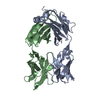
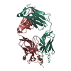
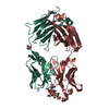
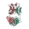
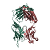
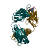
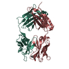
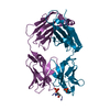
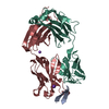
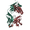
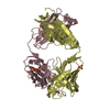
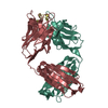
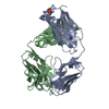
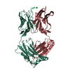
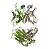
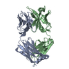
| Title | CRYSTAL STRUCTURES OF AN ANTIBODY TO A PEPTIDE AND ITS COMPLEX WITH PEPTIDE ANTIGEN AT 2.8 ANGSTROMS | ||||||
 Components Components |
| ||||||
 Keywords Keywords | IMMUNOGLOBULIN | ||||||
| Function / homology |  Function and homology information Function and homology information | ||||||
| Biological species |  | ||||||
| Method |  X-RAY DIFFRACTION / Resolution: 2.8 Å X-RAY DIFFRACTION / Resolution: 2.8 Å | ||||||
 Authors Authors | Stanfield, R.L. / Wilson, I.A. | ||||||
 Citation Citation | Journal: Science / Year: 1990 Title: Crystal structures of an antibody to a peptide and its complex with peptide antigen at 2.8 A. Authors: Stanfield, R.L. / Fieser, T.M. / Lerner, R.A. / Wilson, I.A. #1: Journal: J.Biol.Chem. / Year: 1989Title: Preliminary Crystallographic Data and Primary Sequence for Anti-Peptide Fab' B13I2 and its Complex with the C-Helix Peptide from Myohemerythrin Authors: Stura, E.A. / Stanfield, R.L. / Fieser, T.M. / Balderas, R.S. / Smith, L.R. / Lerner, R.A. / Wilson, I.A. | ||||||
| History |
|
- Structure visualization
| Structure viewer | Molecule: MolmilJmol/JSmol |
|---|
- Downloads & links
-Download
| PDBx/mmCIF format | 2igf.cif.gz | 100.9 KB | Display | PDBx/mmCIF format |
|---|---|---|---|---|
| PDB format | pdb2igf.ent.gz | 76 KB | Display | PDB format |
| PDBx/mmJSON format | 2igf.json.gz | Tree view | PDBx/mmJSON format | |
| Others |  Other downloads Other downloads |
-Validation report
| Arichive directory | https://data.pdbj.org/pub/pdb/validation_reports/ig/2igfftp://data.pdbj.org/pub/pdb/validation_reports/ig/2igf | HTTPS FTP |
|---|
-Related structure data


-Links
 PDBj
PDBj

- Assembly
| Deposited unit | 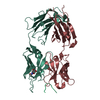
| ||||||||
|---|---|---|---|---|---|---|---|---|---|
| 1 |
| ||||||||
| 2 | 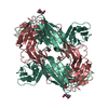
| ||||||||
| Unit cell |
| ||||||||
| Atom site foot note | 1: RESIDUES 8, 95, 141 OF THE *L* CHAIN AND 149, 151 OF THE *H* CHAIN ARE CIS PROLINES. 2: NAG 600 IS LINKED TO ASN L 26. |
-Components
| #1: Antibody | Mass: 24148.803 Da / Num. of mol.: 1 Source method: isolated from a genetically manipulated source Source: (gene. exp.) |
|---|---|
| #2: Antibody | Mass: 23807.738 Da / Num. of mol.: 1 Source method: isolated from a genetically manipulated source Source: (gene. exp.) |
| #3: Protein/peptide | Mass: 2211.666 Da / Num. of mol.: 1 Source method: isolated from a genetically manipulated source References: UniProt: P02247 |
| #4: Sugar | ChemComp-NAG /   Type: D-saccharide, beta linking / Mass: 221.208 Da / Num. of mol.: 1 Type: D-saccharide, beta linking / Mass: 221.208 Da / Num. of mol.: 1Source method: isolated from a genetically manipulated source Formula: C8H15NO6 |
| Has protein modification | Y |
| Sequence details | THE FAB' FRAGMENT IS NUMBERED BY THE CONVENTION OF E.KABAT (E.A.KABAT,T.T.WU,M.REID-MILLER,H.M. ...THE FAB' FRAGMENT IS NUMBERED BY THE CONVENTION |
-Experimental details
-Experiment
| Experiment | Method: X-RAY DIFFRACTION |
|---|
- Sample preparation
| Crystal | Density Matthews: 2.96 Å3/Da / Density % sol: 58.49 % | ||||||||||||||||||||||||||||||
|---|---|---|---|---|---|---|---|---|---|---|---|---|---|---|---|---|---|---|---|---|---|---|---|---|---|---|---|---|---|---|---|
| Crystal grow | *PLUS pH: 5.75 / Method: vapor diffusion, sitting dropDetails: taken from Stura, E.A. et al(1989). J. Biol. Chem., 264, 15721-15725. | ||||||||||||||||||||||||||||||
| Components of the solutions | *PLUS
|
-Data collection
| Reflection | *PLUS Highest resolution: 2.8 Å / Num. obs: 26851 / Observed criterion σ(I): 2 / Num. measured all: 77906 / Rmerge(I) obs: 0.142 |
|---|
- Processing
| Software | Name: X-PLOR / Classification: refinement | ||||||||||||||||||||||||||||||||||||||||||||||||||||||||||||
|---|---|---|---|---|---|---|---|---|---|---|---|---|---|---|---|---|---|---|---|---|---|---|---|---|---|---|---|---|---|---|---|---|---|---|---|---|---|---|---|---|---|---|---|---|---|---|---|---|---|---|---|---|---|---|---|---|---|---|---|---|---|
| Refinement | Resolution: 2.8→8 Å / Rfactor Rwork: 0.22 | ||||||||||||||||||||||||||||||||||||||||||||||||||||||||||||
| Refinement step | Cycle: LAST / Resolution: 2.8→8 Å
| ||||||||||||||||||||||||||||||||||||||||||||||||||||||||||||
| Refine LS restraints |
| ||||||||||||||||||||||||||||||||||||||||||||||||||||||||||||
| Refinement | *PLUS Highest resolution: 2.8 Å / Lowest resolution: 8 Å / Rfactor all: 0.22 | ||||||||||||||||||||||||||||||||||||||||||||||||||||||||||||
| Solvent computation | *PLUS | ||||||||||||||||||||||||||||||||||||||||||||||||||||||||||||
| Displacement parameters | *PLUS | ||||||||||||||||||||||||||||||||||||||||||||||||||||||||||||
| Refine LS restraints | *PLUS Type: x_angle_d / Dev ideal: 3.98 |
