 Movie
Movie Controller

[English] 日本語
 Yorodumi
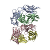
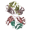
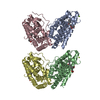
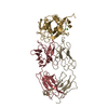
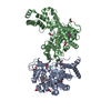
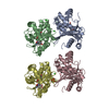
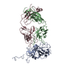
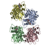
- PDB-1igf: CRYSTAL STRUCTURES OF AN ANTIBODY TO A PEPTIDE AND ITS COMPLEX WI... -
+ Open data

- Basic information
| Entry | Database: PDB / ID: 1igf | ||||||
|---|---|---|---|---|---|---|---|
| Title | CRYSTAL STRUCTURES OF AN ANTIBODY TO A PEPTIDE AND ITS COMPLEX WITH PEPTIDE ANTIGEN AT 2.8 ANGSTROMS | ||||||
 Components Components |
| ||||||
 Keywords Keywords | IMMUNOGLOBULIN | ||||||
| Function / homology | Immunoglobulins / Immunoglobulin-like / Sandwich / Mainly Beta / : / :  Function and homology information Function and homology information | ||||||
| Biological species |  | ||||||
| Method |  X-RAY DIFFRACTION / Resolution: 2.8 Å X-RAY DIFFRACTION / Resolution: 2.8 Å | ||||||
 Authors Authors | Stanfield, R.L. / Wilson, I.A. | ||||||
 Citation Citation | Journal: Science / Year: 1990 Title: Crystal structures of an antibody to a peptide and its complex with peptide antigen at 2.8 A. Authors: Stanfield, R.L. / Fieser, T.M. / Lerner, R.A. / Wilson, I.A. #1: Journal: J.Biol.Chem. / Year: 1989Title: Preliminary Crystallographic Data and Primary Sequence for Anti-Peptide Fab' B13I2 and its Complex with the C-Helix Peptide from Myohemerythrin Authors: Stura, E.A. / Stanfield, R.L. / Fieser, T.M. / Balderas, R.S. / Smith, L.R. / Lerner, R.A. / Wilson, I.A. | ||||||
| History |
|
- Structure visualization
| Structure viewer | Molecule: MolmilJmol/JSmol |
|---|
- Downloads & links
-Download
| PDBx/mmCIF format | 1igf.cif.gz | 179.2 KB | Display | PDBx/mmCIF format |
|---|---|---|---|---|
| PDB format | pdb1igf.ent.gz | 140.9 KB | Display | PDB format |
| PDBx/mmJSON format | 1igf.json.gz | Tree view | PDBx/mmJSON format | |
| Others |  Other downloads Other downloads |
-Validation report
| Arichive directory | https://data.pdbj.org/pub/pdb/validation_reports/ig/1igfftp://data.pdbj.org/pub/pdb/validation_reports/ig/1igf | HTTPS FTP |
|---|
-Related structure data


-Links
 PDBj
PDBj

- Assembly
| Deposited unit | 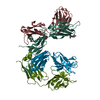
| ||||||||
|---|---|---|---|---|---|---|---|---|---|
| 1 |
| ||||||||
| Unit cell |
| ||||||||
| Atom site foot note | 1: RESIDUES 8, 95, AND 141 OF THE *L* AND *M* CHAINS AND RESIDUES 149 AND 151 OF THE *H* AND *J* CHAINS ARE CIS PROLINES. | ||||||||
| Noncrystallographic symmetry (NCS) | NCS oper: (Code: given Matrix: (0.246, 0.12614, -0.96103), Vector: Details | THE TWO FAB' HEAVY CHAINS (RESIDUES 1-227) OF THE ASYMMETRIC UNIT HAVE BEEN ASSIGNED CHAIN INDICATORS *H* AND *J*. THERE ARE TWO FAB MOLECULES PER ASYMMETRIC UNIT. THE NON-CRYSTALLOGRAPHIC TRANSFORMATION PRESENTED ON THE *MTRIX* RECORDS BELOW YIELDS APPROXIMATE COORDINATES FOR MOLECULE 2 WHEN APPLIED TO MOLECULE 1 (COORDINATES OF CHAIN *M* FROM CHAIN *L* AND COORDINATES OF CHAIN *J* FROM CHAIN *H*). | |
-Components
| #1: Antibody | Mass: 24148.803 Da / Num. of mol.: 2 Source method: isolated from a genetically manipulated source Source: (gene. exp.) #2: Antibody | Mass: 23807.738 Da / Num. of mol.: 2 Source method: isolated from a genetically manipulated source Source: (gene. exp.) #3: Sugar | ChemComp-NAG / |   Type: D-saccharide, beta linking / Mass: 221.208 Da / Num. of mol.: 1 Type: D-saccharide, beta linking / Mass: 221.208 Da / Num. of mol.: 1Source method: isolated from a genetically manipulated source Formula: C8H15NO6 Has protein modification | Y | Nonpolymer details | NAG N 901 IS LINKED TO ASN L 26 OF THE FIRST FAB' FRAGMENT IN THE ASYMMETRIC UNIT. BECAUSE DENSITY ...NAG N 901 IS LINKED TO ASN L 26 OF THE FIRST FAB' FRAGMENT IN THE ASYMMETRIC | Sequence details | THE FAB' FRAGMENT IS NUMBERED BY THE CONVENTION OF E.KABAT (E.A.KABAT,T.T.WU,M.REID-MILLER,H.M. ...THE FAB' FRAGMENT IS NUMBERED BY THE CONVENTION | |
|---|
-Experimental details
-Experiment
| Experiment | Method: X-RAY DIFFRACTION |
|---|
- Sample preparation
| Crystal | Density Matthews: 3.13 Å3/Da / Density % sol: 60.69 % | ||||||||||||||||||||
|---|---|---|---|---|---|---|---|---|---|---|---|---|---|---|---|---|---|---|---|---|---|
| Crystal grow | *PLUS pH: 6 / Method: vapor diffusion, sitting dropDetails: taken from Stura, E.A. et al(1989). J. Biol. Chem., 264, 15721-15725. | ||||||||||||||||||||
| Components of the solutions | *PLUS
|
-Data collection
| Radiation | Scattering type: x-ray |
|---|---|
| Radiation wavelength | Relative weight: 1 |
- Processing
| Software | Name: X-PLOR / Classification: refinement | ||||||||||||||||||||||||||||||||||||||||||||||||||||||||||||
|---|---|---|---|---|---|---|---|---|---|---|---|---|---|---|---|---|---|---|---|---|---|---|---|---|---|---|---|---|---|---|---|---|---|---|---|---|---|---|---|---|---|---|---|---|---|---|---|---|---|---|---|---|---|---|---|---|---|---|---|---|---|
| Refinement | Resolution: 2.8→8 Å / Rfactor Rwork: 0.18 | ||||||||||||||||||||||||||||||||||||||||||||||||||||||||||||
| Refinement step | Cycle: LAST / Resolution: 2.8→8 Å
| ||||||||||||||||||||||||||||||||||||||||||||||||||||||||||||
| Refine LS restraints |
| ||||||||||||||||||||||||||||||||||||||||||||||||||||||||||||
| Refinement | *PLUS Highest resolution: 2.8 Å / Lowest resolution: 8 Å / Rfactor obs: 0.18 | ||||||||||||||||||||||||||||||||||||||||||||||||||||||||||||
| Solvent computation | *PLUS | ||||||||||||||||||||||||||||||||||||||||||||||||||||||||||||
| Displacement parameters | *PLUS | ||||||||||||||||||||||||||||||||||||||||||||||||||||||||||||
| Refine LS restraints | *PLUS Type: x_angle_d / Dev ideal: 3.71 |
