 Movie
Movie Controller


+ Open data

- Basic information
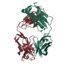
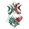
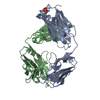
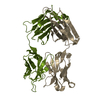
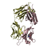
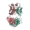
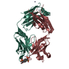
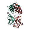
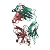
| Entry | Database: PDB / ID: 1xgy | ||||||
|---|---|---|---|---|---|---|---|
| Title | Crystal Structure of Anti-Meta I Rhodopsin Fab Fragment K42-41L | ||||||
 Components Components |
| ||||||
 Keywords Keywords | IMMUNE SYSTEM / Meta-I / Rhodopsin / Fab / Igg / K42-41L / Phage display / antibody / immunoglobulin / antibody imprinting / peptide mimetics | ||||||
| Function / homology |  Function and homology information Function and homology information: / Immunoglobulin V-Type / Immunoglobulin V-set domain / Immunoglobulin V-set domain / Immunoglobulin/major histocompatibility complex, conserved site / Immunoglobulins and major histocompatibility complex proteins signature. / Immunoglobulin subtype / Immunoglobulin / Immunoglobulin C-Type / Immunoglobulin C1-set ...: / Immunoglobulin V-Type / Immunoglobulin V-set domain / Immunoglobulin V-set domain / Immunoglobulin/major histocompatibility complex, conserved site / Immunoglobulins and major histocompatibility complex proteins signature. / Immunoglobulin subtype / Immunoglobulin / Immunoglobulin C-Type / Immunoglobulin C1-set / Immunoglobulin C1-set domain / Ig-like domain profile. / Immunoglobulin-like domain / Immunoglobulin-like domain superfamily / Immunoglobulin-like fold / Immunoglobulins / Immunoglobulin-like / Sandwich / Mainly Beta Similarity search - Domain/homology | ||||||
| Biological species |  | ||||||
| Method |  X-RAY DIFFRACTION / MOLECULAR REPLACEMENT / Resolution: 2.71 Å X-RAY DIFFRACTION / MOLECULAR REPLACEMENT / Resolution: 2.71 Å | ||||||
 Authors Authors | Piscitelli, C.L. / Angel, T.E. / Bailey, B.W. / Lawerence, C.M. | ||||||
 Citation Citation | Journal: J.Biol.Chem. / Year: 2006 Title: Equilibrium between metarhodopsin-I and metarhodopsin-II is dependent on the conformation of the third cytoplasmic loop. Authors: Piscitelli, C.L. / Angel, T.E. / Bailey, B.W. / Hargrave, P. / Dratz, E.A. / Lawrence, C.M. | ||||||
| History |
| ||||||
| Remark 999 | SEQUENCE Suitable sequence database reference not available |
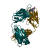
- Structure visualization
| Structure viewer | Molecule: MolmilJmol/JSmol |
|---|
- Downloads & links
-Download
| PDBx/mmCIF format | 1xgy.cif.gz | 175.9 KB | Display | PDBx/mmCIF format |
|---|---|---|---|---|
| PDB format | pdb1xgy.ent.gz | 139.9 KB | Display | PDB format |
| PDBx/mmJSON format | 1xgy.json.gz | Tree view | PDBx/mmJSON format | |
| Others |  Other downloads Other downloads |
-Validation report
| Arichive directory | https://data.pdbj.org/pub/pdb/validation_reports/xg/1xgyftp://data.pdbj.org/pub/pdb/validation_reports/xg/1xgy | HTTPS FTP |
|---|
-Related structure data


-Links
 PDBj
PDBj


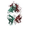
- Assembly
| Deposited unit | 
| ||||||||
|---|---|---|---|---|---|---|---|---|---|
| 1 | 
| ||||||||
| 2 | 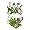
| ||||||||
| Unit cell |
| ||||||||
| Components on special symmetry positions |
|
-Components
| #1: Antibody | Mass: 23807.518 Da / Num. of mol.: 2 / Fragment: Anitgen Binding Fragment, Fab / Source method: isolated from a natural source Details: SP2/0 mouse myeloma cells fused with immunized mouse splenocytes Source: (natural) #2: Antibody | Mass: 23644.568 Da / Num. of mol.: 2 / Fragment: Antigen Binding Fragment, Fab / Source method: isolated from a natural source Details: SP2/0 mouse myeloma cells fused with immunized mouse splenocytes Source: (natural) #3: Protein/peptide | Mass: 991.100 Da / Num. of mol.: 2 / Fragment: Phage Display Consensus Peptide / Source method: obtained synthetically / Details: Chemically synthesized. #4: Water | ChemComp-HOH / |  Mass: 18.015 Da / Num. of mol.: 75 / Source method: isolated from a natural source / Formula: H2O Mass: 18.015 Da / Num. of mol.: 75 / Source method: isolated from a natural source / Formula: H2OHas protein modification | Y | |
|---|
-Experimental details
-Experiment
| Experiment | Method: X-RAY DIFFRACTION / Number of used crystals: 1 |
|---|
- Sample preparation
| Crystal | Density Matthews: 2.46 Å3/Da / Density % sol: 49.96 % |
|---|---|
| Crystal grow | Temperature: 291 K / Method: vapor diffusion, hanging drop / pH: 6.5 Details: peg mme 5000, MES, Ammonium Sulfate, Peptide TGALQERSK, pH 6.5, VAPOR DIFFUSION, HANGING DROP, temperature 291K |
-Data collection
| Diffraction | Mean temperature: 100 K |
|---|---|
| Diffraction source | Source: ROTATING ANODE / Type: RIGAKU RUH3R / Wavelength: 1.5418 Å |
| Detector | Type: MAR scanner 345 mm plate / Detector: IMAGE PLATE / Date: Feb 28, 2003 / Details: OSMIC BLUE |
| Radiation | Monochromator: NI MIRROR / Protocol: SINGLE WAVELENGTH / Monochromatic (M) / Laue (L): M / Scattering type: x-ray |
| Radiation wavelength | Wavelength: 1.5418 Å / Relative weight: 1 |
| Reflection | Resolution: 2.71→98.32 Å / Num. all: 21152 / Num. obs: 21152 / % possible obs: 78.2 % / Observed criterion σ(F): 0 / Redundancy: 4.2 % / Biso Wilson estimate: 41.4 Å2 / Rsym value: 0.083 / Net I/σ(I): 12.7 |
| Reflection shell | Resolution: 2.71→2.83 Å / Redundancy: 1.2 % / Rmerge(I) obs: 0.234 / Mean I/σ(I) obs: 2.4 / Num. unique all: 1122 / % possible all: 43.2 |
- Processing
| Software |
| ||||||||||||||||||||||||||||||||||||
|---|---|---|---|---|---|---|---|---|---|---|---|---|---|---|---|---|---|---|---|---|---|---|---|---|---|---|---|---|---|---|---|---|---|---|---|---|---|
| Refinement | Method to determine structure: MOLECULAR REPLACEMENT Starting model: PDB ENTRIES 1N6Q, 1FAI Resolution: 2.71→19.89 Å / Rfactor Rfree error: 0.008 / Data cutoff high absF: 1635857.51 / Data cutoff low absF: 0 / Isotropic thermal model: RESTRAINED / Cross valid method: THROUGHOUT / σ(F): 0 / Stereochemistry target values: Engh & Huber
| ||||||||||||||||||||||||||||||||||||
| Solvent computation | Solvent model: FLAT MODEL / Bsol: 15.9482 Å2 / ksol: 0.323055 e/Å3 | ||||||||||||||||||||||||||||||||||||
| Displacement parameters | Biso mean: 36.3 Å2
| ||||||||||||||||||||||||||||||||||||
| Refine analyze |
| ||||||||||||||||||||||||||||||||||||
| Refinement step | Cycle: LAST / Resolution: 2.71→19.89 Å
| ||||||||||||||||||||||||||||||||||||
| Refine LS restraints |
| ||||||||||||||||||||||||||||||||||||
| LS refinement shell | Resolution: 2.71→2.83 Å / Rfactor Rfree error: 0.054 / Total num. of bins used: 6
| ||||||||||||||||||||||||||||||||||||
| Xplor file |
|
