 Movie
Movie Controller

[English] 日本語
 Yorodumi
- PDB-1cbv: AN AUTOANTIBODY TO SINGLE-STRANDED DNA: COMPARISON OF THE THREE-D... -
+ Open data

- Basic information
| Entry | Database: PDB / ID: 1cbv | ||||||
|---|---|---|---|---|---|---|---|
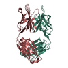
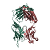
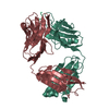
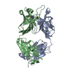
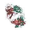
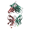
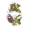
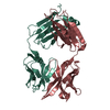
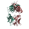
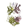
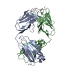
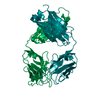
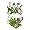
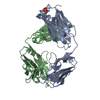
| Title | AN AUTOANTIBODY TO SINGLE-STRANDED DNA: COMPARISON OF THE THREE-DIMENSIONAL STRUCTURES OF THE UNLIGANDED FAB AND A DEOXYNUCLEOTIDE-FAB COMPLEX | ||||||
 Components Components |
| ||||||
 Keywords Keywords | IMMUNE SYSTEM/DNA / PROTEIN-DNA COMPLEX / SINGLE STRAND / IMMUNE SYSTEM-DNA COMPLEX | ||||||
| Function / homology |  Function and homology information Function and homology informationClassical antibody-mediated complement activation / FCGR activation / Role of phospholipids in phagocytosis / Regulation of Complement cascade / positive regulation of type IIa hypersensitivity / humoral immune response mediated by circulating immunoglobulin / phagocytosis, recognition / Regulation of actin dynamics for phagocytic cup formation / positive regulation of type I hypersensitivity / Fc-gamma receptor I complex binding ...Classical antibody-mediated complement activation / FCGR activation / Role of phospholipids in phagocytosis / Regulation of Complement cascade / positive regulation of type IIa hypersensitivity / humoral immune response mediated by circulating immunoglobulin / phagocytosis, recognition / Regulation of actin dynamics for phagocytic cup formation / positive regulation of type I hypersensitivity / Fc-gamma receptor I complex binding / immunoglobulin complex, circulating / phagocytosis, engulfment / immunoglobulin receptor binding / IgG immunoglobulin complex / immunoglobulin mediated immune response / complement activation, classical pathway / immunoglobulin complex / antigen binding / positive regulation of phagocytosis / positive regulation of immune response / antibacterial humoral response / adaptive immune response / : / extracellular region / metal ion binding / plasma membrane Similarity search - Function | ||||||
| Biological species |  | ||||||
| Method |  X-RAY DIFFRACTION / Resolution: 2.66 Å X-RAY DIFFRACTION / Resolution: 2.66 Å | ||||||
 Authors Authors | Herron, J.N. / He, X.M. / Ballard, D.W. / Blier, P.R. / Pace, P.E. / Bothwell, A.L.M. / Voss Junior, E.W. / Edmundson, A.B. | ||||||
 Citation Citation | Journal: Proteins / Year: 1991 Title: An autoantibody to single-stranded DNA: comparison of the three-dimensional structures of the unliganded Fab and a deoxynucleotide-Fab complex. Authors: Herron, J.N. / He, X.M. / Ballard, D.W. / Blier, P.R. / Pace, P.E. / Bothwell, A.L. / Voss Jr., E.W. / Edmundson, A.B. #1: Journal: Mol.Immunol. / Year: 1985Title: Crystallographic Characterization of the FAB Fragment of a Monoclonal Anti-SS- DNA Antibody Authors: Gibson, A.L. / Herron, J.N. / Ballard, D.W. / Voss, E.W. / He, X.M. / Patrick, V.A. / Edmundson, A.B. | ||||||
| History |
|
- Structure visualization
| Structure viewer | Molecule: MolmilJmol/JSmol |
|---|
- Downloads & links
-Download
| PDBx/mmCIF format | 1cbv.cif.gz | 96.6 KB | Display | PDBx/mmCIF format |
|---|---|---|---|---|
| PDB format | pdb1cbv.ent.gz | 71.9 KB | Display | PDB format |
| PDBx/mmJSON format | 1cbv.json.gz | Tree view | PDBx/mmJSON format | |
| Others |  Other downloads Other downloads |
-Validation report
| Arichive directory | https://data.pdbj.org/pub/pdb/validation_reports/cb/1cbvftp://data.pdbj.org/pub/pdb/validation_reports/cb/1cbv | HTTPS FTP |
|---|
-Related structure data


-Links
 PDBj
PDBj


- Assembly
| Deposited unit | 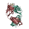
| ||||||||
|---|---|---|---|---|---|---|---|---|---|
| 1 |
| ||||||||
| Unit cell |
|
-Components
| #1: DNA chain | Mass: 867.621 Da / Num. of mol.: 1 / Source method: obtained synthetically |
|---|---|
| #2: Antibody | Mass: 24109.773 Da / Num. of mol.: 1 Source method: isolated from a genetically manipulated source Source: (gene. exp.) |
| #3: Antibody | Mass: 23553.316 Da / Num. of mol.: 1 Source method: isolated from a genetically manipulated source Source: (gene. exp.) |
| Compound details | TURNS L2, L4, AND L10 CONTAIN ONLY 3 RESIDUES. |
| Has protein modification | Y |
| Sequence details | THE PROTEIN WAS SEQUENCED BY D. W. BALLARD, P. R. BLIER, P. E. PACE, AND A. L. M. BOTHWELL. SEE ...THE PROTEIN WAS SEQUENCED BY D. W. BALLARD, P. R. BLIER, P. E. PACE, AND A. L. M. BOTHWELL. SEE REFERENCE 1 ABOVE. |
-Experimental details
-Experiment
| Experiment | Method: X-RAY DIFFRACTION |
|---|
- Sample preparation
| Crystal | Density Matthews: 2.36 Å3/Da / Density % sol: 47.97 % |
|---|---|
| Crystal grow | *PLUS Temperature: 15 ℃ / pH: 7.6 / Method: unknown |
| Components of the solutions | *PLUS Conc.: 1.7 M / Common name: ammonium sulfate |
-Data collection
| Radiation | Protocol: SINGLE WAVELENGTH / Monochromatic (M) / Laue (L): M / Scattering type: x-ray |
|---|---|
| Radiation wavelength | Relative weight: 1 |
| Reflection | *PLUS Highest resolution: 2.66 Å / Num. obs: 8624 / % possible obs: 64 % / Observed criterion σ(I): 1.5 / Num. measured all: 13413 |
- Processing
| Software |
| ||||||||||||||||||||||||||||||||||||||||||||||||||||||||||||||||||||||||||||||||||||||||||||||||||||||||||||||||||||||||||||||||||||||||||||||||||||||||||
|---|---|---|---|---|---|---|---|---|---|---|---|---|---|---|---|---|---|---|---|---|---|---|---|---|---|---|---|---|---|---|---|---|---|---|---|---|---|---|---|---|---|---|---|---|---|---|---|---|---|---|---|---|---|---|---|---|---|---|---|---|---|---|---|---|---|---|---|---|---|---|---|---|---|---|---|---|---|---|---|---|---|---|---|---|---|---|---|---|---|---|---|---|---|---|---|---|---|---|---|---|---|---|---|---|---|---|---|---|---|---|---|---|---|---|---|---|---|---|---|---|---|---|---|---|---|---|---|---|---|---|---|---|---|---|---|---|---|---|---|---|---|---|---|---|---|---|---|---|---|---|---|---|---|---|---|
| Refinement | Resolution: 2.66→6 Å /
| ||||||||||||||||||||||||||||||||||||||||||||||||||||||||||||||||||||||||||||||||||||||||||||||||||||||||||||||||||||||||||||||||||||||||||||||||||||||||||
| Refinement step | Cycle: LAST / Resolution: 2.66→6 Å
| ||||||||||||||||||||||||||||||||||||||||||||||||||||||||||||||||||||||||||||||||||||||||||||||||||||||||||||||||||||||||||||||||||||||||||||||||||||||||||
| Refine LS restraints |
|