 Movie
Movie Controller

[English] 日本語
 Yorodumi
- PDB-1jfq: ANTIGEN-BINDING FRAGMENT OF THE MURINE ANTI-PHENYLARSONATE ANTIBO... -
+ Open data

- Basic information
| Entry | Database: PDB / ID: 1jfq | ||||||
|---|---|---|---|---|---|---|---|
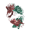
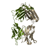
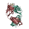
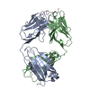
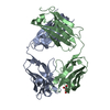
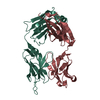
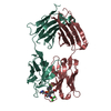
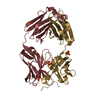
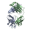
| Title | ANTIGEN-BINDING FRAGMENT OF THE MURINE ANTI-PHENYLARSONATE ANTIBODY 36-71, "FAB 36-71" | ||||||
 Components Components | (ANTIGEN-BINDING FRAGMENT OF ANTI-PHENYLARSONATE ANTIBODY) x 2 | ||||||
 Keywords Keywords | IMMUNE SYSTEM / immunoglobulin | ||||||
| Function / homology |  Function and homology information Function and homology informationimmunoglobulin mediated immune response / immunoglobulin complex / antigen binding / adaptive immune response / immune response / extracellular region / plasma membrane Similarity search - Function | ||||||
| Biological species |  | ||||||
| Method |  X-RAY DIFFRACTION / MOLECULAR REPLACEMENT / Resolution: 1.9 Å X-RAY DIFFRACTION / MOLECULAR REPLACEMENT / Resolution: 1.9 Å | ||||||
 Authors Authors | Parhami-Seren, B. / Viswanathan, M. / Strong, R.K. / Margolies, M.N. | ||||||
 Citation Citation | Journal: J.Immunol. / Year: 2001 Title: Structural analysis of mutants of high-affinity and low-affinity p-azophenylarsonate-specific antibodies generated by alanine scanning of heavy chain complementarity-determining region 2. Authors: Parhami-Seren, B. / Viswanathan, M. / Strong, R.K. / Margolies, M.N. | ||||||
| History |
| ||||||
| Remark 999 | SEQUENCE The sequence database reference for the molecules in the this entry is not available |
- Structure visualization
| Structure viewer | Molecule: MolmilJmol/JSmol |
|---|
- Downloads & links
-Download
| PDBx/mmCIF format | 1jfq.cif.gz | 97.4 KB | Display | PDBx/mmCIF format |
|---|---|---|---|---|
| PDB format | pdb1jfq.ent.gz | 74.1 KB | Display | PDB format |
| PDBx/mmJSON format | 1jfq.json.gz | Tree view | PDBx/mmJSON format | |
| Others |  Other downloads Other downloads |
-Validation report
| Arichive directory | https://data.pdbj.org/pub/pdb/validation_reports/jf/1jfqftp://data.pdbj.org/pub/pdb/validation_reports/jf/1jfq | HTTPS FTP |
|---|
-Related structure data
| Related structure data | 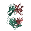 6fabS S: Starting model for refinement |
|---|---|
| Similar structure data |


-Links
 PDBj
PDBj

- Assembly
| Deposited unit | 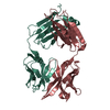
| ||||||||
|---|---|---|---|---|---|---|---|---|---|
| 1 |
| ||||||||
| Unit cell |
|
-Components
| #1: Antibody | Mass: 23715.094 Da / Num. of mol.: 1 / Fragment: LIGHT CHAIN Source method: isolated from a genetically manipulated source Source: (gene. exp.) |
|---|---|
| #2: Antibody | Mass: 23857.559 Da / Num. of mol.: 1 / Fragment: HEAVY CHAIN Source method: isolated from a genetically manipulated source Source: (gene. exp.) |
| #3: Water | ChemComp-HOH /  Mass: 18.015 Da / Num. of mol.: 112 / Source method: isolated from a natural source / Formula: H2O Mass: 18.015 Da / Num. of mol.: 112 / Source method: isolated from a natural source / Formula: H2O |
| Has protein modification | Y |
-Experimental details
-Experiment
| Experiment | Method: X-RAY DIFFRACTION / Number of used crystals: 1 |
|---|
- Sample preparation
| Crystal | Density Matthews: 2.33 Å3/Da / Density % sol: 47.29 % | ||||||||||||||||||||||||||||||||||||||||||||||||||||||||
|---|---|---|---|---|---|---|---|---|---|---|---|---|---|---|---|---|---|---|---|---|---|---|---|---|---|---|---|---|---|---|---|---|---|---|---|---|---|---|---|---|---|---|---|---|---|---|---|---|---|---|---|---|---|---|---|---|---|
| Crystal grow | Temperature: 298 K / Method: vapor diffusion, hanging drop / pH: 7.5 Details: PEG 8000, pH 7.5, VAPOR DIFFUSION, HANGING DROP, temperature 298K | ||||||||||||||||||||||||||||||||||||||||||||||||||||||||
| Crystal grow | *PLUS Method: vapor diffusionDetails: Rose, D.R., (1990) Proc.Natl.Acad.Sci.USA, 87, 338. | ||||||||||||||||||||||||||||||||||||||||||||||||||||||||
| Components of the solutions | *PLUS
|
-Data collection
| Diffraction | Mean temperature: 298 K |
|---|---|
| Diffraction source | Source: ROTATING ANODE / Type: RIGAKU RU200 / Wavelength: 1.5418 |
| Detector | Type: XUONG-HAMLIN MULTIWIRE / Detector: AREA DETECTOR |
| Radiation | Monochromator: graphite / Protocol: SINGLE WAVELENGTH / Monochromatic (M) / Laue (L): M / Scattering type: x-ray |
| Radiation wavelength | Wavelength: 1.5418 Å / Relative weight: 1 |
| Reflection | Resolution: 1.9→50 Å / Num. obs: 28861 |
- Processing
| Software |
| |||||||||||||||
|---|---|---|---|---|---|---|---|---|---|---|---|---|---|---|---|---|
| Refinement | Method to determine structure: MOLECULAR REPLACEMENT Starting model: PDB ENTRY 6FAB Resolution: 1.9→6 Å / Isotropic thermal model: Isotropic / Cross valid method: not applied / σ(F): 2 / Stereochemistry target values: Engh & Huber /
| |||||||||||||||
| Refinement step | Cycle: LAST / Resolution: 1.9→6 Å
| |||||||||||||||
| Refine LS restraints |
| |||||||||||||||
| LS refinement shell | Resolution: 1.9→1.98 Å /
| |||||||||||||||
| Software | *PLUS Name: X-PLOR / Classification: refinement | |||||||||||||||
| Refinement | *PLUS Lowest resolution: 6 Å / σ(F): 2 | |||||||||||||||
| Solvent computation | *PLUS | |||||||||||||||
| Displacement parameters | *PLUS | |||||||||||||||
| Refine LS restraints | *PLUS
| |||||||||||||||
| LS refinement shell | *PLUS Rfactor Rwork: 0.276 / Rfactor obs: 0.276 |
