 Movie
Movie Controller


+ Open data

- Basic information
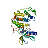
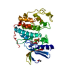
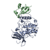
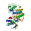
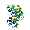
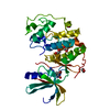
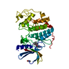
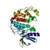
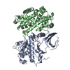
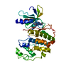
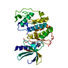
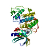
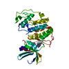
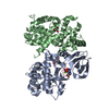
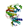
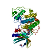
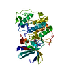
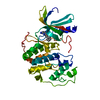
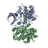
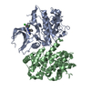
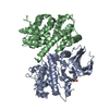
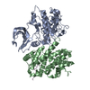
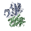
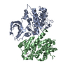
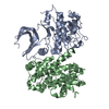
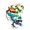
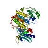
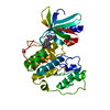
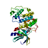
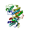
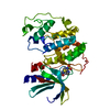
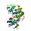
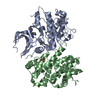
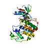
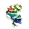
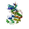
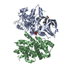
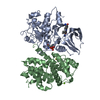
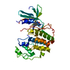
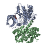
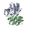
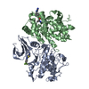
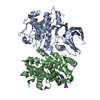
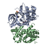
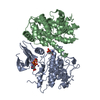
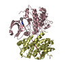
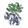
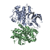
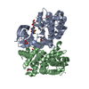
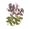
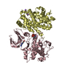
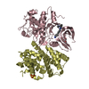
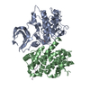
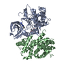
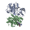
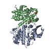
| Entry | Database: PDB / ID: 1okv | ||||||
|---|---|---|---|---|---|---|---|
| Title | Cyclin A binding groove inhibitor H-Arg-Arg-Leu-Ile-Phe-NH2 | ||||||
 Components Components |
| ||||||
 Keywords Keywords | TRANSFERASE/TRANSFERASE INHIBITOR / KINASE-CYCLIN COMPLEX / CYCLIN A / INHIBITOR / LIGAND EXCHANGE / DRUG DESIGN / PEPTIDOMIMETICS / CELL CYCLE / KINASE / TRANSFERASE-TRANSFERASE INHIBITOR COMPLEX | ||||||
| Function / homology |  Function and homology information Function and homology informationcyclin A2-CDK1 complex / cell cycle G1/S phase transition / cellular response to luteinizing hormone stimulus / G2/M DNA replication checkpoint / Transcription of E2F targets under negative control by p107 (RBL1) and p130 (RBL2) in complex with HDAC1 / cellular response to leptin stimulus / male pronucleus / female pronucleus / cellular response to cocaine / response to glucagon ...cyclin A2-CDK1 complex / cell cycle G1/S phase transition / cellular response to luteinizing hormone stimulus / G2/M DNA replication checkpoint / Transcription of E2F targets under negative control by p107 (RBL1) and p130 (RBL2) in complex with HDAC1 / cellular response to leptin stimulus / male pronucleus / female pronucleus / cellular response to cocaine / response to glucagon / cyclin-dependent protein serine/threonine kinase regulator activity / positive regulation of DNA biosynthetic process / cellular response to insulin-like growth factor stimulus / cyclin A1-CDK2 complex / cyclin E2-CDK2 complex / regulation of heterochromatin organization / cyclin E1-CDK2 complex / cyclin A2-CDK2 complex / positive regulation of DNA-templated DNA replication initiation / G2 Phase / Y chromosome / cyclin-dependent protein kinase activity / Phosphorylation of proteins involved in G1/S transition by active Cyclin E:Cdk2 complexes / positive regulation of heterochromatin formation / p53-Dependent G1 DNA Damage Response / X chromosome / PTK6 Regulates Cell Cycle / regulation of anaphase-promoting complex-dependent catabolic process / Defective binding of RB1 mutants to E2F1,(E2F2, E2F3) / centriole replication / Regulation of APC/C activators between G1/S and early anaphase / telomere maintenance in response to DNA damage / regulation of DNA replication / microtubule organizing center / centrosome duplication / G0 and Early G1 / cochlea development / animal organ regeneration / Telomere Extension By Telomerase / Activation of the pre-replicative complex / cyclin-dependent kinase / cyclin-dependent protein serine/threonine kinase activity / TP53 Regulates Transcription of Genes Involved in G1 Cell Cycle Arrest / Activation of ATR in response to replication stress / Regulation of MITF-M-dependent genes involved in cell cycle and proliferation / Cyclin E associated events during G1/S transition / Cajal body / Cyclin A:Cdk2-associated events at S phase entry / Cyclin A/B1/B2 associated events during G2/M transition / cyclin-dependent protein kinase holoenzyme complex / Chk1/Chk2(Cds1) mediated inactivation of Cyclin B:Cdk1 complex / regulation of G2/M transition of mitotic cell cycle / cellular response to platelet-derived growth factor stimulus / condensed chromosome / negative regulation of protein localization to chromatin / mitotic G1 DNA damage checkpoint signaling / cellular response to nitric oxide / post-translational protein modification / regulation of mitotic cell cycle / cyclin binding / positive regulation of DNA replication / male germ cell nucleus / meiotic cell cycle / potassium ion transport / cellular response to estradiol stimulus / Cdc20:Phospho-APC/C mediated degradation of Cyclin A / peptidyl-serine phosphorylation / G1/S transition of mitotic cell cycle / DNA Damage/Telomere Stress Induced Senescence / Meiotic recombination / CDK-mediated phosphorylation and removal of Cdc6 / G2/M transition of mitotic cell cycle / SCF(Skp2)-mediated degradation of p27/p21 / positive regulation of fibroblast proliferation / Transcriptional regulation of granulopoiesis / Orc1 removal from chromatin / Cyclin D associated events in G1 / cellular senescence / Regulation of TP53 Degradation / nuclear envelope / Factors involved in megakaryocyte development and platelet production / regulation of gene expression / Processing of DNA double-strand break ends / Senescence-Associated Secretory Phenotype (SASP) / transcription regulator complex / cellular response to hypoxia / Regulation of TP53 Activity through Phosphorylation / Ras protein signal transduction / DNA replication / protein phosphorylation / chromosome, telomeric region / endosome / Ub-specific processing proteases / chromatin remodeling / protein domain specific binding / cell division / protein serine kinase activity / DNA repair / protein serine/threonine kinase activity / positive regulation of cell population proliferation Similarity search - Function | ||||||
| Biological species |  HOMO SAPIENS (human) HOMO SAPIENS (human)SYNTHETIC CONSTRUCT (others) | ||||||
| Method |  X-RAY DIFFRACTION / SYNCHROTRON / MOLECULAR REPLACEMENT / Resolution: 2.4 Å X-RAY DIFFRACTION / SYNCHROTRON / MOLECULAR REPLACEMENT / Resolution: 2.4 Å | ||||||
 Authors Authors | Kontopidis, G. / Andrews, M. / McInnes, C. / Cowan, A. / Powers, H. / Innes, L. / Plater, A. / Griffiths, G. / Paterson, D. / Zheleva, D. ...Kontopidis, G. / Andrews, M. / McInnes, C. / Cowan, A. / Powers, H. / Innes, L. / Plater, A. / Griffiths, G. / Paterson, D. / Zheleva, D. / Lane, D. / Green, S. / Walkinshaw, M. / Fischer, P. | ||||||
 Citation Citation | Journal: Structure / Year: 2003 Title: Insights Into Cyclin Groove Recognition. Complex Crystal Structures and Inhibitor Design Through Ligand Exchange Authors: Kontopidis, G. / Andrews, M. / Mcinnes, C. / Cowan, A. / Powers, H. / Innes, L. / Plater, A. / Griffiths, G. / Paterson, D. / Zheleva, D. / Lane, D. / Green, S. / Walkinshaw, M. / Fischer, P. | ||||||
| History |
|
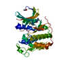
- Structure visualization
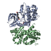
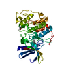
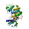
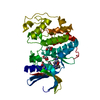
| Structure viewer | Molecule: MolmilJmol/JSmol |
|---|
- Downloads & links
-Download
| PDBx/mmCIF format | 1okv.cif.gz | 249.5 KB | Display | PDBx/mmCIF format |
|---|---|---|---|---|
| PDB format | pdb1okv.ent.gz | 202.5 KB | Display | PDB format |
| PDBx/mmJSON format | 1okv.json.gz | Tree view | PDBx/mmJSON format | |
| Others |  Other downloads Other downloads |
-Validation report
| Arichive directory | https://data.pdbj.org/pub/pdb/validation_reports/ok/1okvftp://data.pdbj.org/pub/pdb/validation_reports/ok/1okv | HTTPS FTP |
|---|
-Related structure data
| Related structure data | 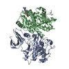 1okwC  1ol1C 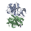 1ol2C 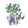 1finS  1h0u S: Starting model for refinement C: citing same article ( |
|---|---|
| Similar structure data |


-Links
 PDBj
PDBj


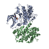
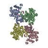
- Assembly
| Deposited unit | 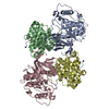
| ||||||||
|---|---|---|---|---|---|---|---|---|---|
| 1 | 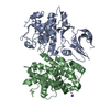
| ||||||||
| 2 | 
| ||||||||
| Unit cell |
|
-Components
| #1: Protein | Mass: 33976.488 Da / Num. of mol.: 2 Source method: isolated from a genetically manipulated source Source: (gene. exp.) HOMO SAPIENS (human) / Production host:   SPODOPTERA FRUGIPERDA (fall armyworm) / References: UniProt: P24941, EC: 2.7.1.37 SPODOPTERA FRUGIPERDA (fall armyworm) / References: UniProt: P24941, EC: 2.7.1.37#2: Protein | Mass: 29867.512 Da / Num. of mol.: 2 / Fragment: RESIDUES 173 - 432 Source method: isolated from a genetically manipulated source Source: (gene. exp.) HOMO SAPIENS (human) / Plasmid: PET3A / Production host:  #3: Protein/peptide | Mass: 703.900 Da / Num. of mol.: 2 / Source method: obtained synthetically Details: CYCLIN GROOVE-BOUND PEPTIDE H-ARG-ARG-LEU-ILE-PHE-NH2 Source: (synth.) SYNTHETIC CONSTRUCT (others) #4: Water | ChemComp-HOH / |  Mass: 18.015 Da / Num. of mol.: 757 / Source method: isolated from a natural source / Formula: H2O Mass: 18.015 Da / Num. of mol.: 757 / Source method: isolated from a natural source / Formula: H2OHas protein modification | Y | Sequence details | 1OKV B SWS P20248 1 - 172 NOT IN ATOMS LIST 1OKV D SWS P20248 1 - 172 NOT IN ATOMS LIST | |
|---|
-Experimental details
-Experiment
| Experiment | Method: X-RAY DIFFRACTION / Number of used crystals: 1 |
|---|
- Sample preparation
| Crystal | Density Matthews: 2.7 Å3/Da / Density % sol: 54 % | |||||||||||||||||||||||||||||||||||||||||||||||||
|---|---|---|---|---|---|---|---|---|---|---|---|---|---|---|---|---|---|---|---|---|---|---|---|---|---|---|---|---|---|---|---|---|---|---|---|---|---|---|---|---|---|---|---|---|---|---|---|---|---|---|
| Crystal grow | pH: 7.8 / Details: 22% PEG 3350, 0.1M NA3-CIT, PH 7.80 | |||||||||||||||||||||||||||||||||||||||||||||||||
| Crystal grow | *PLUS pH: 7 / Method: vapor diffusion, hanging drop | |||||||||||||||||||||||||||||||||||||||||||||||||
| Components of the solutions | *PLUS
|
-Data collection
| Diffraction | Mean temperature: 100 K |
|---|---|
| Diffraction source | Source: SYNCHROTRON / Site: SRS  / Beamline: PX14.2 / Wavelength: 0.98 / Beamline: PX14.2 / Wavelength: 0.98 |
| Detector | Type: ADSC CCD / Detector: CCD / Date: Mar 15, 2002 |
| Radiation | Protocol: SINGLE WAVELENGTH / Monochromatic (M) / Laue (L): M / Scattering type: x-ray |
| Radiation wavelength | Wavelength: 0.98 Å / Relative weight: 1 |
| Reflection | Resolution: 2.4→24 Å / Num. obs: 574133 / % possible obs: 96.7 % / Redundancy: 10.7 % / Rmerge(I) obs: 0.11 / Net I/σ(I): 5.9 |
| Reflection shell | Resolution: 2.4→2.46 Å / Rmerge(I) obs: 0.65 / Mean I/σ(I) obs: 1.3 / % possible all: 84.6 |
| Reflection | *PLUS Highest resolution: 2.4 Å / Num. all: 563579 / Num. obs: 52345 / Rmerge(I) obs: 0.11 |
| Reflection shell | *PLUS Rmerge(I) obs: 0.65 / Mean I/σ(I) obs: 1.2 |
- Processing
| Software |
| ||||||||||||||||||||||||||||||||||||||||||||||||||||||||||||||||||||||||||||||||||||||||||||||||||||||||||||||||||||||||||||||||||||||||||||||||||||||||||||||||||||||||||||||||||||||
|---|---|---|---|---|---|---|---|---|---|---|---|---|---|---|---|---|---|---|---|---|---|---|---|---|---|---|---|---|---|---|---|---|---|---|---|---|---|---|---|---|---|---|---|---|---|---|---|---|---|---|---|---|---|---|---|---|---|---|---|---|---|---|---|---|---|---|---|---|---|---|---|---|---|---|---|---|---|---|---|---|---|---|---|---|---|---|---|---|---|---|---|---|---|---|---|---|---|---|---|---|---|---|---|---|---|---|---|---|---|---|---|---|---|---|---|---|---|---|---|---|---|---|---|---|---|---|---|---|---|---|---|---|---|---|---|---|---|---|---|---|---|---|---|---|---|---|---|---|---|---|---|---|---|---|---|---|---|---|---|---|---|---|---|---|---|---|---|---|---|---|---|---|---|---|---|---|---|---|---|---|---|---|---|
| Refinement | Method to determine structure: MOLECULAR REPLACEMENT Starting model: PDB ENTRY 1FIN Resolution: 2.4→17 Å / Cor.coef. Fo:Fc: 0.942 / Cor.coef. Fo:Fc free: 0.892 / SU B: 15.583 / SU ML: 0.363 / Cross valid method: THROUGHOUT / ESU R: 0.519 / ESU R Free: 0.311 / Stereochemistry target values: MAXIMUM LIKELIHOOD / Details: HYDROGENS HAVE BEEN ADDED IN THE RIDING POSITIONS
| ||||||||||||||||||||||||||||||||||||||||||||||||||||||||||||||||||||||||||||||||||||||||||||||||||||||||||||||||||||||||||||||||||||||||||||||||||||||||||||||||||||||||||||||||||||||
| Solvent computation | Ion probe radii: 0.8 Å / Shrinkage radii: 0.8 Å / VDW probe radii: 1.4 Å / Solvent model: BABINET MODEL WITH MASK | ||||||||||||||||||||||||||||||||||||||||||||||||||||||||||||||||||||||||||||||||||||||||||||||||||||||||||||||||||||||||||||||||||||||||||||||||||||||||||||||||||||||||||||||||||||||
| Displacement parameters | Biso mean: 31.8 Å2
| ||||||||||||||||||||||||||||||||||||||||||||||||||||||||||||||||||||||||||||||||||||||||||||||||||||||||||||||||||||||||||||||||||||||||||||||||||||||||||||||||||||||||||||||||||||||
| Refinement step | Cycle: LAST / Resolution: 2.4→17 Å
| ||||||||||||||||||||||||||||||||||||||||||||||||||||||||||||||||||||||||||||||||||||||||||||||||||||||||||||||||||||||||||||||||||||||||||||||||||||||||||||||||||||||||||||||||||||||
| Refine LS restraints |
|