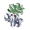
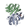
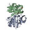
cyclin A2-CDK1 complex / cell cycle G1/S phase transition / cellular response to luteinizing hormone stimulus / G2/M DNA replication checkpoint / Transcription of E2F targets under negative control by p107 (RBL1) and p130 (RBL2) in complex with HDAC1 / cellular response to leptin stimulus / male pronucleus / female pronucleus / cellular response to cocaine / response to glucagon ...cyclin A2-CDK1 complex / cell cycle G1/S phase transition / cellular response to luteinizing hormone stimulus / G2/M DNA replication checkpoint / Transcription of E2F targets under negative control by p107 (RBL1) and p130 (RBL2) in complex with HDAC1 / cellular response to leptin stimulus / male pronucleus / female pronucleus / cellular response to cocaine / response to glucagon / cyclin-dependent protein serine/threonine kinase regulator activity / positive regulation of DNA biosynthetic process / cyclin A1-CDK2 complex / cyclin E2-CDK2 complex / cyclin E1-CDK2 complex / cellular response to insulin-like growth factor stimulus / cyclin A2-CDK2 complex / positive regulation of DNA-templated DNA replication initiation / G2 Phase / Y chromosome / cyclin-dependent protein kinase activity / regulation of heterochromatin organization / Phosphorylation of proteins involved in G1/S transition by active Cyclin E:Cdk2 complexes / positive regulation of heterochromatin formation / p53-Dependent G1 DNA Damage Response / X chromosome / PTK6 Regulates Cell Cycle / regulation of anaphase-promoting complex-dependent catabolic process / Defective binding of RB1 mutants to E2F1,(E2F2, E2F3) / centriole replication / Regulation of APC/C activators between G1/S and early anaphase / telomere maintenance in response to DNA damage / regulation of DNA replication / centrosome duplication / microtubule organizing center / G0 and Early G1 / cochlea development / animal organ regeneration / Telomere Extension By Telomerase / Activation of the pre-replicative complex / cyclin-dependent kinase / cyclin-dependent protein serine/threonine kinase activity / TP53 Regulates Transcription of Genes Involved in G1 Cell Cycle Arrest / Activation of ATR in response to replication stress / Regulation of MITF-M-dependent genes involved in cell cycle and proliferation / Cajal body / Cyclin E associated events during G1/S transition / Cyclin A:Cdk2-associated events at S phase entry / cyclin-dependent protein kinase holoenzyme complex / Cyclin A/B1/B2 associated events during G2/M transition / Chk1/Chk2(Cds1) mediated inactivation of Cyclin B:Cdk1 complex / regulation of G2/M transition of mitotic cell cycle / cellular response to platelet-derived growth factor stimulus / condensed chromosome / mitotic G1 DNA damage checkpoint signaling / negative regulation of protein localization to chromatin / cellular response to nitric oxide / post-translational protein modification / regulation of mitotic cell cycle / cyclin binding / positive regulation of DNA replication / peptidyl-serine phosphorylation / male germ cell nucleus / meiotic cell cycle / cellular response to estradiol stimulus / potassium ion transport / G1/S transition of mitotic cell cycle / Cdc20:Phospho-APC/C mediated degradation of Cyclin A / DNA Damage/Telomere Stress Induced Senescence / Meiotic recombination / G2/M transition of mitotic cell cycle / positive regulation of fibroblast proliferation / CDK-mediated phosphorylation and removal of Cdc6 / Transcriptional regulation of granulopoiesis / SCF(Skp2)-mediated degradation of p27/p21 / Orc1 removal from chromatin / cellular senescence / Cyclin D associated events in G1 / Regulation of TP53 Degradation / nuclear envelope / Factors involved in megakaryocyte development and platelet production / regulation of gene expression / Processing of DNA double-strand break ends / Senescence-Associated Secretory Phenotype (SASP) / transcription regulator complex / cellular response to hypoxia / Regulation of TP53 Activity through Phosphorylation / protein phosphorylation / Ras protein signal transduction / DNA replication / chromosome, telomeric region / endosome / Ub-specific processing proteases / chromatin remodeling / protein domain specific binding / protein serine kinase activity / cell division / DNA repair / protein serine/threonine kinase activity / positive regulation of cell population proliferation 類似検索 - 分子機能
Cyclin-A, N-terminal APC/C binding region / Cyclin-A N-terminal APC/C binding region / : / Cyclin, C-terminal domain / : / Cyclins signature. / Cyclin / Cyclin-like / Cyclin, C-terminal domain / Cyclin_C ...Cyclin-A, N-terminal APC/C binding region / Cyclin-A N-terminal APC/C binding region / : / Cyclin, C-terminal domain / : / Cyclins signature. / Cyclin / Cyclin-like / Cyclin, C-terminal domain / Cyclin_C / Cyclin A; domain 1 / Cyclin, N-terminal / Cyclin, N-terminal domain / Cyclin-like / domain present in cyclins, TFIIB and Retinoblastoma / Cyclin-like superfamily / : / Phosphorylase Kinase; domain 1 / Phosphorylase Kinase; domain 1 / Transferase(Phosphotransferase) domain 1 / Transferase(Phosphotransferase); domain 1 / Serine/threonine-protein kinase, active site / Serine/Threonine protein kinases active-site signature. / Protein kinase domain / Serine/Threonine protein kinases, catalytic domain / Protein kinase, ATP binding site / Protein kinases ATP-binding region signature. / Protein kinase domain profile. / Protein kinase domain / Protein kinase-like domain superfamily / 2-Layer Sandwich / Orthogonal Bundle / Mainly Alpha / Alpha Beta 類似検索 - ドメイン・相同性
 ムービー
ムービー コントローラー


 データを開く
 基本情報
基本情報 要素
要素 キーワード
キーワード 機能・相同性情報
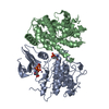
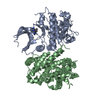
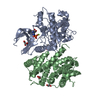
 HOMO SAPIENS (ヒト)
 X線回折 /
  データ登録者
 引用
引用 構造の表示
構造の表示 ダウンロードとリンク
 その他のダウンロード
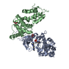


 PDBj
PDBj


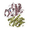
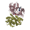
 集合体
集合体


 分子量: 420.534 Da / 分子数: 2 / 由来タイプ: 合成 / 式: C21H24N8S
 分子量: 96.063 Da / 分子数: 2 / 由来タイプ: 合成 / 式: SO4
 分子量: 108.159 Da / 分子数: 3 / 由来タイプ: 合成 / 式: C3H8O2S
 試料調製
試料調製 / ビームライン: I03 / 波長: 0.9763
  解析
解析