 Movie
Movie Controller


+ Open data

- Basic information
| Entry | Database: PDB / ID: 3tnw | ||||||
|---|---|---|---|---|---|---|---|
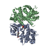
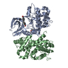
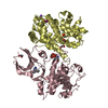
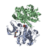
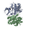
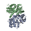
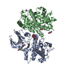
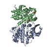
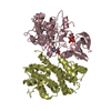
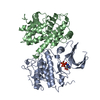
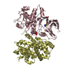
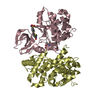
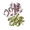
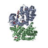
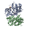
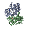
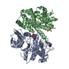
| Title | Structure of CDK2/cyclin A in complex with CAN508 | ||||||
 Components Components |
| ||||||
 Keywords Keywords | TRANSFERASE/TRANSFERASE INHIBITOR / cyclin dependent kinase / kinase cyclin / phosphotransferase / cyclin A / phosphorylation at CDK2 threonine 160 / TRANSFERASE-TRANSFERASE INHIBITOR complex | ||||||
| Function / homology |  Function and homology information Function and homology informationcell cycle G1/S phase transition / cyclin-dependent protein serine/threonine kinase regulator activity / cyclin A1-CDK2 complex / cyclin E2-CDK2 complex / cyclin E1-CDK2 complex / cyclin A2-CDK2 complex / positive regulation of DNA-templated DNA replication initiation / G2 Phase / Y chromosome / cyclin-dependent protein kinase activity ...cell cycle G1/S phase transition / cyclin-dependent protein serine/threonine kinase regulator activity / cyclin A1-CDK2 complex / cyclin E2-CDK2 complex / cyclin E1-CDK2 complex / cyclin A2-CDK2 complex / positive regulation of DNA-templated DNA replication initiation / G2 Phase / Y chromosome / cyclin-dependent protein kinase activity / regulation of heterochromatin organization / Phosphorylation of proteins involved in G1/S transition by active Cyclin E:Cdk2 complexes / positive regulation of heterochromatin formation / p53-Dependent G1 DNA Damage Response / X chromosome / PTK6 Regulates Cell Cycle / regulation of anaphase-promoting complex-dependent catabolic process / Defective binding of RB1 mutants to E2F1,(E2F2, E2F3) / centriole replication / Regulation of APC/C activators between G1/S and early anaphase / telomere maintenance in response to DNA damage / regulation of DNA replication / centrosome duplication / microtubule organizing center / G0 and Early G1 / Telomere Extension By Telomerase / Activation of the pre-replicative complex / cyclin-dependent kinase / cyclin-dependent protein serine/threonine kinase activity / TP53 Regulates Transcription of Genes Involved in G1 Cell Cycle Arrest / Activation of ATR in response to replication stress / Regulation of MITF-M-dependent genes involved in cell cycle and proliferation / Cajal body / Cyclin E associated events during G1/S transition / regulation of G2/M transition of mitotic cell cycle / Cyclin A:Cdk2-associated events at S phase entry / Cyclin A/B1/B2 associated events during G2/M transition / cyclin-dependent protein kinase holoenzyme complex / condensed chromosome / mitotic G1 DNA damage checkpoint signaling / cellular response to nitric oxide / negative regulation of protein localization to chromatin / regulation of mitotic cell cycle / post-translational protein modification / cyclin binding / positive regulation of DNA replication / male germ cell nucleus / potassium ion transport / peptidyl-serine phosphorylation / G1/S transition of mitotic cell cycle / meiotic cell cycle / DNA Damage/Telomere Stress Induced Senescence / Meiotic recombination / G2/M transition of mitotic cell cycle / CDK-mediated phosphorylation and removal of Cdc6 / cellular senescence / Transcriptional regulation of granulopoiesis / SCF(Skp2)-mediated degradation of p27/p21 / Orc1 removal from chromatin / Cyclin D associated events in G1 / Regulation of TP53 Degradation / nuclear envelope / Factors involved in megakaryocyte development and platelet production / regulation of gene expression / Processing of DNA double-strand break ends / Senescence-Associated Secretory Phenotype (SASP) / transcription regulator complex / Regulation of TP53 Activity through Phosphorylation / protein phosphorylation / Ras protein signal transduction / chromosome, telomeric region / DNA replication / endosome / chromatin remodeling / protein domain specific binding / protein serine kinase activity / cell division / DNA repair / protein serine/threonine kinase activity / centrosome / positive regulation of cell population proliferation / protein kinase binding / positive regulation of DNA-templated transcription / magnesium ion binding / negative regulation of transcription by RNA polymerase II / signal transduction / DNA-templated transcription / nucleoplasm / ATP binding / nucleus / cytosol / cytoplasm Similarity search - Function | ||||||
| Biological species |  Homo sapiens (human) Homo sapiens (human) | ||||||
| Method |  X-RAY DIFFRACTION / SYNCHROTRON / MOLECULAR REPLACEMENT / Resolution: 2 Å X-RAY DIFFRACTION / SYNCHROTRON / MOLECULAR REPLACEMENT / Resolution: 2 Å | ||||||
 Authors Authors | Baumli, S. / Hole, A.J. / Endicott, J.A. | ||||||
 Citation Citation | Journal: Acs Chem.Biol. / Year: 2012 Title: The CDK9 C-helix Exhibits Conformational Plasticity That May Explain the Selectivity of CAN508. Authors: Baumli, S. / Hole, A.J. / Noble, M.E. / Endicott, J.A. | ||||||
| History |
|
- Structure visualization
| Structure viewer | Molecule: MolmilJmol/JSmol |
|---|
- Downloads & links
-Download
| PDBx/mmCIF format | 3tnw.cif.gz | 254 KB | Display | PDBx/mmCIF format |
|---|---|---|---|---|
| PDB format | pdb3tnw.ent.gz | 201.8 KB | Display | PDB format |
| PDBx/mmJSON format | 3tnw.json.gz | Tree view | PDBx/mmJSON format | |
| Others |  Other downloads Other downloads |
-Validation report
| Arichive directory | https://data.pdbj.org/pub/pdb/validation_reports/tn/3tnwftp://data.pdbj.org/pub/pdb/validation_reports/tn/3tnw | HTTPS FTP |
|---|
-Related structure data
| Related structure data |  3tn8C  3tnhC  3tniC  3bhtS S: Starting model for refinement C: citing same article ( |
|---|---|
| Similar structure data |


-Links
 PDBj
PDBj


- Assembly
| Deposited unit | 
| ||||||||
|---|---|---|---|---|---|---|---|---|---|
| 1 | 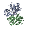
| ||||||||
| 2 | 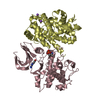
| ||||||||
| Unit cell |
|
-Components
| #1: Protein | Mass: 34200.602 Da / Num. of mol.: 2 Source method: isolated from a genetically manipulated source Source: (gene. exp.) Homo sapiens (human) / Gene: CDK2 / Production host:  #2: Protein | Mass: 30018.631 Da / Num. of mol.: 2 / Fragment: cyclin boxes, UNP resdiues 169-430 Source method: isolated from a genetically manipulated source Source: (gene. exp.) #3: Chemical |   Mass: 218.215 Da / Num. of mol.: 2 / Source method: obtained synthetically / Formula: C9H10N6O Mass: 218.215 Da / Num. of mol.: 2 / Source method: obtained synthetically / Formula: C9H10N6O#4: Chemical |   Mass: 22.990 Da / Num. of mol.: 2 / Source method: obtained synthetically / Formula: Na Mass: 22.990 Da / Num. of mol.: 2 / Source method: obtained synthetically / Formula: Na#5: Water | ChemComp-HOH / |  Mass: 18.015 Da / Num. of mol.: 912 / Source method: isolated from a natural source / Formula: H2O Mass: 18.015 Da / Num. of mol.: 912 / Source method: isolated from a natural source / Formula: H2OHas protein modification | Y | |
|---|
-Experimental details
-Experiment
| Experiment | Method: X-RAY DIFFRACTION / Number of used crystals: 1 |
|---|
- Sample preparation
| Crystal | Density Matthews: 2.88 Å3/Da / Density % sol: 57.23 % |
|---|---|
| Crystal grow | Temperature: 277 K / Method: vapor diffusion, hanging drop / pH: 7 Details: 1.1 M ammonium sulphate, 100 mM HEPES, 5 mM DTT, pH 7.0, VAPOR DIFFUSION, HANGING DROP, temperature 277K |
-Data collection
| Diffraction | Mean temperature: 100 K | ||||||||||||||||||
|---|---|---|---|---|---|---|---|---|---|---|---|---|---|---|---|---|---|---|---|
| Diffraction source | Source: SYNCHROTRON / Site: Diamond  / Beamline: I02 / Wavelength: 0.9795 Å / Beamline: I02 / Wavelength: 0.9795 Å | ||||||||||||||||||
| Detector | Type: ADSC QUANTUM 315r / Detector: CCD / Date: Dec 6, 2009 | ||||||||||||||||||
| Radiation | Monochromator: double crystal monochromator / Protocol: SINGLE WAVELENGTH / Monochromatic (M) / Laue (L): M / Scattering type: x-ray | ||||||||||||||||||
| Radiation wavelength | Wavelength: 0.9795 Å / Relative weight: 1 | ||||||||||||||||||
| Reflection | Resolution: 2→44.86 Å / Num. all: 100622 / Num. obs: 100421 / % possible obs: 99.8 % / Observed criterion σ(F): 0 / Observed criterion σ(I): 0 / Rmerge(I) obs: 0.103 / Net I/σ(I): 4.4 | ||||||||||||||||||
| Reflection shell |
|
- Processing
| Software |
| |||||||||||||||||||||||||||||||||||||||||||||||||||||||||||||||||||||||||||||||||||||||||||||||||||||||||||||||||||||||||||||||||||||||||||||||||||||||||||||||||||||||||||||||||||||||||||||||||||||||||||||||||||||||||
|---|---|---|---|---|---|---|---|---|---|---|---|---|---|---|---|---|---|---|---|---|---|---|---|---|---|---|---|---|---|---|---|---|---|---|---|---|---|---|---|---|---|---|---|---|---|---|---|---|---|---|---|---|---|---|---|---|---|---|---|---|---|---|---|---|---|---|---|---|---|---|---|---|---|---|---|---|---|---|---|---|---|---|---|---|---|---|---|---|---|---|---|---|---|---|---|---|---|---|---|---|---|---|---|---|---|---|---|---|---|---|---|---|---|---|---|---|---|---|---|---|---|---|---|---|---|---|---|---|---|---|---|---|---|---|---|---|---|---|---|---|---|---|---|---|---|---|---|---|---|---|---|---|---|---|---|---|---|---|---|---|---|---|---|---|---|---|---|---|---|---|---|---|---|---|---|---|---|---|---|---|---|---|---|---|---|---|---|---|---|---|---|---|---|---|---|---|---|---|---|---|---|---|---|---|---|---|---|---|---|---|---|---|---|---|---|---|---|---|
| Refinement | Method to determine structure: MOLECULAR REPLACEMENT Starting model: 3BHT, omitting the ligand Resolution: 2→42.935 Å / Occupancy max: 1 / Occupancy min: 0.16 / SU ML: 0.55 / σ(F): 1.34 / Phase error: 21.68 / Stereochemistry target values: ML
| |||||||||||||||||||||||||||||||||||||||||||||||||||||||||||||||||||||||||||||||||||||||||||||||||||||||||||||||||||||||||||||||||||||||||||||||||||||||||||||||||||||||||||||||||||||||||||||||||||||||||||||||||||||||||
| Solvent computation | Shrinkage radii: 0.83 Å / VDW probe radii: 1.1 Å / Solvent model: FLAT BULK SOLVENT MODEL / Bsol: 40.258 Å2 / ksol: 0.356 e/Å3 | |||||||||||||||||||||||||||||||||||||||||||||||||||||||||||||||||||||||||||||||||||||||||||||||||||||||||||||||||||||||||||||||||||||||||||||||||||||||||||||||||||||||||||||||||||||||||||||||||||||||||||||||||||||||||
| Displacement parameters |
| |||||||||||||||||||||||||||||||||||||||||||||||||||||||||||||||||||||||||||||||||||||||||||||||||||||||||||||||||||||||||||||||||||||||||||||||||||||||||||||||||||||||||||||||||||||||||||||||||||||||||||||||||||||||||
| Refinement step | Cycle: LAST / Resolution: 2→42.935 Å
| |||||||||||||||||||||||||||||||||||||||||||||||||||||||||||||||||||||||||||||||||||||||||||||||||||||||||||||||||||||||||||||||||||||||||||||||||||||||||||||||||||||||||||||||||||||||||||||||||||||||||||||||||||||||||
| Refine LS restraints |
| |||||||||||||||||||||||||||||||||||||||||||||||||||||||||||||||||||||||||||||||||||||||||||||||||||||||||||||||||||||||||||||||||||||||||||||||||||||||||||||||||||||||||||||||||||||||||||||||||||||||||||||||||||||||||
| LS refinement shell |
|
