 Movie
Movie Controller

[English] 日本語
 Yorodumi
- PDB-1h1p: Structure of human Thr160-phospho CDK2/cyclin A complexed with th... -
+ Open data

- Basic information
| Entry | Database: PDB / ID: 1h1p | ||||||
|---|---|---|---|---|---|---|---|
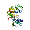
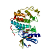
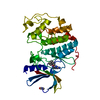
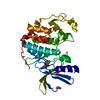
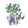
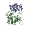
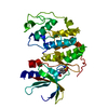
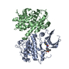
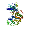
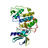
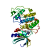
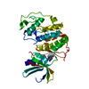
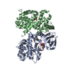
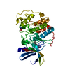
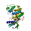
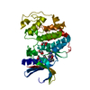
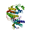
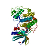
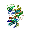
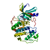
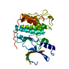
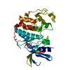
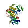
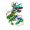
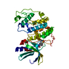
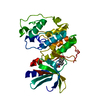
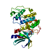
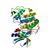
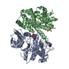
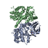
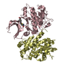
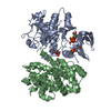
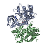
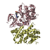
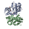
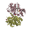
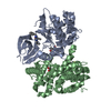
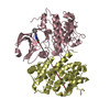
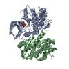
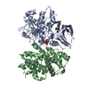
| Title | Structure of human Thr160-phospho CDK2/cyclin A complexed with the inhibitor NU2058 | ||||||
 Components Components |
| ||||||
 Keywords Keywords | TRANSFERASE / KINASE / SERINE/THREONINE-PROTEIN KINASE / ATP-BINDING / CELL CYCLE / CELL DIVISION / MITOSIS / PHOSPHORYLATION | ||||||
| Function / homology |  Function and homology information Function and homology informationcyclin A2-CDK1 complex / cell cycle G1/S phase transition / cellular response to luteinizing hormone stimulus / G2/M DNA replication checkpoint / Transcription of E2F targets under negative control by p107 (RBL1) and p130 (RBL2) in complex with HDAC1 / cellular response to leptin stimulus / male pronucleus / female pronucleus / cellular response to cocaine / response to glucagon ...cyclin A2-CDK1 complex / cell cycle G1/S phase transition / cellular response to luteinizing hormone stimulus / G2/M DNA replication checkpoint / Transcription of E2F targets under negative control by p107 (RBL1) and p130 (RBL2) in complex with HDAC1 / cellular response to leptin stimulus / male pronucleus / female pronucleus / cellular response to cocaine / response to glucagon / cyclin-dependent protein serine/threonine kinase regulator activity / positive regulation of DNA biosynthetic process / cyclin A1-CDK2 complex / cyclin E2-CDK2 complex / cyclin E1-CDK2 complex / cyclin A2-CDK2 complex / cellular response to insulin-like growth factor stimulus / positive regulation of DNA-templated DNA replication initiation / G2 Phase / Y chromosome / cyclin-dependent protein kinase activity / regulation of heterochromatin organization / Phosphorylation of proteins involved in G1/S transition by active Cyclin E:Cdk2 complexes / positive regulation of heterochromatin formation / p53-Dependent G1 DNA Damage Response / X chromosome / PTK6 Regulates Cell Cycle / regulation of anaphase-promoting complex-dependent catabolic process / Defective binding of RB1 mutants to E2F1,(E2F2, E2F3) / centriole replication / Regulation of APC/C activators between G1/S and early anaphase / telomere maintenance in response to DNA damage / centrosome duplication / regulation of DNA replication / microtubule organizing center / G0 and Early G1 / cochlea development / animal organ regeneration / Telomere Extension By Telomerase / Activation of the pre-replicative complex / cyclin-dependent kinase / cyclin-dependent protein serine/threonine kinase activity / TP53 Regulates Transcription of Genes Involved in G1 Cell Cycle Arrest / Regulation of MITF-M-dependent genes involved in cell cycle and proliferation / Activation of ATR in response to replication stress / Cajal body / Cyclin E associated events during G1/S transition / Cyclin A:Cdk2-associated events at S phase entry / cyclin-dependent protein kinase holoenzyme complex / Cyclin A/B1/B2 associated events during G2/M transition / Chk1/Chk2(Cds1) mediated inactivation of Cyclin B:Cdk1 complex / regulation of G2/M transition of mitotic cell cycle / condensed chromosome / cellular response to platelet-derived growth factor stimulus / mitotic G1 DNA damage checkpoint signaling / negative regulation of protein localization to chromatin / cellular response to nitric oxide / post-translational protein modification / regulation of mitotic cell cycle / cyclin binding / positive regulation of DNA replication / peptidyl-serine phosphorylation / male germ cell nucleus / meiotic cell cycle / cellular response to estradiol stimulus / potassium ion transport / Cdc20:Phospho-APC/C mediated degradation of Cyclin A / G1/S transition of mitotic cell cycle / DNA Damage/Telomere Stress Induced Senescence / Meiotic recombination / G2/M transition of mitotic cell cycle / CDK-mediated phosphorylation and removal of Cdc6 / positive regulation of fibroblast proliferation / Transcriptional regulation of granulopoiesis / SCF(Skp2)-mediated degradation of p27/p21 / Orc1 removal from chromatin / cellular senescence / Cyclin D associated events in G1 / Regulation of TP53 Degradation / nuclear envelope / Factors involved in megakaryocyte development and platelet production / regulation of gene expression / Processing of DNA double-strand break ends / Senescence-Associated Secretory Phenotype (SASP) / transcription regulator complex / cellular response to hypoxia / Regulation of TP53 Activity through Phosphorylation / protein phosphorylation / Ras protein signal transduction / DNA replication / chromosome, telomeric region / endosome / Ub-specific processing proteases / chromatin remodeling / protein domain specific binding / protein serine kinase activity / cell division / DNA repair / protein serine/threonine kinase activity / positive regulation of cell population proliferation Similarity search - Function | ||||||
| Biological species |  HOMO SAPIENS (human) HOMO SAPIENS (human) | ||||||
| Method |  X-RAY DIFFRACTION / SYNCHROTRON / MOLECULAR REPLACEMENT / Resolution: 2.1 Å X-RAY DIFFRACTION / SYNCHROTRON / MOLECULAR REPLACEMENT / Resolution: 2.1 Å | ||||||
 Authors Authors | Davies, T.G. / Noble, M.E.M. / Endicott, J.A. / Johnson, L.N. | ||||||
 Citation Citation | Journal: Nat.Struct.Biol. / Year: 2002 Title: Structure-Based Design of a Potent Purine-Based Cyclin-Dependent Kinase Inhibitor Authors: Davies, T.G. / Bentley, J. / Arris, C.E. / Boyle, F.T. / Curtin, N.J. / Endicott, J.A. / Gibson, A.E. / Golding, B.T. / Griffin, R.J. / Hardcastle, I.R. / Jewsbury, P. / Johnson, L.N. / ...Authors: Davies, T.G. / Bentley, J. / Arris, C.E. / Boyle, F.T. / Curtin, N.J. / Endicott, J.A. / Gibson, A.E. / Golding, B.T. / Griffin, R.J. / Hardcastle, I.R. / Jewsbury, P. / Johnson, L.N. / Mesguiche, V. / Newell, D.R. / Noble, M.E.M. / Tucker, J.A. / Wang, L. / Whitfield, H.J. | ||||||
| History |
|
- Structure visualization
| Structure viewer | Molecule: MolmilJmol/JSmol |
|---|
- Downloads & links
-Download
| PDBx/mmCIF format | 1h1p.cif.gz | 241.7 KB | Display | PDBx/mmCIF format |
|---|---|---|---|---|
| PDB format | pdb1h1p.ent.gz | 193.8 KB | Display | PDB format |
| PDBx/mmJSON format | 1h1p.json.gz | Tree view | PDBx/mmJSON format | |
| Others |  Other downloads Other downloads |
-Validation report
| Arichive directory | https://data.pdbj.org/pub/pdb/validation_reports/h1/1h1pftp://data.pdbj.org/pub/pdb/validation_reports/h1/1h1p | HTTPS FTP |
|---|
-Related structure data
| Related structure data |  1h1qC 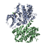 1h1rC 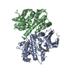 1h1sC  1h0u C: citing same article ( |
|---|---|
| Similar structure data |


-Links
 PDBj
PDBj


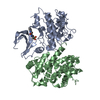
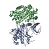
- Assembly
| Deposited unit | 
| ||||||||
|---|---|---|---|---|---|---|---|---|---|
| 1 | 
| ||||||||
| 2 | 
| ||||||||
| Unit cell |
|
-Components
| #1: Protein | Mass: 34467.926 Da / Num. of mol.: 2 Source method: isolated from a genetically manipulated source Details: PHOSPHORYLATED ON THR160 / Source: (gene. exp.) HOMO SAPIENS (human) / Production host:  References: UniProt: P24941, Transferases; Transferring phosphorus-containing groups; Phosphotransferases with an alcohol group as acceptor #2: Protein | Mass: 29624.297 Da / Num. of mol.: 2 Source method: isolated from a genetically manipulated source Source: (gene. exp.) HOMO SAPIENS (human) / Production host: #3: Chemical |   Mass: 247.296 Da / Num. of mol.: 2 / Source method: obtained synthetically / Formula: C12H17N5O Mass: 247.296 Da / Num. of mol.: 2 / Source method: obtained synthetically / Formula: C12H17N5O#4: Water | ChemComp-HOH / |  Mass: 18.015 Da / Num. of mol.: 410 / Source method: isolated from a natural source / Formula: H2O Mass: 18.015 Da / Num. of mol.: 410 / Source method: isolated from a natural source / Formula: H2OHas protein modification | Y | |
|---|
-Experimental details
-Experiment
| Experiment | Method: X-RAY DIFFRACTION / Number of used crystals: 1 |
|---|
- Sample preparation
| Crystal | Density Matthews: 3.69 Å3/Da / Density % sol: 66.69 % | ||||||||||||||||||||||||||||||||||||||||||||||||||||||||
|---|---|---|---|---|---|---|---|---|---|---|---|---|---|---|---|---|---|---|---|---|---|---|---|---|---|---|---|---|---|---|---|---|---|---|---|---|---|---|---|---|---|---|---|---|---|---|---|---|---|---|---|---|---|---|---|---|---|
| Crystal grow | pH: 7 / Details: pH 7.00 | ||||||||||||||||||||||||||||||||||||||||||||||||||||||||
| Crystal grow | *PLUS Method: vapor diffusion, hanging drop | ||||||||||||||||||||||||||||||||||||||||||||||||||||||||
| Components of the solutions | *PLUS
|
-Data collection
| Diffraction | Mean temperature: 100 K |
|---|---|
| Diffraction source | Source: SYNCHROTRON / Site: ESRF  / Beamline: ID14-4 / Wavelength: 0.9789 / Beamline: ID14-4 / Wavelength: 0.9789 |
| Radiation | Protocol: SINGLE WAVELENGTH / Monochromatic (M) / Laue (L): M / Scattering type: x-ray |
| Radiation wavelength | Wavelength: 0.9789 Å / Relative weight: 1 |
| Reflection | Resolution: 2.1→20 Å / Num. obs: 110338 / % possible obs: 92.5 % / Rmerge(I) obs: 0.083 / Net I/σ(I): 5.5 |
| Reflection shell | Resolution: 2.1→2.15 Å / Rmerge(I) obs: 0.406 / Mean I/σ(I) obs: 1.7 / % possible all: 82.7 |
| Reflection | *PLUS Lowest resolution: 20 Å / Num. measured all: 245195 |
| Reflection shell | *PLUS Highest resolution: 2.1 Å / % possible obs: 82.7 % |
- Processing
| Software |
| ||||||||||||||||||||
|---|---|---|---|---|---|---|---|---|---|---|---|---|---|---|---|---|---|---|---|---|---|
| Refinement | Method to determine structure: MOLECULAR REPLACEMENT / Resolution: 2.1→20 Å / Cross valid method: THROUGHOUT
| ||||||||||||||||||||
| Refinement step | Cycle: LAST / Resolution: 2.1→20 Å
| ||||||||||||||||||||
| Refinement | *PLUS Lowest resolution: 20 Å / % reflection Rfree: 5 % | ||||||||||||||||||||
| Solvent computation | *PLUS | ||||||||||||||||||||
| Displacement parameters | *PLUS |
