 Movie
Movie Controller

[English] 日本語
 Yorodumi
- PDB-1g5s: CRYSTAL STRUCTURE OF HUMAN CYCLIN DEPENDENT KINASE 2 (CDK2) IN CO... -
+ Open data

- Basic information
| Entry | Database: PDB / ID: 1g5s | ||||||
|---|---|---|---|---|---|---|---|
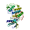
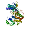
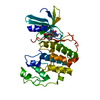
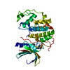
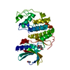
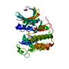
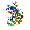
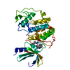
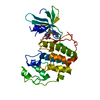
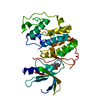
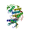
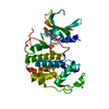
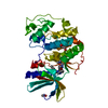
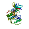
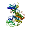
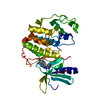
| Title | CRYSTAL STRUCTURE OF HUMAN CYCLIN DEPENDENT KINASE 2 (CDK2) IN COMPLEX WITH THE INHIBITOR H717 | ||||||
 Components Components | CELL DIVISION PROTEIN KINASE 2 | ||||||
 Keywords Keywords | CELL CYCLE/TRANSFERASE / protein-inhibitor complex / CELL CYCLE-TRANSFERASE COMPLEX | ||||||
| Function / homology |  Function and homology information Function and homology informationcyclin A1-CDK2 complex / cyclin E2-CDK2 complex / cyclin E1-CDK2 complex / cyclin A2-CDK2 complex / positive regulation of DNA-templated DNA replication initiation / G2 Phase / Y chromosome / cyclin-dependent protein kinase activity / regulation of heterochromatin organization / Phosphorylation of proteins involved in G1/S transition by active Cyclin E:Cdk2 complexes ...cyclin A1-CDK2 complex / cyclin E2-CDK2 complex / cyclin E1-CDK2 complex / cyclin A2-CDK2 complex / positive regulation of DNA-templated DNA replication initiation / G2 Phase / Y chromosome / cyclin-dependent protein kinase activity / regulation of heterochromatin organization / Phosphorylation of proteins involved in G1/S transition by active Cyclin E:Cdk2 complexes / positive regulation of heterochromatin formation / p53-Dependent G1 DNA Damage Response / X chromosome / PTK6 Regulates Cell Cycle / regulation of anaphase-promoting complex-dependent catabolic process / Defective binding of RB1 mutants to E2F1,(E2F2, E2F3) / centriole replication / Regulation of APC/C activators between G1/S and early anaphase / telomere maintenance in response to DNA damage / centrosome duplication / G0 and Early G1 / Telomere Extension By Telomerase / Activation of the pre-replicative complex / cyclin-dependent kinase / cyclin-dependent protein serine/threonine kinase activity / TP53 Regulates Transcription of Genes Involved in G1 Cell Cycle Arrest / Activation of ATR in response to replication stress / Regulation of MITF-M-dependent genes involved in cell cycle and proliferation / Cajal body / Cyclin E associated events during G1/S transition / regulation of G2/M transition of mitotic cell cycle / Cyclin A:Cdk2-associated events at S phase entry / Cyclin A/B1/B2 associated events during G2/M transition / cyclin-dependent protein kinase holoenzyme complex / condensed chromosome / mitotic G1 DNA damage checkpoint signaling / cellular response to nitric oxide / negative regulation of protein localization to chromatin / regulation of mitotic cell cycle / post-translational protein modification / cyclin binding / positive regulation of DNA replication / male germ cell nucleus / potassium ion transport / peptidyl-serine phosphorylation / G1/S transition of mitotic cell cycle / meiotic cell cycle / DNA Damage/Telomere Stress Induced Senescence / Meiotic recombination / G2/M transition of mitotic cell cycle / CDK-mediated phosphorylation and removal of Cdc6 / cellular senescence / Transcriptional regulation of granulopoiesis / SCF(Skp2)-mediated degradation of p27/p21 / Orc1 removal from chromatin / Cyclin D associated events in G1 / Regulation of TP53 Degradation / nuclear envelope / Factors involved in megakaryocyte development and platelet production / regulation of gene expression / Processing of DNA double-strand break ends / Senescence-Associated Secretory Phenotype (SASP) / transcription regulator complex / Regulation of TP53 Activity through Phosphorylation / protein phosphorylation / Ras protein signal transduction / chromosome, telomeric region / DNA replication / endosome / chromatin remodeling / protein domain specific binding / protein serine kinase activity / cell division / DNA repair / protein serine/threonine kinase activity / centrosome / positive regulation of cell population proliferation / positive regulation of DNA-templated transcription / magnesium ion binding / negative regulation of transcription by RNA polymerase II / signal transduction / DNA-templated transcription / nucleoplasm / ATP binding / nucleus / cytosol / cytoplasm Similarity search - Function | ||||||
| Biological species |  Homo sapiens (human) Homo sapiens (human) | ||||||
| Method |  X-RAY DIFFRACTION / MOLECULAR REPLACEMENT / Resolution: 2.61 Å X-RAY DIFFRACTION / MOLECULAR REPLACEMENT / Resolution: 2.61 Å | ||||||
 Authors Authors | Dreyer, M.K. / Borcherding, D.R. / Dumont, J.A. / Peet, N.P. / Tsay, J.T. / Wright, P.S. / Bitonti, A.J. / Shen, J. / Kim, S.-H. | ||||||
 Citation Citation | Journal: J.Med.Chem. / Year: 2001 Title: Crystal structure of human cyclin-dependent kinase 2 in complex with the adenine-derived inhibitor H717. Authors: Dreyer, M.K. / Borcherding, D.R. / Dumont, J.A. / Peet, N.P. / Tsay, J.T. / Wright, P.S. / Bitonti, A.J. / Shen, J. / Kim, S.H. #1: Journal: J.Mol.Biol. / Year: 1993Title: Purification and crystallization of human cyclin-dependent kinase 2 Authors: Rosenblatt, J. / DeBondt, H. / Jancarik, J. / Morgan, D.O. / Kim, S.-H. | ||||||
| History |
|
- Structure visualization
| Structure viewer | Molecule: MolmilJmol/JSmol |
|---|
- Downloads & links
-Download
| PDBx/mmCIF format | 1g5s.cif.gz | 63.5 KB | Display | PDBx/mmCIF format |
|---|---|---|---|---|
| PDB format | pdb1g5s.ent.gz | 46.8 KB | Display | PDB format |
| PDBx/mmJSON format | 1g5s.json.gz | Tree view | PDBx/mmJSON format | |
| Others |  Other downloads Other downloads |
-Validation report
| Arichive directory | https://data.pdbj.org/pub/pdb/validation_reports/g5/1g5sftp://data.pdbj.org/pub/pdb/validation_reports/g5/1g5s | HTTPS FTP |
|---|
-Related structure data
| Related structure data |  3bmpS S: Starting model for refinement |
|---|---|
| Similar structure data |


-Links
 PDBj
PDBj


- Assembly
| Deposited unit | 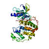
| ||||||||
|---|---|---|---|---|---|---|---|---|---|
| 1 |
| ||||||||
| Unit cell |
|
-Components
| #1: Protein | Mass: 33976.488 Da / Num. of mol.: 1 Source method: isolated from a genetically manipulated source Source: (gene. exp.) Homo sapiens (human) / Gene: CDK2 / Production host:   Spodoptera frugiperda (fall armyworm) Spodoptera frugiperda (fall armyworm)References: UniProt: P24941, Transferases; Transferring phosphorus-containing groups; Phosphotransferases with an alcohol group as acceptor |
|---|---|
| #2: Chemical | ChemComp-I17 /   Mass: 405.539 Da / Num. of mol.: 1 / Source method: obtained synthetically / Formula: C23H31N7 Mass: 405.539 Da / Num. of mol.: 1 / Source method: obtained synthetically / Formula: C23H31N7 |
| #3: Water | ChemComp-HOH /  Mass: 18.015 Da / Num. of mol.: 30 / Source method: isolated from a natural source / Formula: H2O Mass: 18.015 Da / Num. of mol.: 30 / Source method: isolated from a natural source / Formula: H2O |
-Experimental details
-Experiment
| Experiment | Method: X-RAY DIFFRACTION / Number of used crystals: 1 |
|---|
- Sample preparation
| Crystal | Density Matthews: 2.13 Å3/Da / Density % sol: 42.31 % |
|---|---|
| Crystal grow | *PLUS Method: other / Details: Rosenblatt, J., (1993) J. Mol. Biol., 230, 1317. |
-Data collection
| Diffraction | Mean temperature: 277 K |
|---|---|
| Diffraction source | Source: ROTATING ANODE / Type: RIGAKU RU200 / Wavelength: 1.5418 |
| Detector | Type: RIGAKU RAXIS IIC / Detector: IMAGE PLATE / Date: Aug 28, 1997 / Details: mirrors |
| Radiation | Protocol: SINGLE WAVELENGTH / Monochromatic (M) / Laue (L): M / Scattering type: x-ray |
| Radiation wavelength | Wavelength: 1.5418 Å / Relative weight: 1 |
| Reflection | *PLUS Highest resolution: 2.61 Å / Num. obs: 10442 / % possible obs: 99.8 % / Num. measured all: 47878 / Rmerge(I) obs: 0.048 |
| Reflection shell | *PLUS % possible obs: 99.9 % / Rmerge(I) obs: 0.333 |
- Processing
| Software |
| ||||||||||||||||||||
|---|---|---|---|---|---|---|---|---|---|---|---|---|---|---|---|---|---|---|---|---|---|
| Refinement | Method to determine structure: MOLECULAR REPLACEMENT Starting model: PDB ENTRY 3BMP Resolution: 2.61→50 Å / Isotropic thermal model: Isotropic / Cross valid method: THROUGHOUT / σ(F): 0 / σ(I): 0 / Stereochemistry target values: Engh & Huber / Details: grouped B-factors only
| ||||||||||||||||||||
| Displacement parameters | Biso mean: 41 Å2 | ||||||||||||||||||||
| Refinement step | Cycle: LAST / Resolution: 2.61→50 Å
| ||||||||||||||||||||
| Refine LS restraints |
| ||||||||||||||||||||
| Software | *PLUS Name: CNS / Classification: refinement | ||||||||||||||||||||
| Refinement | *PLUS Lowest resolution: 50 Å / σ(F): 0 / Rfactor obs: 0.201 | ||||||||||||||||||||
| Solvent computation | *PLUS | ||||||||||||||||||||
| Displacement parameters | *PLUS Biso mean: 41 Å2 | ||||||||||||||||||||
| Refine LS restraints | *PLUS Type: c_angle_deg / Dev ideal: 1.6 |
