 Movie
Movie Controller

[English] 日本語
 Yorodumi
- PDB-4bco: Structure of CDK2 in complex with cyclin A and a 2-amino-4-hetero... -
+ Open data

- Basic information
| Entry | Database: PDB / ID: 4bco | ||||||
|---|---|---|---|---|---|---|---|
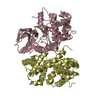
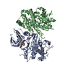
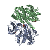
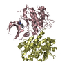
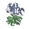
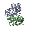
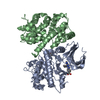
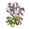
| Title | Structure of CDK2 in complex with cyclin A and a 2-amino-4-heteroaryl- pyrimidine inhibitor | ||||||
 Components Components |
| ||||||
 Keywords Keywords | TRANSFERASE/CELL CYCLE / TRANSFERASE-CELL CYCLE COMPLEX / CDK-CYCLIN COMPLEX / CYCLIN-INHIBITOR / STRUCTURE-BASED DRUG DESIGN | ||||||
| Function / homology |  Function and homology information Function and homology informationcell cycle G1/S phase transition / cyclin-dependent protein serine/threonine kinase regulator activity / cyclin A1-CDK2 complex / cyclin E2-CDK2 complex / cyclin E1-CDK2 complex / cyclin A2-CDK2 complex / positive regulation of DNA-templated DNA replication initiation / G2 Phase / Y chromosome / cyclin-dependent protein kinase activity ...cell cycle G1/S phase transition / cyclin-dependent protein serine/threonine kinase regulator activity / cyclin A1-CDK2 complex / cyclin E2-CDK2 complex / cyclin E1-CDK2 complex / cyclin A2-CDK2 complex / positive regulation of DNA-templated DNA replication initiation / G2 Phase / Y chromosome / cyclin-dependent protein kinase activity / regulation of heterochromatin organization / Phosphorylation of proteins involved in G1/S transition by active Cyclin E:Cdk2 complexes / positive regulation of heterochromatin formation / p53-Dependent G1 DNA Damage Response / X chromosome / PTK6 Regulates Cell Cycle / regulation of anaphase-promoting complex-dependent catabolic process / Defective binding of RB1 mutants to E2F1,(E2F2, E2F3) / centriole replication / Regulation of APC/C activators between G1/S and early anaphase / telomere maintenance in response to DNA damage / centrosome duplication / regulation of DNA replication / microtubule organizing center / G0 and Early G1 / Telomere Extension By Telomerase / Activation of the pre-replicative complex / cyclin-dependent kinase / cyclin-dependent protein serine/threonine kinase activity / TP53 Regulates Transcription of Genes Involved in G1 Cell Cycle Arrest / Regulation of MITF-M-dependent genes involved in cell cycle and proliferation / Activation of ATR in response to replication stress / Cajal body / Cyclin E associated events during G1/S transition / Cyclin A:Cdk2-associated events at S phase entry / cyclin-dependent protein kinase holoenzyme complex / Cyclin A/B1/B2 associated events during G2/M transition / regulation of G2/M transition of mitotic cell cycle / condensed chromosome / mitotic G1 DNA damage checkpoint signaling / negative regulation of protein localization to chromatin / cellular response to nitric oxide / post-translational protein modification / regulation of mitotic cell cycle / cyclin binding / positive regulation of DNA replication / peptidyl-serine phosphorylation / male germ cell nucleus / meiotic cell cycle / potassium ion transport / G1/S transition of mitotic cell cycle / DNA Damage/Telomere Stress Induced Senescence / Meiotic recombination / G2/M transition of mitotic cell cycle / CDK-mediated phosphorylation and removal of Cdc6 / Transcriptional regulation of granulopoiesis / SCF(Skp2)-mediated degradation of p27/p21 / Orc1 removal from chromatin / cellular senescence / Cyclin D associated events in G1 / Regulation of TP53 Degradation / nuclear envelope / Factors involved in megakaryocyte development and platelet production / regulation of gene expression / Processing of DNA double-strand break ends / Senescence-Associated Secretory Phenotype (SASP) / transcription regulator complex / Regulation of TP53 Activity through Phosphorylation / protein phosphorylation / Ras protein signal transduction / DNA replication / chromosome, telomeric region / endosome / chromatin remodeling / protein domain specific binding / protein serine kinase activity / cell division / DNA repair / protein serine/threonine kinase activity / positive regulation of cell population proliferation / centrosome / protein kinase binding / positive regulation of DNA-templated transcription / DNA-templated transcription / magnesium ion binding / negative regulation of transcription by RNA polymerase II / signal transduction / nucleoplasm / ATP binding / nucleus / cytoplasm / cytosol Similarity search - Function | ||||||
| Biological species |  HOMO SAPIENS (human) HOMO SAPIENS (human) | ||||||
| Method |  X-RAY DIFFRACTION / SYNCHROTRON / MOLECULAR REPLACEMENT / Resolution: 2.05 Å X-RAY DIFFRACTION / SYNCHROTRON / MOLECULAR REPLACEMENT / Resolution: 2.05 Å | ||||||
 Authors Authors | Hole, A.J. / Baumli, S. / Wang, S. / Endicott, J.A. / Noble, M.E.M. | ||||||
 Citation Citation | Journal: J.Med.Chem. / Year: 2013 Title: Comparative Structural and Functional Studies of 4-(Thiazol- 5-Yl)-2-(Phenylamino)Pyrimidine-5-Carbonitrile Cdk9 Inhibitors Suggest the Basis for Isotype Selectivity. Authors: Hole, A.J. / Baumli, S. / Shao, H. / Shi, S. / Pepper, C. / Fischer, P.M. / Wang, S. / Endicott, J.A. / Noble, M.E.M. | ||||||
| History |
|
- Structure visualization
| Structure viewer | Molecule: MolmilJmol/JSmol |
|---|
- Downloads & links
-Download
| PDBx/mmCIF format | 4bco.cif.gz | 455.3 KB | Display | PDBx/mmCIF format |
|---|---|---|---|---|
| PDB format | pdb4bco.ent.gz | 374.8 KB | Display | PDB format |
| PDBx/mmJSON format | 4bco.json.gz | Tree view | PDBx/mmJSON format | |
| Others |  Other downloads Other downloads |
-Validation report
| Arichive directory | https://data.pdbj.org/pub/pdb/validation_reports/bc/4bcoftp://data.pdbj.org/pub/pdb/validation_reports/bc/4bco | HTTPS FTP |
|---|
-Related structure data
| Related structure data |  4bcfC  4bchC  4bciC  4bcjC  4bckC  4bcmC  4bcnC  4bcqC C: citing same article ( |
|---|---|
| Similar structure data |


-Links
 PDBj
PDBj


- Assembly
| Deposited unit | 
| ||||||||
|---|---|---|---|---|---|---|---|---|---|
| 1 | 
| ||||||||
| 2 | 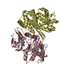
| ||||||||
| Unit cell |
|
-Components
-Protein , 2 types, 4 molecules ACBD
| #1: Protein | Mass: 34200.602 Da / Num. of mol.: 2 Source method: isolated from a genetically manipulated source Source: (gene. exp.) HOMO SAPIENS (human) / Production host:  #2: Protein | Mass: 30067.744 Da / Num. of mol.: 2 Source method: isolated from a genetically manipulated source Source: (gene. exp.) |
|---|
-Non-polymers , 4 types, 527 molecules 


| #3: Chemical |  Mass: 462.571 Da / Num. of mol.: 2 / Source method: obtained synthetically / Formula: C23H26N8OS Mass: 462.571 Da / Num. of mol.: 2 / Source method: obtained synthetically / Formula: C23H26N8OS#4: Chemical | ChemComp-SGM /  Mass: 108.159 Da / Num. of mol.: 4 / Source method: obtained synthetically / Formula: C3H8O2S Mass: 108.159 Da / Num. of mol.: 4 / Source method: obtained synthetically / Formula: C3H8O2S#5: Chemical |  Mass: 96.063 Da / Num. of mol.: 2 / Source method: obtained synthetically / Formula: SO4 Mass: 96.063 Da / Num. of mol.: 2 / Source method: obtained synthetically / Formula: SO4#6: Water | ChemComp-HOH / | Mass: 18.015 Da / Num. of mol.: 519 / Source method: isolated from a natural source / Formula: H2O |
|---|
-Details
| Has protein modification | Y |
|---|
-Experimental details
-Experiment
| Experiment | Method: X-RAY DIFFRACTION / Number of used crystals: 1 |
|---|
- Sample preparation
| Crystal | Density Matthews: 2.9 Å3/Da / Density % sol: 58 % / Description: NONE |
|---|---|
| Crystal grow | pH: 7 Details: COCRYSTALS WERE GROWN IN 1-1.25M AMMONIUM SULFATE, 0.5-0.9M KCL, 100MM HEPES, PH 7.0, 5MM DTT AND IN THE PRESENCE OF COMPOUND. |
-Data collection
| Diffraction | Mean temperature: 100 K |
|---|---|
| Diffraction source | Source: SYNCHROTRON / Site: Diamond  / Beamline: I03 / Wavelength: 0.9763 / Beamline: I03 / Wavelength: 0.9763 |
| Detector | Type: ADSC CCD / Detector: CCD |
| Radiation | Protocol: SINGLE WAVELENGTH / Monochromatic (M) / Laue (L): M / Scattering type: x-ray |
| Radiation wavelength | Wavelength: 0.9763 Å / Relative weight: 1 |
| Reflection | Resolution: 1.58→52.36 Å / Num. obs: 92057 / % possible obs: 99.2 % / Redundancy: 3.22 % / Biso Wilson estimate: 32.61 Å2 / Rmerge(I) obs: 0.08 / Net I/σ(I): 7.16 |
| Reflection shell | Resolution: 2.05→2.16 Å / Redundancy: 3.29 % / Rmerge(I) obs: 0.54 / Mean I/σ(I) obs: 1.41 / % possible all: 99.4 |
- Processing
| Software |
| |||||||||||||||||||||||||||||||||||||||||||||||||||||||||||||||||||||||||||||||||||||||||||||||||||||||||||||||||||||||||||||||||||||||||||||||||||||||||||||||||||||||||||||||||||||||||||||||||||||||||||||||||||||||||||||||||
|---|---|---|---|---|---|---|---|---|---|---|---|---|---|---|---|---|---|---|---|---|---|---|---|---|---|---|---|---|---|---|---|---|---|---|---|---|---|---|---|---|---|---|---|---|---|---|---|---|---|---|---|---|---|---|---|---|---|---|---|---|---|---|---|---|---|---|---|---|---|---|---|---|---|---|---|---|---|---|---|---|---|---|---|---|---|---|---|---|---|---|---|---|---|---|---|---|---|---|---|---|---|---|---|---|---|---|---|---|---|---|---|---|---|---|---|---|---|---|---|---|---|---|---|---|---|---|---|---|---|---|---|---|---|---|---|---|---|---|---|---|---|---|---|---|---|---|---|---|---|---|---|---|---|---|---|---|---|---|---|---|---|---|---|---|---|---|---|---|---|---|---|---|---|---|---|---|---|---|---|---|---|---|---|---|---|---|---|---|---|---|---|---|---|---|---|---|---|---|---|---|---|---|---|---|---|---|---|---|---|---|---|---|---|---|---|---|---|---|---|---|---|---|---|---|---|---|
| Refinement | Method to determine structure: MOLECULAR REPLACEMENT / Resolution: 2.05→49.599 Å / SU ML: 0.64 / σ(F): 0 / Phase error: 21.08 / Stereochemistry target values: ML
| |||||||||||||||||||||||||||||||||||||||||||||||||||||||||||||||||||||||||||||||||||||||||||||||||||||||||||||||||||||||||||||||||||||||||||||||||||||||||||||||||||||||||||||||||||||||||||||||||||||||||||||||||||||||||||||||||
| Solvent computation | Shrinkage radii: 0.95 Å / VDW probe radii: 1.2 Å / Solvent model: FLAT BULK SOLVENT MODEL / Bsol: 44.553 Å2 / ksol: 0.389 e/Å3 | |||||||||||||||||||||||||||||||||||||||||||||||||||||||||||||||||||||||||||||||||||||||||||||||||||||||||||||||||||||||||||||||||||||||||||||||||||||||||||||||||||||||||||||||||||||||||||||||||||||||||||||||||||||||||||||||||
| Displacement parameters |
| |||||||||||||||||||||||||||||||||||||||||||||||||||||||||||||||||||||||||||||||||||||||||||||||||||||||||||||||||||||||||||||||||||||||||||||||||||||||||||||||||||||||||||||||||||||||||||||||||||||||||||||||||||||||||||||||||
| Refinement step | Cycle: LAST / Resolution: 2.05→49.599 Å
| |||||||||||||||||||||||||||||||||||||||||||||||||||||||||||||||||||||||||||||||||||||||||||||||||||||||||||||||||||||||||||||||||||||||||||||||||||||||||||||||||||||||||||||||||||||||||||||||||||||||||||||||||||||||||||||||||
| Refine LS restraints |
| |||||||||||||||||||||||||||||||||||||||||||||||||||||||||||||||||||||||||||||||||||||||||||||||||||||||||||||||||||||||||||||||||||||||||||||||||||||||||||||||||||||||||||||||||||||||||||||||||||||||||||||||||||||||||||||||||
| LS refinement shell |
| |||||||||||||||||||||||||||||||||||||||||||||||||||||||||||||||||||||||||||||||||||||||||||||||||||||||||||||||||||||||||||||||||||||||||||||||||||||||||||||||||||||||||||||||||||||||||||||||||||||||||||||||||||||||||||||||||
| Refinement TLS params. | Method: refined / Refine-ID: X-RAY DIFFRACTION
| |||||||||||||||||||||||||||||||||||||||||||||||||||||||||||||||||||||||||||||||||||||||||||||||||||||||||||||||||||||||||||||||||||||||||||||||||||||||||||||||||||||||||||||||||||||||||||||||||||||||||||||||||||||||||||||||||
| Refinement TLS group |
|
