 Movie
Movie Controller


+ Open data

- Basic information
| Entry | Database: PDB / ID: 1jsu | ||||||
|---|---|---|---|---|---|---|---|
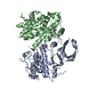
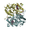
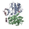
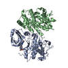
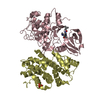
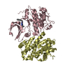
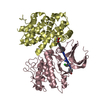
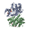
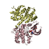
| Title | P27(KIP1)/CYCLIN A/CDK2 COMPLEX | ||||||
 Components Components |
| ||||||
 Keywords Keywords | COMPLEX (TRANSFERASE/CYCLIN/INHIBITOR) / COMPLEX (TRANSFERASE-CYCLIN-INHIBITOR) / KINASE / CELL CYCLE / CELL DIVISION / CDK / CYCLIN / INHIBITOR / COMPLEX (TRANSFERASE-CYCLIN-INHIBITOR) complex | ||||||
| Function / homology |  Function and homology information Function and homology informationcyclin-dependent protein kinase regulator activity / regulation of lens fiber cell differentiation / negative regulation of cardiac muscle tissue regeneration / autophagic cell death / FOXO-mediated transcription of cell cycle genes / negative regulation of epithelial cell proliferation involved in prostate gland development / cyclin A2-CDK1 complex / regulation of cell cycle G1/S phase transition / epithelial cell proliferation involved in prostate gland development / cell cycle G1/S phase transition ...cyclin-dependent protein kinase regulator activity / regulation of lens fiber cell differentiation / negative regulation of cardiac muscle tissue regeneration / autophagic cell death / FOXO-mediated transcription of cell cycle genes / negative regulation of epithelial cell proliferation involved in prostate gland development / cyclin A2-CDK1 complex / regulation of cell cycle G1/S phase transition / epithelial cell proliferation involved in prostate gland development / cell cycle G1/S phase transition / cellular response to luteinizing hormone stimulus / negative regulation of epithelial cell apoptotic process / regulation of exit from mitosis / G2/M DNA replication checkpoint / cyclin-dependent protein serine/threonine kinase inhibitor activity / negative regulation of mitotic cell cycle / Transcription of E2F targets under negative control by p107 (RBL1) and p130 (RBL2) in complex with HDAC1 / cellular response to leptin stimulus / regulation of cyclin-dependent protein serine/threonine kinase activity / male pronucleus / female pronucleus / nuclear export / RHO GTPases activate CIT / cellular response to cocaine / epithelial cell apoptotic process / AKT phosphorylates targets in the cytosol / response to glucagon / ubiquitin ligase activator activity / cyclin-dependent protein serine/threonine kinase regulator activity / cellular response to lithium ion / molecular function inhibitor activity / positive regulation of DNA biosynthetic process / cyclin A1-CDK2 complex / cyclin E2-CDK2 complex / cyclin E1-CDK2 complex / cellular response to insulin-like growth factor stimulus / cyclin A2-CDK2 complex / positive regulation of DNA-templated DNA replication initiation / G2 Phase / Y chromosome / cyclin-dependent protein kinase activity / regulation of heterochromatin organization / Phosphorylation of proteins involved in G1/S transition by active Cyclin E:Cdk2 complexes / positive regulation of heterochromatin formation / p53-Dependent G1 DNA Damage Response / X chromosome / PTK6 Regulates Cell Cycle / Constitutive Signaling by AKT1 E17K in Cancer / inner ear development / regulation of anaphase-promoting complex-dependent catabolic process / cellular response to antibiotic / Cul4A-RING E3 ubiquitin ligase complex / Defective binding of RB1 mutants to E2F1,(E2F2, E2F3) / centriole replication / Regulation of APC/C activators between G1/S and early anaphase / protein kinase inhibitor activity / telomere maintenance in response to DNA damage / ciliary transition zone / regulation of DNA replication / centrosome duplication / animal organ regeneration / microtubule organizing center / G0 and Early G1 / negative regulation of vascular associated smooth muscle cell proliferation / cochlea development / Telomere Extension By Telomerase / Activation of the pre-replicative complex / Estrogen-dependent nuclear events downstream of ESR-membrane signaling / cyclin-dependent kinase / cyclin-dependent protein serine/threonine kinase activity / TP53 Regulates Transcription of Genes Involved in G1 Cell Cycle Arrest / Notch signaling pathway / Activation of ATR in response to replication stress / Regulation of MITF-M-dependent genes involved in cell cycle and proliferation / Cajal body / Cyclin E associated events during G1/S transition / regulation of G2/M transition of mitotic cell cycle / Cyclin A:Cdk2-associated events at S phase entry / cellular response to platelet-derived growth factor stimulus / Cyclin A/B1/B2 associated events during G2/M transition / Chk1/Chk2(Cds1) mediated inactivation of Cyclin B:Cdk1 complex / cyclin-dependent protein kinase holoenzyme complex / positive regulation of microtubule polymerization / regulation of G1/S transition of mitotic cell cycle / condensed chromosome / mitotic G1 DNA damage checkpoint signaling / cellular response to nitric oxide / negative regulation of protein localization to chromatin / FLT3 Signaling / regulation of mitotic cell cycle / post-translational protein modification / placenta development / regulation of cell migration / cyclin binding / positive regulation of DNA replication / male germ cell nucleus / potassium ion transport / peptidyl-serine phosphorylation / DNA damage response, signal transduction by p53 class mediator / cellular response to estradiol stimulus Similarity search - Function | ||||||
| Biological species |  Homo sapiens (human) Homo sapiens (human) | ||||||
| Method |  X-RAY DIFFRACTION / Resolution: 2.3 Å X-RAY DIFFRACTION / Resolution: 2.3 Å | ||||||
 Authors Authors | Russo, A.A. / Jeffrey, P.D. / Pavletich, N.P. | ||||||
 Citation Citation | Journal: Nature / Year: 1996 Title: Crystal structure of the p27Kip1 cyclin-dependent-kinase inhibitor bound to the cyclin A-Cdk2 complex. Authors: Russo, A.A. / Jeffrey, P.D. / Patten, A.K. / Massague, J. / Pavletich, N.P. | ||||||
| History |
|
- Structure visualization
| Structure viewer | Molecule: MolmilJmol/JSmol |
|---|
- Downloads & links
-Download
| PDBx/mmCIF format | 1jsu.cif.gz | 139.8 KB | Display | PDBx/mmCIF format |
|---|---|---|---|---|
| PDB format | pdb1jsu.ent.gz | 108.5 KB | Display | PDB format |
| PDBx/mmJSON format | 1jsu.json.gz | Tree view | PDBx/mmJSON format | |
| Others |  Other downloads Other downloads |
-Validation report
| Arichive directory | https://data.pdbj.org/pub/pdb/validation_reports/js/1jsuftp://data.pdbj.org/pub/pdb/validation_reports/js/1jsu | HTTPS FTP |
|---|
-Related structure data
| Similar structure data |
|---|

-Links
 PDBj
PDBj


- Assembly
| Deposited unit | 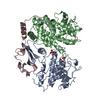
| ||||||||
|---|---|---|---|---|---|---|---|---|---|
| 1 |
| ||||||||
| Unit cell |
|
-Components
| #1: Protein | Mass: 34056.469 Da / Num. of mol.: 1 / Mutation: PHOSPHORYLATED AT THR A 160 Source method: isolated from a genetically manipulated source Source: (gene. exp.) Homo sapiens (human)Description: CYCLIN A-BOUND FORM PHOSPHORYLATED ON THR 160 IN VITRO USING A CDK-ACTIVATING KINASE CONSISTING OF THE CYCLINH-CDK7 COMPLEX; Cell line: SF9 / Plasmid: PET3A / Cell line (production host): SF9 / Production host:   Spodoptera frugiperda (fall armyworm) Spodoptera frugiperda (fall armyworm)References: UniProt: P24941, Transferases; Transferring phosphorus-containing groups; Phosphotransferases with an alcohol group as acceptor |
|---|---|
| #2: Protein | Mass: 29867.512 Da / Num. of mol.: 1 / Fragment: RESIDUES 173 - 432 Source method: isolated from a genetically manipulated source Source: (gene. exp.) Homo sapiens (human)Description: THE FRAGMENT USED IN THE CRYSTALLIZATION WAS PRODUCED BY THE CLEAVAGE OF FULL-LENGTH CYCLIN A BY SUBTILISIN; Cell line: SF9 / Plasmid: PET3A / Production host:  |
| #3: Protein | Mass: 9970.153 Da / Num. of mol.: 1 / Fragment: RESIDUES 22 - 106 Source method: isolated from a genetically manipulated source Source: (gene. exp.) Homo sapiens (human) / Cell line: SF9 / Plasmid: PET3A / Production host: |
| #4: Chemical | ChemComp-SO4 /   Mass: 96.063 Da / Num. of mol.: 1 / Source method: obtained synthetically / Formula: SO4 Mass: 96.063 Da / Num. of mol.: 1 / Source method: obtained synthetically / Formula: SO4 |
| #5: Water | ChemComp-HOH /  Mass: 18.015 Da / Num. of mol.: 197 / Source method: isolated from a natural source / Formula: H2O Mass: 18.015 Da / Num. of mol.: 197 / Source method: isolated from a natural source / Formula: H2O |
| Has protein modification | Y |
-Experimental details
-Experiment
| Experiment | Method: X-RAY DIFFRACTION |
|---|
- Sample preparation
| Crystal | Density Matthews: 2.68 Å3/Da / Density % sol: 54.12 % | ||||||||||||||||||||||||||||||||||||||||||||||||
|---|---|---|---|---|---|---|---|---|---|---|---|---|---|---|---|---|---|---|---|---|---|---|---|---|---|---|---|---|---|---|---|---|---|---|---|---|---|---|---|---|---|---|---|---|---|---|---|---|---|
| Crystal grow | *PLUS Temperature: 4 ℃ / pH: 7 / Method: vapor diffusion, hanging dropDetails: drop solution was mixed with an equal volume of reservoir solution | ||||||||||||||||||||||||||||||||||||||||||||||||
| Components of the solutions | *PLUS
|
-Data collection
| Diffraction source | Wavelength: 1.5418 |
|---|---|
| Detector | Type: RIGAKU / Detector: IMAGE PLATE / Date: Feb 20, 1996 |
| Radiation | Monochromatic (M) / Laue (L): M / Scattering type: x-ray |
| Radiation wavelength | Wavelength: 1.5418 Å / Relative weight: 1 |
| Reflection | Num. obs: 162339 / % possible obs: 96.1 % / Redundancy: 4.7 % / Rmerge(I) obs: 0.056 |
| Reflection | *PLUS Highest resolution: 2.3 Å / Num. obs: 34683 / Num. measured all: 162339 |
- Processing
| Software |
| ||||||||||||||||||||||||||||||
|---|---|---|---|---|---|---|---|---|---|---|---|---|---|---|---|---|---|---|---|---|---|---|---|---|---|---|---|---|---|---|---|
| Refinement | Resolution: 2.3→7 Å / σ(F): 2
| ||||||||||||||||||||||||||||||
| Refinement step | Cycle: LAST / Resolution: 2.3→7 Å
| ||||||||||||||||||||||||||||||
| Refine LS restraints |
| ||||||||||||||||||||||||||||||
| Software | *PLUS Name: TNT / Classification: refinement | ||||||||||||||||||||||||||||||
| Refinement | *PLUS Rfactor obs: 0.192 | ||||||||||||||||||||||||||||||
| Solvent computation | *PLUS | ||||||||||||||||||||||||||||||
| Displacement parameters | *PLUS |
