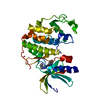
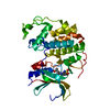
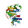
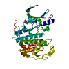
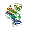
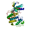
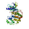
- PDB-1jvp: Crystal structure of human CDK2 (unphosphorylated) in complex wit... -
+
Open data
ID or keywords:
Loading...
-
Basic information
Entry
Database: PDB / ID: 1jvp
Title
Crystal structure of human CDK2 (unphosphorylated) in complex with PKF049-365
Components
Cell division protein kinase 2
Keywords
TRANSFERASE / Protein Kinase / Cell Division / Cell cycle / Enzyme Inhibitors / Drug design
Function / homology
Function and homology information
cyclin A1-CDK2 complex / cyclin E2-CDK2 complex / cyclin E1-CDK2 complex / cyclin A2-CDK2 complex / positive regulation of DNA-templated DNA replication initiation / G2 Phase / Y chromosome / cyclin-dependent protein kinase activity / regulation of heterochromatin organization / Phosphorylation of proteins involved in G1/S transition by active Cyclin E:Cdk2 complexes ...cyclin A1-CDK2 complex / cyclin E2-CDK2 complex / cyclin E1-CDK2 complex / cyclin A2-CDK2 complex / positive regulation of DNA-templated DNA replication initiation / G2 Phase / Y chromosome / cyclin-dependent protein kinase activity / regulation of heterochromatin organization / Phosphorylation of proteins involved in G1/S transition by active Cyclin E:Cdk2 complexes / positive regulation of heterochromatin formation / p53-Dependent G1 DNA Damage Response / X chromosome / PTK6 Regulates Cell Cycle / regulation of anaphase-promoting complex-dependent catabolic process / Defective binding of RB1 mutants to E2F1,(E2F2, E2F3) / centriole replication / Regulation of APC/C activators between G1/S and early anaphase / telomere maintenance in response to DNA damage / centrosome duplication / G0 and Early G1 / Telomere Extension By Telomerase / Activation of the pre-replicative complex / cyclin-dependent kinase / cyclin-dependent protein serine/threonine kinase activity / TP53 Regulates Transcription of Genes Involved in G1 Cell Cycle Arrest / Regulation of MITF-M-dependent genes involved in cell cycle and proliferation / Activation of ATR in response to replication stress / Cajal body / Cyclin E associated events during G1/S transition / Cyclin A:Cdk2-associated events at S phase entry / cyclin-dependent protein kinase holoenzyme complex / Cyclin A/B1/B2 associated events during G2/M transition / regulation of G2/M transition of mitotic cell cycle / condensed chromosome / mitotic G1 DNA damage checkpoint signaling / negative regulation of protein localization to chromatin / cellular response to nitric oxide / post-translational protein modification / regulation of mitotic cell cycle / cyclin binding / positive regulation of DNA replication / peptidyl-serine phosphorylation / male germ cell nucleus / meiotic cell cycle / potassium ion transport / G1/S transition of mitotic cell cycle / DNA Damage/Telomere Stress Induced Senescence / Meiotic recombination / G2/M transition of mitotic cell cycle / CDK-mediated phosphorylation and removal of Cdc6 / Transcriptional regulation of granulopoiesis / SCF(Skp2)-mediated degradation of p27/p21 / Orc1 removal from chromatin / cellular senescence / Cyclin D associated events in G1 / Regulation of TP53 Degradation / nuclear envelope / Factors involved in megakaryocyte development and platelet production / regulation of gene expression / Processing of DNA double-strand break ends / Senescence-Associated Secretory Phenotype (SASP) / transcription regulator complex / Regulation of TP53 Activity through Phosphorylation / protein phosphorylation / Ras protein signal transduction / DNA replication / chromosome, telomeric region / endosome / chromatin remodeling / protein domain specific binding / protein serine kinase activity / cell division / DNA repair / protein serine/threonine kinase activity / positive regulation of cell population proliferation / centrosome / positive regulation of DNA-templated transcription / DNA-templated transcription / magnesium ion binding / negative regulation of transcription by RNA polymerase II / signal transduction / nucleoplasm / ATP binding / nucleus / cytoplasm / cytosol Similarity search - Function
: / Phosphorylase Kinase; domain 1 / Phosphorylase Kinase; domain 1 / Transferase(Phosphotransferase) domain 1 / Transferase(Phosphotransferase); domain 1 / Serine/threonine-protein kinase, active site / Serine/Threonine protein kinases active-site signature. / Protein kinase domain / Serine/Threonine protein kinases, catalytic domain / Protein kinase, ATP binding site ...: / Phosphorylase Kinase; domain 1 / Phosphorylase Kinase; domain 1 / Transferase(Phosphotransferase) domain 1 / Transferase(Phosphotransferase); domain 1 / Serine/threonine-protein kinase, active site / Serine/Threonine protein kinases active-site signature. / Protein kinase domain / Serine/Threonine protein kinases, catalytic domain / Protein kinase, ATP binding site / Protein kinases ATP-binding region signature. / Protein kinase domain profile. / Protein kinase domain / Protein kinase-like domain superfamily / 2-Layer Sandwich / Orthogonal Bundle / Mainly Alpha / Alpha Beta Similarity search - Domain/homology
Group: Data collection / Category: chem_comp_atom / chem_comp_bond
Remark 600
HETEROGEN THERE ARE TWO TAUTOMERIC FORMS OF THE COMPOUND (LIG) WHICH CORRESPONDS TO THE TWO ...HETEROGEN THERE ARE TWO TAUTOMERIC FORMS OF THE COMPOUND (LIG) WHICH CORRESPONDS TO THE TWO ALTERNATE CONFROMATIONS LABELLED (1) AND (2), RESPECTIVELY, IN THE PDB FILE.
Resolution: 1.53→1.62 Å / Redundancy: 3.6 % / Rmerge(I) obs: 0.316 / Mean I/σ(I) obs: 2 / Num. unique all: 4990 / % possible all: 75.4
Reflection
*PLUS
Num. measured all: 179814
-
Processing
Software
Name
Version
Classification
AMoRE
phasing
X-PLOR
3.851
refinement
DENZO
datareduction
SCALEPACK
datascaling
Refinement
Method to determine structure: MOLECULAR REPLACEMENT / Resolution: 1.53→30 Å / Rfactor Rfree error: 0.004 / Data cutoff high absF: 1000000 / Data cutoff low absF: 0.001 / Isotropic thermal model: RESTRAINED / Cross valid method: THROUGHOUT / σ(F): 2 / Stereochemistry target values: Engh & Huber Details: two alternate conformations built for ligand and also for kinase residues 81 to 85 which make non-bonded contacts to the ligand
Rfactor
Num. reflection
% reflection
Selection details
Rfree
0.253
3864
10.1 %
RANDOM
Rwork
0.206
-
-
-
all
0.215
38946
-
-
obs
0.21
38298
89.5 %
-
Displacement parameters
Biso mean: 24.3 Å2
Baniso -1
Baniso -2
Baniso -3
1-
0 Å2
0 Å2
0 Å2
2-
-
0 Å2
0 Å2
3-
-
-
0 Å2
Refine analyze
Free
Obs
Luzzati coordinate error
0.22 Å
0.2 Å
Luzzati d res low
-
5 Å
Luzzati sigma a
0.2 Å
0.22 Å
Refinement step
Cycle: LAST / Resolution: 1.53→30 Å
Protein
Nucleic acid
Ligand
Solvent
Total
Num. atoms
2409
0
36
264
2709
Refine LS restraints
Refine-ID
Type
Dev ideal
Dev ideal target
X-RAY DIFFRACTION
x_bond_d
0.008
X-RAY DIFFRACTION
x_angle_deg
1.7
X-RAY DIFFRACTION
x_dihedral_angle_d
25
X-RAY DIFFRACTION
x_improper_angle_d
0.69
X-RAY DIFFRACTION
x_mcbond_it
1.45
1.5
X-RAY DIFFRACTION
x_mcangle_it
2.39
2
X-RAY DIFFRACTION
x_scbond_it
2.33
2
X-RAY DIFFRACTION
x_scangle_it
3.62
2.5
LS refinement shell
Resolution: 1.53→1.63 Å / Rfactor Rfree error: 0.014 / Total num. of bins used: 6
Rfactor
Num. reflection
% reflection
Rfree
0.322
503
9.9 %
Rwork
0.31
4573
-
obs
-
-
72.3 %
Xplor file
Refine-ID
Serial no
Param file
Topol file
X-RAY DIFFRACTION
1
PROTEIN_REP.PARAM
TOPHCSDX.PRO
X-RAY DIFFRACTION
2
TOPH19.SOL
Refinement
*PLUS
Rfactor obs: 0.206
Solvent computation
*PLUS
Displacement parameters
*PLUS
+
About Yorodumi
-
News
-
Feb 9, 2022. New format data for meta-information of EMDB entries
New format data for meta-information of EMDB entries
Version 3 of the EMDB header file is now the official format.
The previous official version 1.9 will be removed from the archive.
In the structure databanks used in Yorodumi, some data are registered as the other names, "COVID-19 virus" and "2019-nCoV". Here are the details of the virus and the list of structure data.
Jan 31, 2019. EMDB accession codes are about to change! (news from PDBe EMDB page)
EMDB accession codes are about to change! (news from PDBe EMDB page)
The allocation of 4 digits for EMDB accession codes will soon come to an end. Whilst these codes will remain in use, new EMDB accession codes will include an additional digit and will expand incrementally as the available range of codes is exhausted. The current 4-digit format prefixed with “EMD-” (i.e. EMD-XXXX) will advance to a 5-digit format (i.e. EMD-XXXXX), and so on. It is currently estimated that the 4-digit codes will be depleted around Spring 2019, at which point the 5-digit format will come into force.
The EM Navigator/Yorodumi systems omit the EMD- prefix.
Related info.:Q: What is EMD? / ID/Accession-code notation in Yorodumi/EM Navigator
Yorodumi is a browser for structure data from EMDB, PDB, SASBDB, etc.
This page is also the successor to EM Navigator detail page, and also detail information page/front-end page for Omokage search.
The word "yorodu" (or yorozu) is an old Japanese word meaning "ten thousand". "mi" (miru) is to see.
Related info.:EMDB / PDB / SASBDB / Comparison of 3 databanks / Yorodumi Search / Aug 31, 2016. New EM Navigator & Yorodumi / Yorodumi Papers / Jmol/JSmol / Function and homology information / Changes in new EM Navigator and Yorodumi
 Movie
Movie Controller

 Yorodumi
 Open data
 Basic information
 Components
 Keywords
 Function and homology information
 Homo sapiens (human)
 X-RAY DIFFRACTION /
  Authors
 Citation
 Structure visualization
 Downloads & links
 Other downloads


 PDBj
PDBj


 Assembly

 Spodoptera frugiperda (fall armyworm) / References: UniProt: P24941, EC: 2.7.1.37
 Mass: 233.268 Da / Num. of mol.: 1 / Source method: obtained synthetically / Formula: C15H11N3
 Mass: 18.015 Da / Num. of mol.: 264 / Source method: isolated from a natural source / Formula: H2O
 Sample preparation
 / Beamline: BM1A / Wavelength: 0.873 Å
 Processing