 Movie
Movie Controller

[English] 日本語
 Yorodumi
- PDB-1oir: Imidazopyridines: a potent and selective class of Cyclin-dependen... -
+ Open data

- Basic information
| Entry | Database: PDB / ID: 1oir | ||||||
|---|---|---|---|---|---|---|---|
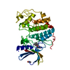
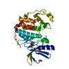
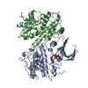
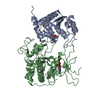
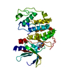
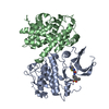
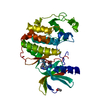
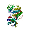
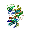
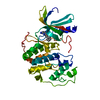
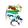
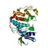
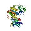
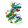
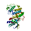
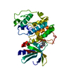
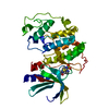
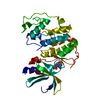
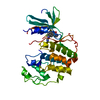
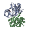
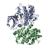
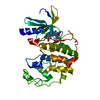
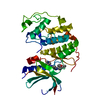
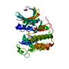
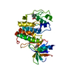
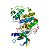
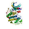
| Title | Imidazopyridines: a potent and selective class of Cyclin-dependent Kinase inhibitors identified through Structure-based hybridisation | ||||||
 Components Components | CELL DIVISION PROTEIN KINASE 2 | ||||||
 Keywords Keywords | KINASE / PROTEIN KINASE | ||||||
| Function / homology |  Function and homology information Function and homology informationcyclin A1-CDK2 complex / cyclin E2-CDK2 complex / cyclin E1-CDK2 complex / cyclin A2-CDK2 complex / positive regulation of DNA-templated DNA replication initiation / G2 Phase / Y chromosome / cyclin-dependent protein kinase activity / regulation of heterochromatin organization / Phosphorylation of proteins involved in G1/S transition by active Cyclin E:Cdk2 complexes ...cyclin A1-CDK2 complex / cyclin E2-CDK2 complex / cyclin E1-CDK2 complex / cyclin A2-CDK2 complex / positive regulation of DNA-templated DNA replication initiation / G2 Phase / Y chromosome / cyclin-dependent protein kinase activity / regulation of heterochromatin organization / Phosphorylation of proteins involved in G1/S transition by active Cyclin E:Cdk2 complexes / positive regulation of heterochromatin formation / p53-Dependent G1 DNA Damage Response / X chromosome / PTK6 Regulates Cell Cycle / regulation of anaphase-promoting complex-dependent catabolic process / Defective binding of RB1 mutants to E2F1,(E2F2, E2F3) / centriole replication / Regulation of APC/C activators between G1/S and early anaphase / telomere maintenance in response to DNA damage / centrosome duplication / G0 and Early G1 / Telomere Extension By Telomerase / Activation of the pre-replicative complex / cyclin-dependent kinase / cyclin-dependent protein serine/threonine kinase activity / TP53 Regulates Transcription of Genes Involved in G1 Cell Cycle Arrest / Regulation of MITF-M-dependent genes involved in cell cycle and proliferation / Activation of ATR in response to replication stress / Cajal body / Cyclin E associated events during G1/S transition / Cyclin A:Cdk2-associated events at S phase entry / cyclin-dependent protein kinase holoenzyme complex / Cyclin A/B1/B2 associated events during G2/M transition / regulation of G2/M transition of mitotic cell cycle / condensed chromosome / mitotic G1 DNA damage checkpoint signaling / negative regulation of protein localization to chromatin / cellular response to nitric oxide / post-translational protein modification / regulation of mitotic cell cycle / cyclin binding / positive regulation of DNA replication / peptidyl-serine phosphorylation / male germ cell nucleus / meiotic cell cycle / potassium ion transport / G1/S transition of mitotic cell cycle / DNA Damage/Telomere Stress Induced Senescence / Meiotic recombination / G2/M transition of mitotic cell cycle / CDK-mediated phosphorylation and removal of Cdc6 / Transcriptional regulation of granulopoiesis / SCF(Skp2)-mediated degradation of p27/p21 / Orc1 removal from chromatin / cellular senescence / Cyclin D associated events in G1 / Regulation of TP53 Degradation / nuclear envelope / Factors involved in megakaryocyte development and platelet production / regulation of gene expression / Processing of DNA double-strand break ends / Senescence-Associated Secretory Phenotype (SASP) / transcription regulator complex / Regulation of TP53 Activity through Phosphorylation / protein phosphorylation / Ras protein signal transduction / DNA replication / chromosome, telomeric region / endosome / chromatin remodeling / protein domain specific binding / protein serine kinase activity / cell division / DNA repair / protein serine/threonine kinase activity / positive regulation of cell population proliferation / centrosome / positive regulation of DNA-templated transcription / DNA-templated transcription / magnesium ion binding / negative regulation of transcription by RNA polymerase II / signal transduction / nucleoplasm / ATP binding / nucleus / cytoplasm / cytosol Similarity search - Function | ||||||
| Biological species |  HOMO SAPIENS (human) HOMO SAPIENS (human) | ||||||
| Method |  X-RAY DIFFRACTION / SYNCHROTRON / MOLECULAR REPLACEMENT / Resolution: 1.91 Å X-RAY DIFFRACTION / SYNCHROTRON / MOLECULAR REPLACEMENT / Resolution: 1.91 Å | ||||||
 Authors Authors | Beattie, J.F. / Breault, G.A. / Byth, K.F. / Culshaw, J.D. / Ellston, R.P.A. / Green, S. / Minshull, C.A. / Norman, R.A. / Pauptit, R.A. / Thomas, A.P. / Jewsbury, P.J. | ||||||
 Citation Citation | Journal: Bioorg.Med.Chem.Lett. / Year: 2003 Title: Imidazo[1,2-A]Pyridines: A Potent and Selective Class of Cyclin-Dependent Kinase Inhibitors Identified Through Structure-Based Hybridisation Authors: Anderson, M. / Beattie, J.F. / Breault, G.A. / Breed, J. / Byth, K.F. / Culshaw, J.D. / Ellston, R.P.A. / Green, S. / Minshull, C.A. / Norman, R.A. / Pauptit, R.A. / Stanway, J. / Thomas, A.P. / Jewsbury, P.J. #1: Journal: J.Med.Chem. / Year: 1996Title: High-Resolution Crystal Structures of Human Cyclin-Dependent Kinase 2 with and without ATP: Bound Waters and Natural Ligand as a Guide for Inhibitor Design Authors: Schulze-Gahmen, U. / De Bondt, H. / Kim, S.-H. | ||||||
| History |
| ||||||
| Remark 700 | SHEET THE SHEET STRUCTURE OF THIS MOLECULE IS BIFURCATED. IN ORDER TO REPRESENT THIS FEATURE IN ... SHEET THE SHEET STRUCTURE OF THIS MOLECULE IS BIFURCATED. IN ORDER TO REPRESENT THIS FEATURE IN THE SHEET RECORDS BELOW, TWO SHEETS ARE DEFINED. |
- Structure visualization
| Structure viewer | Molecule: MolmilJmol/JSmol |
|---|
- Downloads & links
-Download
| PDBx/mmCIF format | 1oir.cif.gz | 74.7 KB | Display | PDBx/mmCIF format |
|---|---|---|---|---|
| PDB format | pdb1oir.ent.gz | 55 KB | Display | PDB format |
| PDBx/mmJSON format | 1oir.json.gz | Tree view | PDBx/mmJSON format | |
| Others |  Other downloads Other downloads |
-Validation report
| Arichive directory | https://data.pdbj.org/pub/pdb/validation_reports/oi/1oirftp://data.pdbj.org/pub/pdb/validation_reports/oi/1oir | HTTPS FTP |
|---|
-Related structure data
| Related structure data |  1oiqC  1oitC  1h0u C: citing same article ( |
|---|---|
| Similar structure data |


-Links
 PDBj
PDBj


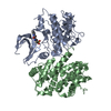
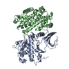
- Assembly
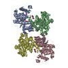
| Deposited unit | 
| ||||||||
|---|---|---|---|---|---|---|---|---|---|
| 1 |
| ||||||||
| Unit cell |
|
-Components
| #1: Protein | Mass: 34045.531 Da / Num. of mol.: 1 Source method: isolated from a genetically manipulated source Source: (gene. exp.) HOMO SAPIENS (human) / Production host:   SPODOPTERA FRUGIPERDA (fall armyworm) / Strain (production host): SF9 / References: UniProt: P24941, EC: 2.7.1.37 SPODOPTERA FRUGIPERDA (fall armyworm) / Strain (production host): SF9 / References: UniProt: P24941, EC: 2.7.1.37 |
|---|---|
| #2: Chemical | ChemComp-HDY /   Mass: 418.492 Da / Num. of mol.: 1 / Source method: obtained synthetically / Formula: C23H26N6O2 Mass: 418.492 Da / Num. of mol.: 1 / Source method: obtained synthetically / Formula: C23H26N6O2 |
| #3: Water | ChemComp-HOH /  Mass: 18.015 Da / Num. of mol.: 123 / Source method: isolated from a natural source / Formula: H2O Mass: 18.015 Da / Num. of mol.: 123 / Source method: isolated from a natural source / Formula: H2O |
| Compound details | FUNCTION: PROBABLY INVOLVED IN THE CONTROL OF THE CELL CYCLE. INTERACTS WITH CYCLINS A, D, OR E. ...FUNCTION: PROBABLY INVOLVED IN THE CONTROL OF THE CELL CYCLE. INTERACTS WITH CYCLINS A, D, OR E. ACTIVITY OF CDK2 IS MAXIMAL DURING S PHASE AND G2. |
| Has protein modification | Y |
-Experimental details
-Experiment
| Experiment | Method: X-RAY DIFFRACTION / Number of used crystals: 1 |
|---|
- Sample preparation
| Crystal | Density Matthews: 2.24 Å3/Da / Density % sol: 40 % | |||||||||||||||||||||||||||||||||||||||||||||||||
|---|---|---|---|---|---|---|---|---|---|---|---|---|---|---|---|---|---|---|---|---|---|---|---|---|---|---|---|---|---|---|---|---|---|---|---|---|---|---|---|---|---|---|---|---|---|---|---|---|---|---|
| Crystal grow | pH: 7 Details: PROTEIN AT 10MG/ML WELL BUFFER CONTAINING 17.5% PEG3350, 200MM HEPES, PH7.0, 100MM AMMONIUM ACETATE, pH 7.00 | |||||||||||||||||||||||||||||||||||||||||||||||||
| Crystal grow | *PLUS Temperature: 20 ℃ / pH: 7.4 / Method: vapor diffusion, sitting drop / Details: Lawrie, A.M., (1997) Nature Struct. Biol., 4, 796. | |||||||||||||||||||||||||||||||||||||||||||||||||
| Components of the solutions | *PLUS
|
-Data collection
| Diffraction | Mean temperature: 100 K |
|---|---|
| Diffraction source | Source: SYNCHROTRON / Site: SRS  / Beamline: PX9.6 / Wavelength: 0.875 / Beamline: PX9.6 / Wavelength: 0.875 |
| Detector | Type: MARRESEARCH / Detector: IMAGE PLATE / Date: Jul 15, 1998 |
| Radiation | Protocol: SINGLE WAVELENGTH / Monochromatic (M) / Laue (L): M / Scattering type: x-ray |
| Radiation wavelength | Wavelength: 0.875 Å / Relative weight: 1 |
| Reflection | Resolution: 1.91→43.03 Å / Num. obs: 20394 / % possible obs: 84.8 % / Redundancy: 2.2 % / Rmerge(I) obs: 0.066 / Net I/σ(I): 21.7 |
| Reflection shell | Resolution: 1.9→2.01 Å / Redundancy: 2 % / Rmerge(I) obs: 0.137 / Mean I/σ(I) obs: 10.3 / % possible all: 73.3 |
| Reflection | *PLUS Highest resolution: 1.91 Å / Num. measured all: 95201 / Rmerge(I) obs: 0.052 |
- Processing
| Software |
| ||||||||||||||||||||
|---|---|---|---|---|---|---|---|---|---|---|---|---|---|---|---|---|---|---|---|---|---|
| Refinement | Method to determine structure: MOLECULAR REPLACEMENT / Resolution: 1.91→43.03 Å / SU B: 3.881 / SU ML: 0.114 / Cross valid method: THROUGHOUT / ESU R: 0.223 / ESU R Free: 0.18 / Details: TLS REFINEMENT CARRIED OUT
| ||||||||||||||||||||
| Displacement parameters | Biso mean: 16.54 Å2
| ||||||||||||||||||||
| Refinement step | Cycle: LAST / Resolution: 1.91→43.03 Å
| ||||||||||||||||||||
| Refinement | *PLUS % reflection Rfree: 5 % / Rfactor Rfree: 0.237 / Rfactor Rwork: 0.197 | ||||||||||||||||||||
| Solvent computation | *PLUS | ||||||||||||||||||||
| Displacement parameters | *PLUS |
