 Movie
Movie Controller

[English] 日本語
 Yorodumi
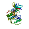
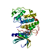
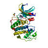
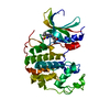
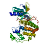
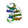
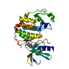
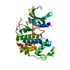
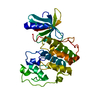
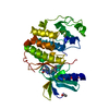
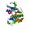
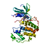
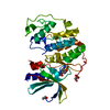
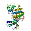
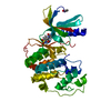
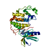
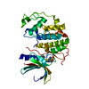
- PDB-5fp5: Structure of cyclin-dependent kinase 2 with small-molecule ligand... -
+ Open data

- Basic information
| Entry | Database: PDB / ID: 5fp5 | |||||||||
|---|---|---|---|---|---|---|---|---|---|---|
| Title | Structure of cyclin-dependent kinase 2 with small-molecule ligand 4- fluorobenzoic acid (AT222) in an alternate binding site. | |||||||||
 Components Components | CYCLIN-DEPENDENT KINASE 2 | |||||||||
 Keywords Keywords | TRANSFERASE / KINASE / MITOSIS / CELL CYCLE / FRAGMENT SCREENING / ALTERNATE BINDING SITE. | |||||||||
| Function / homology |  Function and homology information Function and homology informationcyclin A1-CDK2 complex / cyclin E2-CDK2 complex / cyclin E1-CDK2 complex / cyclin A2-CDK2 complex / positive regulation of DNA-templated DNA replication initiation / G2 Phase / Y chromosome / cyclin-dependent protein kinase activity / regulation of heterochromatin organization / Phosphorylation of proteins involved in G1/S transition by active Cyclin E:Cdk2 complexes ...cyclin A1-CDK2 complex / cyclin E2-CDK2 complex / cyclin E1-CDK2 complex / cyclin A2-CDK2 complex / positive regulation of DNA-templated DNA replication initiation / G2 Phase / Y chromosome / cyclin-dependent protein kinase activity / regulation of heterochromatin organization / Phosphorylation of proteins involved in G1/S transition by active Cyclin E:Cdk2 complexes / positive regulation of heterochromatin formation / p53-Dependent G1 DNA Damage Response / X chromosome / PTK6 Regulates Cell Cycle / regulation of anaphase-promoting complex-dependent catabolic process / Defective binding of RB1 mutants to E2F1,(E2F2, E2F3) / centriole replication / Regulation of APC/C activators between G1/S and early anaphase / telomere maintenance in response to DNA damage / centrosome duplication / G0 and Early G1 / Telomere Extension By Telomerase / Activation of the pre-replicative complex / cyclin-dependent kinase / cyclin-dependent protein serine/threonine kinase activity / TP53 Regulates Transcription of Genes Involved in G1 Cell Cycle Arrest / Activation of ATR in response to replication stress / Regulation of MITF-M-dependent genes involved in cell cycle and proliferation / Cajal body / Cyclin E associated events during G1/S transition / regulation of G2/M transition of mitotic cell cycle / Cyclin A:Cdk2-associated events at S phase entry / Cyclin A/B1/B2 associated events during G2/M transition / cyclin-dependent protein kinase holoenzyme complex / condensed chromosome / mitotic G1 DNA damage checkpoint signaling / cellular response to nitric oxide / negative regulation of protein localization to chromatin / regulation of mitotic cell cycle / post-translational protein modification / cyclin binding / positive regulation of DNA replication / male germ cell nucleus / potassium ion transport / peptidyl-serine phosphorylation / G1/S transition of mitotic cell cycle / meiotic cell cycle / DNA Damage/Telomere Stress Induced Senescence / Meiotic recombination / G2/M transition of mitotic cell cycle / CDK-mediated phosphorylation and removal of Cdc6 / cellular senescence / Transcriptional regulation of granulopoiesis / SCF(Skp2)-mediated degradation of p27/p21 / Orc1 removal from chromatin / Cyclin D associated events in G1 / Regulation of TP53 Degradation / nuclear envelope / Factors involved in megakaryocyte development and platelet production / regulation of gene expression / Processing of DNA double-strand break ends / Senescence-Associated Secretory Phenotype (SASP) / transcription regulator complex / Regulation of TP53 Activity through Phosphorylation / protein phosphorylation / Ras protein signal transduction / chromosome, telomeric region / DNA replication / endosome / chromatin remodeling / protein domain specific binding / protein serine kinase activity / cell division / DNA repair / protein serine/threonine kinase activity / centrosome / positive regulation of cell population proliferation / positive regulation of DNA-templated transcription / magnesium ion binding / negative regulation of transcription by RNA polymerase II / signal transduction / DNA-templated transcription / nucleoplasm / ATP binding / nucleus / cytosol / cytoplasm Similarity search - Function | |||||||||
| Biological species |  HOMO SAPIENS (human) HOMO SAPIENS (human) | |||||||||
| Method |  X-RAY DIFFRACTION / SYNCHROTRON / MOLECULAR REPLACEMENT / Resolution: 2.16 Å X-RAY DIFFRACTION / SYNCHROTRON / MOLECULAR REPLACEMENT / Resolution: 2.16 Å | |||||||||
 Authors Authors | Jhoti, H. / Ludlow, R.F. / O'Reilly, M. / Saini, H.K. / Tickle, I.J. / Verdonk, M. | |||||||||
 Citation Citation | Journal: Proc.Natl.Acad.Sci.USA / Year: 2015 Title: Detection of Secondary Binding Sites in Proteins Using Fragment Screening. Authors: Ludlow, R.F. / Verdonk, M.L. / Saini, H.K. / Tickle, I.J. / Jhoti, H. #1: Journal: J.Med.Chem. / Year: 2008Title: Identification of N-(4-Piperidinyl)-4-(2,6-Dichlorobenzoylamino)-1H-Pyrazole-3-Carboxamide (at7519), a Novel Cyclin Dependent Kinase Inhibitor Using Fragment-Based X-Ray Crystallography and ...Title: Identification of N-(4-Piperidinyl)-4-(2,6-Dichlorobenzoylamino)-1H-Pyrazole-3-Carboxamide (at7519), a Novel Cyclin Dependent Kinase Inhibitor Using Fragment-Based X-Ray Crystallography and Structure Based Drug Design. Authors: Wyatt, P.G. / Woodhead, A.J. / Berdini, V. / Boulstridge, J.A. / Carr, M.G. / Cross, D.M. / Davis, D.J. / Devine, L.A. / Early, T.R. / Feltell, R.E. / Lewis, E.J. / McMenamin, R.L. / ...Authors: Wyatt, P.G. / Woodhead, A.J. / Berdini, V. / Boulstridge, J.A. / Carr, M.G. / Cross, D.M. / Davis, D.J. / Devine, L.A. / Early, T.R. / Feltell, R.E. / Lewis, E.J. / McMenamin, R.L. / Navarro, E.F. / O'Brien, M.A. / O'Reilly, M. / Reule, M. / Saxty, G. / Seavers, L.C.A. / Smith, D. / Squires, M.S. / Trewartha, G. / Walker, M.T. / Woolford, A.J. | |||||||||
| History |
|
- Structure visualization
| Structure viewer | Molecule: MolmilJmol/JSmol |
|---|
- Downloads & links
-Download
| PDBx/mmCIF format | 5fp5.cif.gz | 124.7 KB | Display | PDBx/mmCIF format |
|---|---|---|---|---|
| PDB format | pdb5fp5.ent.gz | 97.1 KB | Display | PDB format |
| PDBx/mmJSON format | 5fp5.json.gz | Tree view | PDBx/mmJSON format | |
| Others |  Other downloads Other downloads |
-Validation report
| Arichive directory | https://data.pdbj.org/pub/pdb/validation_reports/fp/5fp5ftp://data.pdbj.org/pub/pdb/validation_reports/fp/5fp5 | HTTPS FTP |
|---|
-Related structure data
| Related structure data |  5fp6C  5fpdC  5fpeC  5fpmC  5fpnC  5fpoC  5fprC  5fpsC 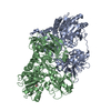 5fptC 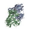 5fpyC  1hckS C: citing same article ( S: Starting model for refinement |
|---|---|
| Similar structure data |


-Links
 PDBj
PDBj


- Assembly
| Deposited unit | 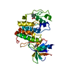
| ||||||||
|---|---|---|---|---|---|---|---|---|---|
| 1 |
| ||||||||
| Unit cell |
|
-Components
| #1: Protein | Mass: 33976.488 Da / Num. of mol.: 1 Source method: isolated from a genetically manipulated source Source: (gene. exp.) HOMO SAPIENS (human) / Plasmid: PFASTBAC1 / Cell line (production host): SF9 / Production host:   SPODOPTERA FRUGIPERDA (fall armyworm) / References: UniProt: P24941, cyclin-dependent kinase SPODOPTERA FRUGIPERDA (fall armyworm) / References: UniProt: P24941, cyclin-dependent kinase | ||||||
|---|---|---|---|---|---|---|---|
| #2: Chemical | 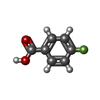  Mass: 140.112 Da / Num. of mol.: 2 / Source method: obtained synthetically / Formula: C7H5FO2 Mass: 140.112 Da / Num. of mol.: 2 / Source method: obtained synthetically / Formula: C7H5FO2#3: Chemical | ChemComp-ACE / |   Mass: 44.053 Da / Num. of mol.: 1 / Source method: obtained synthetically / Formula: C2H4O Mass: 44.053 Da / Num. of mol.: 1 / Source method: obtained synthetically / Formula: C2H4O#4: Water | ChemComp-HOH / |  Mass: 18.015 Da / Num. of mol.: 321 / Source method: isolated from a natural source / Formula: H2O Mass: 18.015 Da / Num. of mol.: 321 / Source method: isolated from a natural source / Formula: H2ONonpolymer details | 4-FLUOROBENZOIC ACID (L02): ASTEX COMPOUND REGISTRY AT222 4-FLUOROBENZOIC ACID (L01): ASTEX ...4-FLUOROBENZ | |
-Experimental details
-Experiment
| Experiment | Method: X-RAY DIFFRACTION / Number of used crystals: 1 |
|---|
- Sample preparation
| Crystal | Density Matthews: 2.35 Å3/Da / Density % sol: 47 % / Description: NONE |
|---|---|
| Crystal grow | pH: 7.4 Details: 10MM HEPES/NAOH PH7.4 15MM NACL PROTEIN CONCENTRATION: 8.6 MG/ML |
-Data collection
| Diffraction | Mean temperature: 93 K |
|---|---|
| Diffraction source | Source: SYNCHROTRON / Site: SRS  / Type: SRS / Wavelength: 1 / Type: SRS / Wavelength: 1 |
| Detector | Type: ADSC CCD / Detector: CCD / Date: May 2, 2002 |
| Radiation | Protocol: SINGLE WAVELENGTH / Monochromatic (M) / Laue (L): M / Scattering type: x-ray |
| Radiation wavelength | Wavelength: 1 Å / Relative weight: 1 |
| Reflection | Resolution: 2.16→30 Å / Num. obs: 14466 / % possible obs: 93.1 % / Observed criterion σ(I): -3.7 / Biso Wilson estimate: 32.2 Å2 |
- Processing
| Software |
| ||||||||||||||||||||||||||||||||||||||||||||||||||||||||||||||||||||||||||||||||||||||||||||||||||||||||||||||||||
|---|---|---|---|---|---|---|---|---|---|---|---|---|---|---|---|---|---|---|---|---|---|---|---|---|---|---|---|---|---|---|---|---|---|---|---|---|---|---|---|---|---|---|---|---|---|---|---|---|---|---|---|---|---|---|---|---|---|---|---|---|---|---|---|---|---|---|---|---|---|---|---|---|---|---|---|---|---|---|---|---|---|---|---|---|---|---|---|---|---|---|---|---|---|---|---|---|---|---|---|---|---|---|---|---|---|---|---|---|---|---|---|---|---|---|---|
| Refinement | Method to determine structure: MOLECULAR REPLACEMENT Starting model: PDB ENTRY 1HCK Resolution: 2.16→30.01 Å / Cor.coef. Fo:Fc: 0.9405 / Cor.coef. Fo:Fc free: 0.9067 / SU R Cruickshank DPI: 0.294 / Cross valid method: THROUGHOUT / σ(F): 0 / SU R Blow DPI: 0.392 / SU Rfree Blow DPI: 0.244 / SU Rfree Cruickshank DPI: 0.229
| ||||||||||||||||||||||||||||||||||||||||||||||||||||||||||||||||||||||||||||||||||||||||||||||||||||||||||||||||||
| Displacement parameters | Biso mean: 32.09 Å2
| ||||||||||||||||||||||||||||||||||||||||||||||||||||||||||||||||||||||||||||||||||||||||||||||||||||||||||||||||||
| Refine analyze | Luzzati coordinate error obs: 0.242 Å | ||||||||||||||||||||||||||||||||||||||||||||||||||||||||||||||||||||||||||||||||||||||||||||||||||||||||||||||||||
| Refinement step | Cycle: LAST / Resolution: 2.16→30.01 Å
| ||||||||||||||||||||||||||||||||||||||||||||||||||||||||||||||||||||||||||||||||||||||||||||||||||||||||||||||||||
| Refine LS restraints |
| ||||||||||||||||||||||||||||||||||||||||||||||||||||||||||||||||||||||||||||||||||||||||||||||||||||||||||||||||||
| LS refinement shell | Resolution: 2.16→2.33 Å / Total num. of bins used: 7
| ||||||||||||||||||||||||||||||||||||||||||||||||||||||||||||||||||||||||||||||||||||||||||||||||||||||||||||||||||
| Refinement TLS params. | Method: refined / Refine-ID: X-RAY DIFFRACTION
| ||||||||||||||||||||||||||||||||||||||||||||||||||||||||||||||||||||||||||||||||||||||||||||||||||||||||||||||||||
| Refinement TLS group | Selection details: CHAIN A |
