 Movie
Movie Controller

[English] 日本語
 Yorodumi
- PDB-5fpt: Structure of hepatitis C virus (HCV) full-length NS3 complex with... -
+ Open data

- Basic information
| Entry | Database: PDB / ID: 5fpt | ||||||
|---|---|---|---|---|---|---|---|
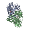
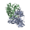
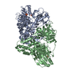
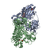
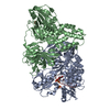
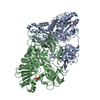
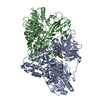
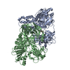
| Title | Structure of hepatitis C virus (HCV) full-length NS3 complex with small-molecule ligand 2-(1-methyl-1H-indol-3-yl)acetic acid (AT3437) in an alternate binding site. | ||||||
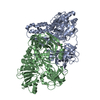
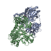
 Components Components | HEPATITIS C VIRUS FULL-LENGTH NS3 COMPLEX | ||||||
 Keywords Keywords | HYDROLASE / HEPATITIS C VIRUS / HCV / NS3 COMPLEX / PROTEASE-HELICASE / PROTEIN-LIGAND COMPLEX / FRAGMENT SCREENING / ALTERNATE BINDING SITE / AT3437. | ||||||
| Function / homology |  Function and homology information Function and homology informationhepacivirin / host cell mitochondrial membrane / host cell lipid droplet / symbiont-mediated transformation of host cell / symbiont-mediated suppression of host TRAF-mediated signal transduction / symbiont-mediated perturbation of host cell cycle G1/S transition checkpoint / symbiont-mediated suppression of host JAK-STAT cascade via inhibition of STAT1 activity / symbiont-mediated suppression of host cytoplasmic pattern recognition receptor signaling pathway via inhibition of MAVS activity / SH3 domain binding / nucleoside-triphosphate phosphatase ...hepacivirin / host cell mitochondrial membrane / host cell lipid droplet / symbiont-mediated transformation of host cell / symbiont-mediated suppression of host TRAF-mediated signal transduction / symbiont-mediated perturbation of host cell cycle G1/S transition checkpoint / symbiont-mediated suppression of host JAK-STAT cascade via inhibition of STAT1 activity / symbiont-mediated suppression of host cytoplasmic pattern recognition receptor signaling pathway via inhibition of MAVS activity / SH3 domain binding / nucleoside-triphosphate phosphatase / viral nucleocapsid / channel activity / monoatomic ion transmembrane transport / clathrin-dependent endocytosis of virus by host cell / Hydrolases; Acting on peptide bonds (peptidases); Cysteine endopeptidases / RNA helicase activity / host cell perinuclear region of cytoplasm / host cell endoplasmic reticulum membrane / RNA helicase / symbiont-mediated suppression of host type I interferon-mediated signaling pathway / ribonucleoprotein complex / serine-type endopeptidase activity / symbiont-mediated activation of host autophagy / RNA-directed RNA polymerase / cysteine-type endopeptidase activity / viral RNA genome replication / RNA-directed RNA polymerase activity / fusion of virus membrane with host endosome membrane / viral envelope / virion attachment to host cell / host cell nucleus / host cell plasma membrane / virion membrane / structural molecule activity / ATP hydrolysis activity / proteolysis / RNA binding / zinc ion binding / ATP binding Similarity search - Function | ||||||
| Biological species |  HEPATITIS C VIRUS HEPATITIS C VIRUS | ||||||
| Method |  X-RAY DIFFRACTION / SYNCHROTRON / MOLECULAR REPLACEMENT / Resolution: 2.72 Å X-RAY DIFFRACTION / SYNCHROTRON / MOLECULAR REPLACEMENT / Resolution: 2.72 Å | ||||||
 Authors Authors | Jhoti, H. / Ludlow, R.F. / Saini, H.K. / Tickle, I.J. / Verdonk, M. / Pathuri, P. / Williams, P.A. | ||||||
 Citation Citation | Journal: Proc.Natl.Acad.Sci.USA / Year: 2015 Title: Detection of Secondary Binding Sites in Proteins Using Fragment Screening. Authors: Ludlow, R.F. / Verdonk, M.L. / Saini, H.K. / Tickle, I.J. / Jhoti, H. #1: Journal: Structure / Year: 1999Title: Molecular Views of Viral Polyprotein Processing Revealed by the Crystal Structure of the Hepatitis C Virus Bifunctional Protease-Helicase. Authors: Yao, N. / Reichert, P. / Taremi, S.S. / Prosise, W.W. / Weber, P.C. #2: Journal: Nat.Chem.Biol. / Year: 2012Title: Discovery of an Allosteric Mechanism for the Regulation of Hcv Ns3 Protein Function. Authors: Saalau-Bethell, S.M. / Woodhead, A.J. / Chessari, G. / Carr, M.G. / Coyle, J. / Graham, B. / Hiscock, S.D. / Murray, C.W. / Pathuri, P. / Rich, S.J. / Richardson, C.J. / Williams, P.A. / Jhoti, H. | ||||||
| History |
| ||||||
| Remark 700 | SHEET DETERMINATION METHOD: DSSP THE SHEETS PRESENTED AS "AB" IN EACH CHAIN ON SHEET RECORDS BELOW ... SHEET DETERMINATION METHOD: DSSP THE SHEETS PRESENTED AS "AB" IN EACH CHAIN ON SHEET RECORDS BELOW IS ACTUALLY AN 6-STRANDED BARREL THIS IS REPRESENTED BY A 7-STRANDED SHEET IN WHICH THE FIRST AND LAST STRANDS ARE IDENTICAL. THE SHEETS PRESENTED AS "BB" IN EACH CHAIN ON SHEET RECORDS BELOW IS ACTUALLY AN 6-STRANDED BARREL THIS IS REPRESENTED BY A 7-STRANDED SHEET IN WHICH THE FIRST AND LAST STRANDS ARE IDENTICAL. |
- Structure visualization
| Structure viewer | Molecule: MolmilJmol/JSmol |
|---|
- Downloads & links
-Download
| PDBx/mmCIF format | 5fpt.cif.gz | 480.1 KB | Display | PDBx/mmCIF format |
|---|---|---|---|---|
| PDB format | pdb5fpt.ent.gz | 393.9 KB | Display | PDB format |
| PDBx/mmJSON format | 5fpt.json.gz | Tree view | PDBx/mmJSON format | |
| Others |  Other downloads Other downloads |
-Validation report
| Arichive directory | https://data.pdbj.org/pub/pdb/validation_reports/fp/5fptftp://data.pdbj.org/pub/pdb/validation_reports/fp/5fpt | HTTPS FTP |
|---|
-Related structure data
| Related structure data |  5fp5C  5fp6C  5fpdC  5fpeC  5fpmC  5fpnC  5fpoC  5fprC  5fpsSC  5fpyC C: citing same article ( S: Starting model for refinement |
|---|---|
| Similar structure data |
-Links
 PDBj
PDBj
- Assembly
| Deposited unit | 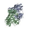
| ||||||||
|---|---|---|---|---|---|---|---|---|---|
| 1 |
| ||||||||
| Unit cell |
| ||||||||
| Noncrystallographic symmetry (NCS) | NCS oper: (Code: given Matrix: (-0.91125, 0.41157, -0.01519), Vector: |
-Components
| #1: Protein | Mass: 70869.391 Da / Num. of mol.: 2 Source method: isolated from a genetically manipulated source Source: (gene. exp.) HEPATITIS C VIRUS (ISOLATE BK) / Strain: GENOTYPE 1B / Plasmid: PET17 / Production host:  References: UniProt: P26663, Hydrolases; Acting on peptide bonds (peptidases); Serine endopeptidases #2: Chemical | 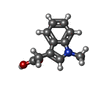  Mass: 189.211 Da / Num. of mol.: 2 / Source method: obtained synthetically / Formula: C11H11NO2 Mass: 189.211 Da / Num. of mol.: 2 / Source method: obtained synthetically / Formula: C11H11NO2#3: Water | ChemComp-HOH / |  Mass: 18.015 Da / Num. of mol.: 463 / Source method: isolated from a natural source / Formula: H2O Mass: 18.015 Da / Num. of mol.: 463 / Source method: isolated from a natural source / Formula: H2ONonpolymer details | 2-(1-METHYL-1H-INDOL-3-YL)ACETIC ACID (3VY): ASTEX COMPOUND REGISTRY AT3437. | Sequence details | N-TERM HIS TAG (37). DELETION 1-2. DELETION 632-686. | |
|---|
-Experimental details
-Experiment
| Experiment | Method: X-RAY DIFFRACTION / Number of used crystals: 1 |
|---|
- Sample preparation
| Crystal | Density Matthews: 2.66 Å3/Da / Density % sol: 53.33 % / Description: NONE |
|---|---|
| Crystal grow | pH: 6.6 Details: 0.2 M 2-(N-MORPHOLINO)ETHANESULFONIC ACID (MES)-NAOH, 18% W/V POLYETHYLENE GLYCOL (PEG) 6000, 10% W/V 2-METHYL-2, 4-PENTANDIOL (MPD). PROTEIN CONC. = 7.5 MG/ML., pH 6.6 |
-Data collection
| Diffraction | Mean temperature: 93 K |
|---|---|
| Diffraction source | Source: SYNCHROTRON / Site: ESRF  / Beamline: ID23-1 / Wavelength: 0.9724 / Beamline: ID23-1 / Wavelength: 0.9724 |
| Detector | Type: ADSC QUANTUM 315 / Detector: CCD / Date: Aug 27, 2010 / Details: MIRRORS |
| Radiation | Protocol: SINGLE WAVELENGTH / Monochromatic (M) / Laue (L): M / Scattering type: x-ray |
| Radiation wavelength | Wavelength: 0.9724 Å / Relative weight: 1 |
| Reflection | Resolution: 2.72→70.55 Å / Num. obs: 39316 / % possible obs: 99 % / Observed criterion σ(I): -3.7 / Redundancy: 3.2 % / Biso Wilson estimate: 61.95 Å2 / Rmerge(I) obs: 0.11 / Net I/σ(I): 10.6 |
| Reflection shell | Resolution: 2.72→2.83 Å / Rmerge(I) obs: 0.73 / Mean I/σ(I) obs: 2 / % possible all: 99.8 |
- Processing
| Software |
| ||||||||||||||||||||||||||||||||||||||||||||||||||||||||||||||||||||||||||||||||||||||||||||||||||||||||||||||||||
|---|---|---|---|---|---|---|---|---|---|---|---|---|---|---|---|---|---|---|---|---|---|---|---|---|---|---|---|---|---|---|---|---|---|---|---|---|---|---|---|---|---|---|---|---|---|---|---|---|---|---|---|---|---|---|---|---|---|---|---|---|---|---|---|---|---|---|---|---|---|---|---|---|---|---|---|---|---|---|---|---|---|---|---|---|---|---|---|---|---|---|---|---|---|---|---|---|---|---|---|---|---|---|---|---|---|---|---|---|---|---|---|---|---|---|---|
| Refinement | Method to determine structure: MOLECULAR REPLACEMENT Starting model: PDB ENTRY 5FPS Resolution: 2.72→70.55 Å / Cor.coef. Fo:Fc: 0.952 / Cor.coef. Fo:Fc free: 0.889 / Rfactor Rfree error: 0 / Cross valid method: THROUGHOUT / σ(F): 0 / SU Rfree Blow DPI: 0.348
| ||||||||||||||||||||||||||||||||||||||||||||||||||||||||||||||||||||||||||||||||||||||||||||||||||||||||||||||||||
| Displacement parameters | Biso mean: 47.16 Å2
| ||||||||||||||||||||||||||||||||||||||||||||||||||||||||||||||||||||||||||||||||||||||||||||||||||||||||||||||||||
| Refine analyze | Luzzati coordinate error obs: 0.27 Å | ||||||||||||||||||||||||||||||||||||||||||||||||||||||||||||||||||||||||||||||||||||||||||||||||||||||||||||||||||
| Refinement step | Cycle: LAST / Resolution: 2.72→70.55 Å
| ||||||||||||||||||||||||||||||||||||||||||||||||||||||||||||||||||||||||||||||||||||||||||||||||||||||||||||||||||
| Refine LS restraints |
| ||||||||||||||||||||||||||||||||||||||||||||||||||||||||||||||||||||||||||||||||||||||||||||||||||||||||||||||||||
| Refinement TLS params. | Method: refined / Refine-ID: X-RAY DIFFRACTION
| ||||||||||||||||||||||||||||||||||||||||||||||||||||||||||||||||||||||||||||||||||||||||||||||||||||||||||||||||||
| Refinement TLS group |
|
