 Movie
Movie Controller

[English] 日本語
 Yorodumi
- PDB-7kkz: Crystal structure of mouse anti-HIV potent neutralizing antibody ... -
+ Open data

- Basic information
| Entry | Database: PDB / ID: 7kkz | ||||||
|---|---|---|---|---|---|---|---|
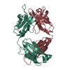
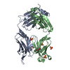
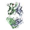
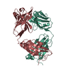
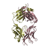
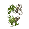
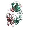
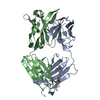
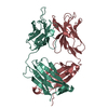
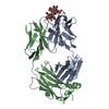
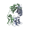
| Title | Crystal structure of mouse anti-HIV potent neutralizing antibody M4H2K1 | ||||||
 Components Components |
| ||||||
 Keywords Keywords | ANTIVIRAL PROTEIN / HIV-1 / ENV / MONOCLONAL ANTIBODY / IMMUNE SYSTEM | ||||||
| Function / homology | Immunoglobulins / Immunoglobulin-like / Sandwich / Mainly Beta Function and homology information Function and homology information | ||||||
| Biological species |  | ||||||
| Method |  X-RAY DIFFRACTION / SYNCHROTRON / MOLECULAR REPLACEMENT / Resolution: 1.5 Å X-RAY DIFFRACTION / SYNCHROTRON / MOLECULAR REPLACEMENT / Resolution: 1.5 Å | ||||||
 Authors Authors | Kumar, S. / Wilson, I.A. | ||||||
| Funding support |  United States, 1items United States, 1items
| ||||||
 Citation Citation | Journal: mBio / Year: 2021 Title: Neutralizing Antibodies Induced by First-Generation gp41-Stabilized HIV-1 Envelope Trimers and Nanoparticles. Authors: Sonu Kumar / Xiaohe Lin / Timothy Ngo / Benjamin Shapero / Cindy Sou / Joel D Allen / Jeffrey Copps / Lei Zhang / Gabriel Ozorowski / Linling He / Max Crispin / Andrew B Ward / Ian A Wilson / Jiang Zhu /  Abstract: The immunogenicity of gp41-stabilized HIV-1 BG505 envelope (Env) trimers and nanoparticles (NPs) was recently assessed in mice and rabbits. Here, we combined Env-specific B-cell sorting and ...The immunogenicity of gp41-stabilized HIV-1 BG505 envelope (Env) trimers and nanoparticles (NPs) was recently assessed in mice and rabbits. Here, we combined Env-specific B-cell sorting and repertoire sequencing to identify neutralizing antibodies (NAbs) from immunized animals. A panel of mouse NAbs was isolated from mice immunized with a 60-meric I3-01 NP presenting 20 stabilized trimers. Three mouse NAbs potently neutralized BG505.T332N by recognizing a glycan epitope centered in the C3/V4 region on BG505 Env, as revealed by electron microscopy (EM), X-ray crystallography, and epitope mapping. A set of rabbit NAbs was isolated from rabbits immunized with a soluble trimer and a 24-meric ferritin NP presenting 8 trimers. Neutralization assays against BG505.T332N variants confirmed that potent rabbit NAbs targeted previously described glycan holes on BG505 Env and accounted for a significant portion of the autologous NAb response in both the trimer and ferritin NP groups. Last, we examined NAb responses that were induced by non-BG505 Env immunogens. We determined a 3.4-Å-resolution crystal structure for the clade C transmitted/founder (T/F) Du172.17 Env with a redesigned heptad repeat 1 (HR1) bend in gp41. This clade C Env, in a soluble trimer form and in a multivalent form with 8 trimers attached to ferritin NP, and the gp41-stabilized clade A Q482-d12 Env trimer elicited distinct NAb responses in rabbits, with notable differences in neutralization breadth. Although eliciting a broad NAb response remains a major challenge, our study provides valuable information on an HIV-1 vaccine design strategy that combines gp41 stabilization and NP display. Self-assembling protein nanoparticles (NPs) presenting BG505 envelope (Env) trimers can elicit tier 2 HIV-1-neutralizing antibody (NAb) responses more effectively than soluble trimers. In the present study, monoclonal NAbs were isolated from previously immunized mice and rabbits for structural and functional analyses, which revealed that potent mouse NAbs recognize the C3/V4 region and small NP-elicited rabbit NAbs primarily target known glycan holes on BG505 Env. This study validates the gp41 stabilization strategy for HIV-1 Env vaccine design and highlights the challenge in eliciting a broad NAb response. | ||||||
| History |
|
- Structure visualization
| Structure viewer | Molecule: MolmilJmol/JSmol |
|---|
- Downloads & links
-Download
| PDBx/mmCIF format | 7kkz.cif.gz | 118 KB | Display | PDBx/mmCIF format |
|---|---|---|---|---|
| PDB format | pdb7kkz.ent.gz | 85.7 KB | Display | PDB format |
| PDBx/mmJSON format | 7kkz.json.gz | Tree view | PDBx/mmJSON format | |
| Others |  Other downloads Other downloads |
-Validation report
| Arichive directory | https://data.pdbj.org/pub/pdb/validation_reports/kk/7kkzftp://data.pdbj.org/pub/pdb/validation_reports/kk/7kkz | HTTPS FTP |
|---|
-Related structure data
| Related structure data |  7klcC  7kmdC 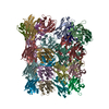 5gs1S S: Starting model for refinement C: citing same article ( |
|---|---|
| Similar structure data |


-Links
 PDBj
PDBj


- Assembly
| Deposited unit | 
| ||||||||||||
|---|---|---|---|---|---|---|---|---|---|---|---|---|---|
| 1 |
| ||||||||||||
| Unit cell |
| ||||||||||||
| Components on special symmetry positions |
|
-Components
| #1: Antibody | Mass: 23805.518 Da / Num. of mol.: 1 Source method: isolated from a genetically manipulated source Source: (gene. exp.)  Homo sapiens (human) Homo sapiens (human) |
|---|---|
| #2: Antibody | Mass: 24216.145 Da / Num. of mol.: 1 Source method: isolated from a genetically manipulated source Source: (gene. exp.) Homo sapiens (human) |
| #3: Chemical | ChemComp-NHE /   Mass: 207.290 Da / Num. of mol.: 1 / Source method: obtained synthetically / Formula: C8H17NO3S / Feature type: SUBJECT OF INVESTIGATION / Comment: pH buffer*YM Mass: 207.290 Da / Num. of mol.: 1 / Source method: obtained synthetically / Formula: C8H17NO3S / Feature type: SUBJECT OF INVESTIGATION / Comment: pH buffer*YM |
| #4: Chemical | ChemComp-EDO /   Mass: 62.068 Da / Num. of mol.: 1 / Source method: obtained synthetically / Formula: C2H6O2 Mass: 62.068 Da / Num. of mol.: 1 / Source method: obtained synthetically / Formula: C2H6O2 |
| #5: Water | ChemComp-HOH /  Mass: 18.015 Da / Num. of mol.: 624 / Source method: isolated from a natural source / Formula: H2O Mass: 18.015 Da / Num. of mol.: 624 / Source method: isolated from a natural source / Formula: H2O |
| Has ligand of interest | Y |
| Has protein modification | Y |
-Experimental details
-Experiment
| Experiment | Method: X-RAY DIFFRACTION / Number of used crystals: 1 |
|---|
- Sample preparation
| Crystal | Density Matthews: 2.6 Å3/Da / Density % sol: 52.62 % |
|---|---|
| Crystal grow | Temperature: 277.15 K / Method: vapor diffusion, sitting drop / Details: 0.1M CHES (pH=9.5), 36% PEG600 |
-Data collection
| Diffraction | Mean temperature: 100 K / Serial crystal experiment: N |
|---|---|
| Diffraction source | Source: SYNCHROTRON / Site: APS / Beamline: 23-ID-D / Wavelength: 1.033 Å |
| Detector | Type: DECTRIS PILATUS3 6M / Detector: PIXEL / Date: Oct 26, 2018 |
| Radiation | Protocol: SINGLE WAVELENGTH / Monochromatic (M) / Laue (L): M / Scattering type: x-ray |
| Radiation wavelength | Wavelength: 1.033 Å / Relative weight: 1 |
| Reflection | Resolution: 1.5→50 Å / Num. obs: 81206 / % possible obs: 99.8 % / Redundancy: 11.4 % / Biso Wilson estimate: 18.56 Å2 / CC1/2: 0.94 / Net I/σ(I): 34.9 |
| Reflection shell | Resolution: 1.5→1.53 Å / Mean I/σ(I) obs: 2 / Num. unique obs: 3998 / CC1/2: 0.73 |
- Processing
| Software |
| ||||||||||||||||||||||||||||||||||||||||||||||||||||||||||||||||||||||||||||||||||||||||||||||||||||||||||||||||||||||||||||||||||||||||||||||||||||||||||||||||||||||||||||||||||||||||||||||||||||||||||||||||||
|---|---|---|---|---|---|---|---|---|---|---|---|---|---|---|---|---|---|---|---|---|---|---|---|---|---|---|---|---|---|---|---|---|---|---|---|---|---|---|---|---|---|---|---|---|---|---|---|---|---|---|---|---|---|---|---|---|---|---|---|---|---|---|---|---|---|---|---|---|---|---|---|---|---|---|---|---|---|---|---|---|---|---|---|---|---|---|---|---|---|---|---|---|---|---|---|---|---|---|---|---|---|---|---|---|---|---|---|---|---|---|---|---|---|---|---|---|---|---|---|---|---|---|---|---|---|---|---|---|---|---|---|---|---|---|---|---|---|---|---|---|---|---|---|---|---|---|---|---|---|---|---|---|---|---|---|---|---|---|---|---|---|---|---|---|---|---|---|---|---|---|---|---|---|---|---|---|---|---|---|---|---|---|---|---|---|---|---|---|---|---|---|---|---|---|---|---|---|---|---|---|---|---|---|---|---|---|---|---|---|---|---|
| Refinement | Method to determine structure: MOLECULAR REPLACEMENT Starting model: 5GS1 Resolution: 1.5→49.86 Å / SU ML: 0.1807 / Cross valid method: FREE R-VALUE / σ(F): 1.34 / Phase error: 22.1798 Stereochemistry target values: GeoStd + Monomer Library + CDL v1.2
| ||||||||||||||||||||||||||||||||||||||||||||||||||||||||||||||||||||||||||||||||||||||||||||||||||||||||||||||||||||||||||||||||||||||||||||||||||||||||||||||||||||||||||||||||||||||||||||||||||||||||||||||||||
| Solvent computation | Shrinkage radii: 0.9 Å / VDW probe radii: 1.11 Å / Solvent model: FLAT BULK SOLVENT MODEL | ||||||||||||||||||||||||||||||||||||||||||||||||||||||||||||||||||||||||||||||||||||||||||||||||||||||||||||||||||||||||||||||||||||||||||||||||||||||||||||||||||||||||||||||||||||||||||||||||||||||||||||||||||
| Displacement parameters | Biso mean: 25.84 Å2 | ||||||||||||||||||||||||||||||||||||||||||||||||||||||||||||||||||||||||||||||||||||||||||||||||||||||||||||||||||||||||||||||||||||||||||||||||||||||||||||||||||||||||||||||||||||||||||||||||||||||||||||||||||
| Refinement step | Cycle: LAST / Resolution: 1.5→49.86 Å
| ||||||||||||||||||||||||||||||||||||||||||||||||||||||||||||||||||||||||||||||||||||||||||||||||||||||||||||||||||||||||||||||||||||||||||||||||||||||||||||||||||||||||||||||||||||||||||||||||||||||||||||||||||
| Refine LS restraints |
| ||||||||||||||||||||||||||||||||||||||||||||||||||||||||||||||||||||||||||||||||||||||||||||||||||||||||||||||||||||||||||||||||||||||||||||||||||||||||||||||||||||||||||||||||||||||||||||||||||||||||||||||||||
| LS refinement shell |
|
