 Movie
Movie Controller

[English] 日本語
 Yorodumi
- PDB-2aab: Structural basis of antigen mimicry in a clinically relevant mela... -
+ Open data

- Basic information
| Entry | Database: PDB / ID: 2aab | ||||||
|---|---|---|---|---|---|---|---|
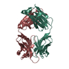
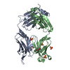
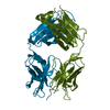
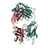
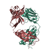
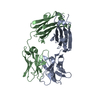
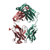
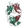
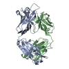
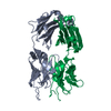
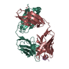
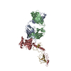
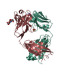
| Title | Structural basis of antigen mimicry in a clinically relevant melanoma antigen system | ||||||
 Components Components |
| ||||||
 Keywords Keywords | IMMUNE SYSTEM / Anti-idiotypic antibody / molecular mimicry / melanoma / immunotherapy | ||||||
| Function / homology | Immunoglobulins / Immunoglobulin-like / Sandwich / Mainly Beta Function and homology information Function and homology information | ||||||
| Biological species |  | ||||||
| Method |  X-RAY DIFFRACTION / MOLECULAR REPLACEMENT / Resolution: 2.5 Å X-RAY DIFFRACTION / MOLECULAR REPLACEMENT / Resolution: 2.5 Å | ||||||
 Authors Authors | Chang, C.-C. / Hernandez-Guzman, F.G. / Luo, W. / Wang, X. / Ferrone, S. / Ghosh, D. | ||||||
 Citation Citation | Journal: J.Biol.Chem. / Year: 2005 Title: Structural basis of antigen mimicry in a clinically relevant melanoma antigen system. Authors: Chang, C.-C. / Hernandez-Guzman, F.G. / Luo, W. / Wang, X. / Ferrone, S. / Ghosh, D. | ||||||
| History |
| ||||||
| Remark 999 | SEQUENCE an appropriate sequence database reference was not available at the time of processing. |
- Structure visualization
| Structure viewer | Molecule: MolmilJmol/JSmol |
|---|
- Downloads & links
-Download
| PDBx/mmCIF format | 2aab.cif.gz | 97.6 KB | Display | PDBx/mmCIF format |
|---|---|---|---|---|
| PDB format | pdb2aab.ent.gz | 73.7 KB | Display | PDB format |
| PDBx/mmJSON format | 2aab.json.gz | Tree view | PDBx/mmJSON format | |
| Others |  Other downloads Other downloads |
-Validation report
| Arichive directory | https://data.pdbj.org/pub/pdb/validation_reports/aa/2aabftp://data.pdbj.org/pub/pdb/validation_reports/aa/2aab | HTTPS FTP |
|---|
-Related structure data
| Related structure data |  2rscS S: Starting model for refinement |
|---|---|
| Similar structure data |


-Links
 PDBj
PDBj

- Assembly
| Deposited unit | 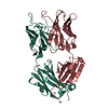
| ||||||||
|---|---|---|---|---|---|---|---|---|---|
| 1 |
| ||||||||
| Unit cell |
| ||||||||
| Details | Monoclonal antibody fragment Fab' consisting of a light chain L and a heavy chain H |
-Components
| #1: Antibody | Mass: 23993.562 Da / Num. of mol.: 1 / Source method: isolated from a natural source / Source: (natural) |
|---|---|
| #2: Antibody | Mass: 26233.570 Da / Num. of mol.: 1 / Fragment: Fab fragment / Source method: isolated from a natural source / Source: (natural) |
| #3: Water | ChemComp-HOH /  Mass: 18.015 Da / Num. of mol.: 66 / Source method: isolated from a natural source / Formula: H2O Mass: 18.015 Da / Num. of mol.: 66 / Source method: isolated from a natural source / Formula: H2O |
| Has protein modification | Y |
-Experimental details
-Experiment
| Experiment | Method: X-RAY DIFFRACTION / Number of used crystals: 1 |
|---|
- Sample preparation
| Crystal | Density Matthews: 2.3 Å3/Da / Density % sol: 50 % |
|---|---|
| Crystal grow | Temperature: 295 K / Method: vapor diffusion, hanging drop / pH: 7.5 Details: PEG 6000, pH 7.5, VAPOR DIFFUSION, HANGING DROP, temperature 295.0K |
-Data collection
| Diffraction | Mean temperature: 100 K |
|---|---|
| Diffraction source | Source: ROTATING ANODE / Type: RIGAKU RU200 / Wavelength: 1.5418 Å |
| Detector | Type: RIGAKU RAXIS IV / Detector: IMAGE PLATE / Date: Jan 1, 2002 / Details: mirrors |
| Radiation | Monochromator: graphite / Protocol: SINGLE WAVELENGTH / Monochromatic (M) / Laue (L): M / Scattering type: x-ray |
| Radiation wavelength | Wavelength: 1.5418 Å / Relative weight: 1 |
| Reflection | Resolution: 2.5→33 Å / Num. all: 17003 / Num. obs: 16656 / % possible obs: 98 % / Observed criterion σ(F): 2 / Observed criterion σ(I): 1 / Redundancy: 5.5 % / Biso Wilson estimate: 45.8 Å2 / Rmerge(I) obs: 0.067 |
| Reflection shell | Resolution: 2.5→2.59 Å / Mean I/σ(I) obs: 4.2 / % possible all: 95 |
- Processing
| Software |
| |||||||||||||||||||||||||
|---|---|---|---|---|---|---|---|---|---|---|---|---|---|---|---|---|---|---|---|---|---|---|---|---|---|---|
| Refinement | Method to determine structure: MOLECULAR REPLACEMENT Starting model: 2RSC Resolution: 2.5→33 Å / σ(F): 2 / Stereochemistry target values: Engh & Huber
| |||||||||||||||||||||||||
| Refine analyze | Luzzati coordinate error obs: 0.34 Å / Luzzati sigma a obs: 0.31 Å | |||||||||||||||||||||||||
| Refinement step | Cycle: LAST / Resolution: 2.5→33 Å
| |||||||||||||||||||||||||
| Refine LS restraints |
|
