 Movie
Movie Controller


+ Open data

- Basic information
| Entry | Database: PDB / ID: 5fhb | ||||||
|---|---|---|---|---|---|---|---|
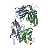
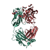
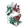
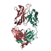
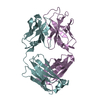
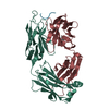
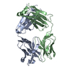
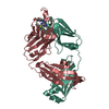
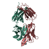
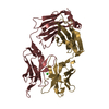
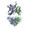
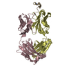
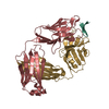
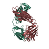
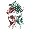
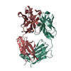
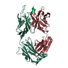
| Title | Crystal Structure of Protective Ebola Virus Antibody 100 | ||||||
 Components Components |
| ||||||
 Keywords Keywords | IMMUNE SYSTEM / Ig domain / Fab | ||||||
| Function / homology | Immunoglobulins / Immunoglobulin-like / Sandwich / Mainly Beta / ACETATE ION Function and homology information Function and homology information | ||||||
| Biological species |  Homo sapiens (human) Homo sapiens (human) | ||||||
| Method |  X-RAY DIFFRACTION / SYNCHROTRON / MOLECULAR REPLACEMENT / molecular replacement / Resolution: 1.973 Å X-RAY DIFFRACTION / SYNCHROTRON / MOLECULAR REPLACEMENT / molecular replacement / Resolution: 1.973 Å | ||||||
 Authors Authors | Gilman, M.S.A. / McLellan, J.S. | ||||||
 Citation Citation | Journal: Science / Year: 2016 Title: Structural and molecular basis for Ebola virus neutralization by protective human antibodies. Authors: John Misasi / Morgan S A Gilman / Masaru Kanekiyo / Miao Gui / Alberto Cagigi / Sabue Mulangu / Davide Corti / Julie E Ledgerwood / Antonio Lanzavecchia / James Cunningham / Jean Jacques ...Authors: John Misasi / Morgan S A Gilman / Masaru Kanekiyo / Miao Gui / Alberto Cagigi / Sabue Mulangu / Davide Corti / Julie E Ledgerwood / Antonio Lanzavecchia / James Cunningham / Jean Jacques Muyembe-Tamfun / Ulrich Baxa / Barney S Graham / Ye Xiang / Nancy J Sullivan / Jason S McLellan /     Abstract: Ebola virus causes hemorrhagic fever with a high case fatality rate for which there is no approved therapy. Two human monoclonal antibodies, mAb100 and mAb114, in combination, protect nonhuman ...Ebola virus causes hemorrhagic fever with a high case fatality rate for which there is no approved therapy. Two human monoclonal antibodies, mAb100 and mAb114, in combination, protect nonhuman primates against all signs of Ebola virus disease, including viremia. Here, we demonstrate that mAb100 recognizes the base of the Ebola virus glycoprotein (GP) trimer, occludes access to the cathepsin-cleavage loop, and prevents the proteolytic cleavage of GP that is required for virus entry. We show that mAb114 interacts with the glycan cap and inner chalice of GP, remains associated after proteolytic removal of the glycan cap, and inhibits binding of cleaved GP to its receptor. These results define the basis of neutralization for two protective antibodies and may facilitate development of therapies and vaccines. | ||||||
| History |
|
- Structure visualization
| Structure viewer | Molecule: MolmilJmol/JSmol |
|---|
- Downloads & links
-Download
| PDBx/mmCIF format | 5fhb.cif.gz | 186 KB | Display | PDBx/mmCIF format |
|---|---|---|---|---|
| PDB format | pdb5fhb.ent.gz | 148.1 KB | Display | PDB format |
| PDBx/mmJSON format | 5fhb.json.gz | Tree view | PDBx/mmJSON format | |
| Others |  Other downloads Other downloads |
-Validation report
| Arichive directory | https://data.pdbj.org/pub/pdb/validation_reports/fh/5fhbftp://data.pdbj.org/pub/pdb/validation_reports/fh/5fhb | HTTPS FTP |
|---|
-Related structure data
| Related structure data |  3310C  3311C 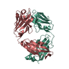 5fhaC  5fhcC 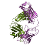 4qhkS C: citing same article ( S: Starting model for refinement |
|---|---|
| Similar structure data |


-Links
 PDBj
PDBj

- Assembly
| Deposited unit | 
| |||||||||
|---|---|---|---|---|---|---|---|---|---|---|
| 1 |
| |||||||||
| Unit cell |
| |||||||||
| Components on special symmetry positions |
|
-Components
| #1: Antibody | Mass: 24337.166 Da / Num. of mol.: 1 Source method: isolated from a genetically manipulated source Source: (gene. exp.) Homo sapiens (human) / Cell: PBMC / Cell line (production host): HEK293 Cells / Production host: Homo sapiens (human) | ||||||
|---|---|---|---|---|---|---|---|
| #2: Antibody | Mass: 22384.715 Da / Num. of mol.: 1 Source method: isolated from a genetically manipulated source Source: (gene. exp.) Homo sapiens (human) / Cell: PBMC / Cell line (production host): HEK293 Cells / Production host: Homo sapiens (human) | ||||||
| #3: Chemical | ChemComp-EDO /   Mass: 62.068 Da / Num. of mol.: 10 / Source method: obtained synthetically / Formula: C2H6O2 Mass: 62.068 Da / Num. of mol.: 10 / Source method: obtained synthetically / Formula: C2H6O2#4: Chemical |   Mass: 59.044 Da / Num. of mol.: 2 / Source method: obtained synthetically / Formula: C2H3O2 Mass: 59.044 Da / Num. of mol.: 2 / Source method: obtained synthetically / Formula: C2H3O2#5: Water | ChemComp-HOH / |  Mass: 18.015 Da / Num. of mol.: 291 / Source method: isolated from a natural source / Formula: H2O Mass: 18.015 Da / Num. of mol.: 291 / Source method: isolated from a natural source / Formula: H2OHas protein modification | Y | |
-Experimental details
-Experiment
| Experiment | Method: X-RAY DIFFRACTION / Number of used crystals: 1 |
|---|
- Sample preparation
| Crystal | Density Matthews: 3.15 Å3/Da / Density % sol: 60.91 % |
|---|---|
| Crystal grow | Temperature: 293 K / Method: vapor diffusion, hanging drop / pH: 5.5 Details: 3.4mg/ml 100 Fab, 38% PEG 400, 4.75% PEG 3350, 0.1M sodium acetate pH 5.5 |
-Data collection
| Diffraction | Mean temperature: 100 K | |||||||||||||||||||||||||||
|---|---|---|---|---|---|---|---|---|---|---|---|---|---|---|---|---|---|---|---|---|---|---|---|---|---|---|---|---|
| Diffraction source | Source: SYNCHROTRON / Site: APS / Beamline: 19-ID / Wavelength: 0.9793 Å | |||||||||||||||||||||||||||
| Detector | Type: ADSC QUANTUM 315r / Detector: CCD / Date: Jul 23, 2015 | |||||||||||||||||||||||||||
| Radiation | Protocol: SINGLE WAVELENGTH / Monochromatic (M) / Laue (L): M / Scattering type: x-ray | |||||||||||||||||||||||||||
| Radiation wavelength | Wavelength: 0.9793 Å / Relative weight: 1 | |||||||||||||||||||||||||||
| Reflection | Resolution: 1.97→33.487 Å / Num. obs: 42605 / % possible obs: 100 % / Redundancy: 10.4 % / Biso Wilson estimate: 24.01 Å2 / CC1/2: 0.997 / Rmerge(I) obs: 0.134 / Rpim(I) all: 0.044 / Net I/σ(I): 11.3 / Num. measured all: 443933 / Scaling rejects: 587 | |||||||||||||||||||||||||||
| Reflection shell | Diffraction-ID: 1 / Rejects: _
|
-Phasing
| Phasing | Method: molecular replacement |
|---|
- Processing
| Software |
| |||||||||||||||||||||||||||||||||||||||||||||||||||||||||||||||||||||||||||||||||||||||||||||||||||||||||||||||||||||||||||||
|---|---|---|---|---|---|---|---|---|---|---|---|---|---|---|---|---|---|---|---|---|---|---|---|---|---|---|---|---|---|---|---|---|---|---|---|---|---|---|---|---|---|---|---|---|---|---|---|---|---|---|---|---|---|---|---|---|---|---|---|---|---|---|---|---|---|---|---|---|---|---|---|---|---|---|---|---|---|---|---|---|---|---|---|---|---|---|---|---|---|---|---|---|---|---|---|---|---|---|---|---|---|---|---|---|---|---|---|---|---|---|---|---|---|---|---|---|---|---|---|---|---|---|---|---|---|---|
| Refinement | Method to determine structure: MOLECULAR REPLACEMENT Starting model: PDB ID: 4QHK Resolution: 1.973→33.487 Å / SU ML: 0.19 / Cross valid method: FREE R-VALUE / σ(F): 1.34 / Phase error: 16.96 / Stereochemistry target values: ML
| |||||||||||||||||||||||||||||||||||||||||||||||||||||||||||||||||||||||||||||||||||||||||||||||||||||||||||||||||||||||||||||
| Solvent computation | Shrinkage radii: 0.9 Å / VDW probe radii: 1.11 Å / Solvent model: FLAT BULK SOLVENT MODEL | |||||||||||||||||||||||||||||||||||||||||||||||||||||||||||||||||||||||||||||||||||||||||||||||||||||||||||||||||||||||||||||
| Refinement step | Cycle: LAST / Resolution: 1.973→33.487 Å
| |||||||||||||||||||||||||||||||||||||||||||||||||||||||||||||||||||||||||||||||||||||||||||||||||||||||||||||||||||||||||||||
| Refine LS restraints |
| |||||||||||||||||||||||||||||||||||||||||||||||||||||||||||||||||||||||||||||||||||||||||||||||||||||||||||||||||||||||||||||
| LS refinement shell |
| |||||||||||||||||||||||||||||||||||||||||||||||||||||||||||||||||||||||||||||||||||||||||||||||||||||||||||||||||||||||||||||
| Refinement TLS params. | Method: refined / Refine-ID: X-RAY DIFFRACTION
| |||||||||||||||||||||||||||||||||||||||||||||||||||||||||||||||||||||||||||||||||||||||||||||||||||||||||||||||||||||||||||||
| Refinement TLS group |
|
