 Movie
Movie Controller

[English] 日本語
 Yorodumi
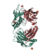
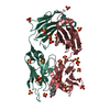
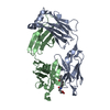
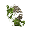
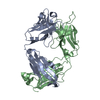
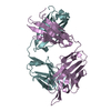
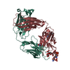
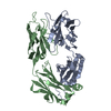
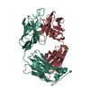
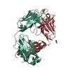
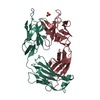
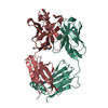
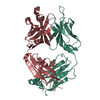
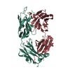
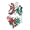
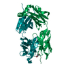
- PDB-2o5y: Crystal structure of the 1E9 LeuH47Trp/ArgH100Trp Fab progesteron... -
+ Open data

- Basic information
| Entry | Database: PDB / ID: 2o5y | ||||||
|---|---|---|---|---|---|---|---|
| Title | Crystal structure of the 1E9 LeuH47Trp/ArgH100Trp Fab progesterone complex | ||||||
 Components Components | (chimeric antibody Fab 1E9-DB3) x 2 | ||||||
 Keywords Keywords | IMMUNE SYSTEM / immunoglobulin / chimeric Fab / antibody engineering / evolution of ligand recognition | ||||||
| Function / homology | Immunoglobulins / Immunoglobulin-like / Sandwich / Mainly Beta / PROGESTERONE Function and homology information Function and homology information | ||||||
| Biological species |   Homo sapiens (human) Homo sapiens (human) | ||||||
| Method |  X-RAY DIFFRACTION / SYNCHROTRON / rigid body refinement / Resolution: 2.85 Å X-RAY DIFFRACTION / SYNCHROTRON / rigid body refinement / Resolution: 2.85 Å | ||||||
 Authors Authors | Verdino, P. / Wilson, I.A. | ||||||
 Citation Citation | Journal: Proc.Natl.Acad.Sci.Usa / Year: 2008 Title: Closely related antibody receptors exploit fundamentally different strategies for steroid recognition. Authors: Verdino, P. / Aldag, C. / Hilvert, D. / Wilson, I.A. | ||||||
| History |
| ||||||
| Remark 999 | SEQUENCE There is no aminoacid sequence database reference available for the protein. The protein ...SEQUENCE There is no aminoacid sequence database reference available for the protein. The protein was designed using variable domains from mouse and the constant domains from humans |
- Structure visualization
| Structure viewer | Molecule: MolmilJmol/JSmol |
|---|
- Downloads & links
-Download
| PDBx/mmCIF format | 2o5y.cif.gz | 100.1 KB | Display | PDBx/mmCIF format |
|---|---|---|---|---|
| PDB format | pdb2o5y.ent.gz | 76.3 KB | Display | PDB format |
| PDBx/mmJSON format | 2o5y.json.gz | Tree view | PDBx/mmJSON format | |
| Others |  Other downloads Other downloads |
-Validation report
| Arichive directory | https://data.pdbj.org/pub/pdb/validation_reports/o5/2o5yftp://data.pdbj.org/pub/pdb/validation_reports/o5/2o5y | HTTPS FTP |
|---|
-Related structure data


-Links
 PDBj
PDBj

- Assembly
| Deposited unit | 
| ||||||||
|---|---|---|---|---|---|---|---|---|---|
| 1 |
| ||||||||
| 2 | 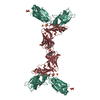
| ||||||||
| 3 | 
| ||||||||
| Unit cell |
|
-Components
| #1: Antibody | Mass: 24143.002 Da / Num. of mol.: 1 / Fragment: light chain / Mutation: G63S Source method: isolated from a genetically manipulated source Source: (gene. exp.) Mus musculus, Homo sapiens / Genus: Mus, Homo / Species: , / Strain: , / Plasmid: p4xH-1E9 / Production host:  | ||||||
|---|---|---|---|---|---|---|---|
| #2: Antibody | Mass: 24425.525 Da / Num. of mol.: 1 / Fragment: heavy chain / Mutation: L47W, M87T, R100W Source method: isolated from a genetically manipulated source Source: (gene. exp.) Mus musculus, Homo sapiens / Genus: Mus, Homo / Species: , / Strain: , / Plasmid: p4xH-1E9 / Production host: | ||||||
| #3: Chemical | ChemComp-SO4 /   Mass: 96.063 Da / Num. of mol.: 13 / Source method: obtained synthetically / Formula: SO4 Mass: 96.063 Da / Num. of mol.: 13 / Source method: obtained synthetically / Formula: SO4#4: Chemical | ChemComp-STR / | 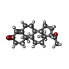  Mass: 314.462 Da / Num. of mol.: 1 / Source method: obtained synthetically / Formula: C21H30O2 Mass: 314.462 Da / Num. of mol.: 1 / Source method: obtained synthetically / Formula: C21H30O2#5: Water | ChemComp-HOH / |  Mass: 18.015 Da / Num. of mol.: 65 / Source method: isolated from a natural source / Formula: H2O Mass: 18.015 Da / Num. of mol.: 65 / Source method: isolated from a natural source / Formula: H2OHas protein modification | Y | |
-Experimental details
-Experiment
| Experiment | Method: X-RAY DIFFRACTION / Number of used crystals: 1 |
|---|
- Sample preparation
| Crystal | Density Matthews: 4.59 Å3/Da / Density % sol: 73.18 % |
|---|---|
| Crystal grow | Temperature: 295 K / Method: vapor diffusion, sitting drop / pH: 7.5 Details: 1.5 M ammonium sulfate, 0.15 M sodium citrate, 0.01% PEG 20000, pH 7.5, VAPOR DIFFUSION, SITTING DROP, temperature 295.0K |
-Data collection
| Diffraction | Mean temperature: 100 K |
|---|---|
| Diffraction source | Source: SYNCHROTRON / Site: ALS  / Beamline: 8.2.1 / Wavelength: 0.99996 Å / Beamline: 8.2.1 / Wavelength: 0.99996 Å |
| Detector | Type: ADSC QUANTUM 210 / Detector: CCD / Date: Nov 11, 2004 |
| Radiation | Protocol: SINGLE WAVELENGTH / Monochromatic (M) / Laue (L): M / Scattering type: x-ray |
| Radiation wavelength | Wavelength: 0.99996 Å / Relative weight: 1 |
| Reflection | Resolution: 2.85→50 Å / Num. all: 21201 / Num. obs: 20438 / % possible obs: 96.4 % / Observed criterion σ(F): 0 / Observed criterion σ(I): 0 / Redundancy: 3.9 % / Biso Wilson estimate: 52.3 Å2 / Rmerge(I) obs: 0.111 / Rsym value: 11.1 / Net I/σ(I): 9.6 |
| Reflection shell | Resolution: 2.85→2.92 Å / Redundancy: 4.1 % / Rmerge(I) obs: 0.382 / Mean I/σ(I) obs: 2.4 / Rsym value: 38.2 / % possible all: 88.3 |
- Processing
| Software |
| ||||||||||||||||||||||||||||||||||||||||||||||||||||||||||||||||||||||||||||||||||||||||||
|---|---|---|---|---|---|---|---|---|---|---|---|---|---|---|---|---|---|---|---|---|---|---|---|---|---|---|---|---|---|---|---|---|---|---|---|---|---|---|---|---|---|---|---|---|---|---|---|---|---|---|---|---|---|---|---|---|---|---|---|---|---|---|---|---|---|---|---|---|---|---|---|---|---|---|---|---|---|---|---|---|---|---|---|---|---|---|---|---|---|---|---|
| Refinement | Method to determine structure: rigid body refinement / Resolution: 2.85→50 Å / Cor.coef. Fo:Fc: 0.94 / Cor.coef. Fo:Fc free: 0.89 / SU B: 24.013 / SU ML: 0.224 / Cross valid method: THROUGHOUT / σ(F): 0 / σ(I): 0 / ESU R: 0.443 / ESU R Free: 0.295 / Stereochemistry target values: MAXIMUM LIKELIHOOD / Details: HYDROGENS HAVE BEEN ADDED IN THE RIDING POSITIONS
| ||||||||||||||||||||||||||||||||||||||||||||||||||||||||||||||||||||||||||||||||||||||||||
| Solvent computation | Ion probe radii: 0.8 Å / Shrinkage radii: 0.8 Å / VDW probe radii: 1.2 Å / Solvent model: BABINET MODEL WITH MASK | ||||||||||||||||||||||||||||||||||||||||||||||||||||||||||||||||||||||||||||||||||||||||||
| Displacement parameters | Biso mean: 45.127 Å2
| ||||||||||||||||||||||||||||||||||||||||||||||||||||||||||||||||||||||||||||||||||||||||||
| Refinement step | Cycle: LAST / Resolution: 2.85→50 Å
| ||||||||||||||||||||||||||||||||||||||||||||||||||||||||||||||||||||||||||||||||||||||||||
| Refine LS restraints |
| ||||||||||||||||||||||||||||||||||||||||||||||||||||||||||||||||||||||||||||||||||||||||||
| LS refinement shell | Resolution: 2.85→2.924 Å / Total num. of bins used: 20
|
