 Movie
Movie Controller

[English] 日本語
 Yorodumi
- PDB-5fhc: Crystal Structure of Protective Human Antibodies 100 and 114 in C... -
+ Open data

- Basic information
| Entry | Database: PDB / ID: 5fhc | ||||||
|---|---|---|---|---|---|---|---|
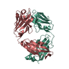
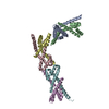
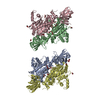
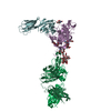
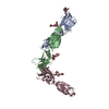
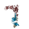
| Title | Crystal Structure of Protective Human Antibodies 100 and 114 in Complex with Ebola Virus Fusion Glycoprotein (GP) | ||||||
 Components Components |
| ||||||
 Keywords Keywords | VIRAL PROTEIN/IMMUNE SYSTEM / Ig domain / Fab / immune system / fusion / ebola virus / glycoprotein / GP / VIRAL PROTEIN-IMMUNE SYSTEM complex | ||||||
| Function / homology |  Function and homology information Function and homology informationsymbiont-mediated killing of host cell / host cell endoplasmic reticulum / viral budding from plasma membrane / symbiont-mediated-mediated suppression of host tetherin activity / clathrin-dependent endocytosis of virus by host cell / host cell cytoplasm / entry receptor-mediated virion attachment to host cell / symbiont-mediated suppression of host innate immune response / membrane raft / fusion of virus membrane with host endosome membrane ...symbiont-mediated killing of host cell / host cell endoplasmic reticulum / viral budding from plasma membrane / symbiont-mediated-mediated suppression of host tetherin activity / clathrin-dependent endocytosis of virus by host cell / host cell cytoplasm / entry receptor-mediated virion attachment to host cell / symbiont-mediated suppression of host innate immune response / membrane raft / fusion of virus membrane with host endosome membrane / viral envelope / symbiont entry into host cell / lipid binding / host cell plasma membrane / virion membrane / extracellular region / identical protein binding Similarity search - Function | ||||||
| Biological species |   Zaire ebolavirus Zaire ebolavirus Homo sapiens (human) Homo sapiens (human) | ||||||
| Method |  X-RAY DIFFRACTION / SYNCHROTRON / MOLECULAR REPLACEMENT / molecular replacement / Resolution: 6.704 Å X-RAY DIFFRACTION / SYNCHROTRON / MOLECULAR REPLACEMENT / molecular replacement / Resolution: 6.704 Å | ||||||
 Authors Authors | Gilman, M.S.A. / McLellan, J.S. | ||||||
 Citation Citation | Journal: Science / Year: 2016 Title: Structural and molecular basis for Ebola virus neutralization by protective human antibodies. Authors: John Misasi / Morgan S A Gilman / Masaru Kanekiyo / Miao Gui / Alberto Cagigi / Sabue Mulangu / Davide Corti / Julie E Ledgerwood / Antonio Lanzavecchia / James Cunningham / Jean Jacques ...Authors: John Misasi / Morgan S A Gilman / Masaru Kanekiyo / Miao Gui / Alberto Cagigi / Sabue Mulangu / Davide Corti / Julie E Ledgerwood / Antonio Lanzavecchia / James Cunningham / Jean Jacques Muyembe-Tamfun / Ulrich Baxa / Barney S Graham / Ye Xiang / Nancy J Sullivan / Jason S McLellan /     Abstract: Ebola virus causes hemorrhagic fever with a high case fatality rate for which there is no approved therapy. Two human monoclonal antibodies, mAb100 and mAb114, in combination, protect nonhuman ...Ebola virus causes hemorrhagic fever with a high case fatality rate for which there is no approved therapy. Two human monoclonal antibodies, mAb100 and mAb114, in combination, protect nonhuman primates against all signs of Ebola virus disease, including viremia. Here, we demonstrate that mAb100 recognizes the base of the Ebola virus glycoprotein (GP) trimer, occludes access to the cathepsin-cleavage loop, and prevents the proteolytic cleavage of GP that is required for virus entry. We show that mAb114 interacts with the glycan cap and inner chalice of GP, remains associated after proteolytic removal of the glycan cap, and inhibits binding of cleaved GP to its receptor. These results define the basis of neutralization for two protective antibodies and may facilitate development of therapies and vaccines. | ||||||
| History |
|
- Structure visualization
| Structure viewer | Molecule: MolmilJmol/JSmol |
|---|
- Downloads & links
-Download
| PDBx/mmCIF format | 5fhc.cif.gz | 480.3 KB | Display | PDBx/mmCIF format |
|---|---|---|---|---|
| PDB format | pdb5fhc.ent.gz | 398.7 KB | Display | PDB format |
| PDBx/mmJSON format | 5fhc.json.gz | Tree view | PDBx/mmJSON format | |
| Others |  Other downloads Other downloads |
-Validation report
| Arichive directory | https://data.pdbj.org/pub/pdb/validation_reports/fh/5fhcftp://data.pdbj.org/pub/pdb/validation_reports/fh/5fhc | HTTPS FTP |
|---|
-Related structure data


-Links
 PDBj
PDBj


- Assembly
| Deposited unit | 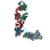
| ||||||||
|---|---|---|---|---|---|---|---|---|---|
| 1 |
| ||||||||
| Unit cell |
|
-Components
-Protein , 2 types, 2 molecules JK
| #1: Protein | Mass: 10968.456 Da / Num. of mol.: 1 / Fragment: UNP residues 502-599 Source method: isolated from a genetically manipulated source Source: (gene. exp.) Zaire ebolavirus (strain Mayinga-76) / Strain: Mayinga-76 / Gene: GP / Cell line (production host): HEK293 Cells / Production host: Homo sapiens (human) / References: UniProt: Q05320 |
|---|---|
| #2: Protein | Mass: 36316.125 Da / Num. of mol.: 1 / Fragment: UNP residues 1-302,UNP residues 1-302 Source method: isolated from a genetically manipulated source Source: (gene. exp.) Zaire ebolavirus / Strain: Mayinga-76 / Gene: GP / Cell line (production host): HEK293 Cells / Production host: Homo sapiens (human) / References: UniProt: Q05320 |
-Antibody , 4 types, 4 molecules ABLH
| #3: Antibody | Mass: 24234.023 Da / Num. of mol.: 1 Source method: isolated from a genetically manipulated source Source: (gene. exp.) Homo sapiens (human) / Cell: PBMC / Cell line (production host): HEK293 Cells / Production host: Homo sapiens (human) |
|---|---|
| #4: Antibody | Mass: 22255.602 Da / Num. of mol.: 1 Source method: isolated from a genetically manipulated source Source: (gene. exp.) Homo sapiens (human) / Cell: PBMC / Cell line (production host): HEK293 Cells / Production host: Homo sapiens (human) |
| #5: Antibody | Mass: 22942.482 Da / Num. of mol.: 1 Source method: isolated from a genetically manipulated source Source: (gene. exp.) Homo sapiens (human) / Cell: PBMC / Cell line (production host): HEK293 Cells / Production host: Homo sapiens (human) |
| #6: Antibody | Mass: 23203.109 Da / Num. of mol.: 1 Source method: isolated from a genetically manipulated source Source: (gene. exp.) Homo sapiens (human) / Cell: PBMC / Cell line (production host): HEK293 Cells / Production host: Homo sapiens (human) |
-Details
| Has protein modification | Y |
|---|
-Experimental details
-Experiment
| Experiment | Method: X-RAY DIFFRACTION / Number of used crystals: 1 |
|---|
- Sample preparation
| Crystal | Density Matthews: 3.88 Å3/Da / Density % sol: 68.32 % |
|---|---|
| Crystal grow | Temperature: 293 K / Method: vapor diffusion, sitting drop / pH: 7.5 Details: Ternary complex of Ebola virus GP (mucin-like domain deleted) with 114 and 100 at 2.9mg/ml, 13.4% PEG 8000, 6.7% isopropanol, 0.2M ammonium sulfate, 0.1M HEPES pH 7.5, 0.01M GSH-GSSG (L- ...Details: Ternary complex of Ebola virus GP (mucin-like domain deleted) with 114 and 100 at 2.9mg/ml, 13.4% PEG 8000, 6.7% isopropanol, 0.2M ammonium sulfate, 0.1M HEPES pH 7.5, 0.01M GSH-GSSG (L-Glutathione reduced-L-Glutathione oxidized) |
-Data collection
| Diffraction | Mean temperature: 100 K | |||||||||||||||||||||||||||
|---|---|---|---|---|---|---|---|---|---|---|---|---|---|---|---|---|---|---|---|---|---|---|---|---|---|---|---|---|
| Diffraction source | Source: SYNCHROTRON / Site: APS / Beamline: 19-ID / Wavelength: 0.9792 Å | |||||||||||||||||||||||||||
| Detector | Type: ADSC QUANTUM 315r / Detector: CCD / Date: Oct 24, 2014 | |||||||||||||||||||||||||||
| Radiation | Protocol: SINGLE WAVELENGTH / Monochromatic (M) / Laue (L): M / Scattering type: x-ray | |||||||||||||||||||||||||||
| Radiation wavelength | Wavelength: 0.9792 Å / Relative weight: 1 | |||||||||||||||||||||||||||
| Reflection | Resolution: 6.7→47.85 Å / Num. obs: 3952 / % possible obs: 99.7 % / Redundancy: 8.9 % / Biso Wilson estimate: 286.238 Å2 / CC1/2: 0.987 / Rmerge(I) obs: 0.301 / Rpim(I) all: 0.106 / Net I/σ(I): 8.9 / Num. measured all: 35041 / Scaling rejects: 12 | |||||||||||||||||||||||||||
| Reflection shell | Diffraction-ID: 1 / Rejects: _
|
-Phasing
| Phasing | Method: molecular replacement |
|---|
- Processing
| Software |
| |||||||||||||||||||||||||||||||||||||||||||||||||||||||||||||||||||||||||||||||||||||||||||||||||||||||||||||||||||||||||||||||||||||||||||||||||||||||||||||||||||||||||||||||
|---|---|---|---|---|---|---|---|---|---|---|---|---|---|---|---|---|---|---|---|---|---|---|---|---|---|---|---|---|---|---|---|---|---|---|---|---|---|---|---|---|---|---|---|---|---|---|---|---|---|---|---|---|---|---|---|---|---|---|---|---|---|---|---|---|---|---|---|---|---|---|---|---|---|---|---|---|---|---|---|---|---|---|---|---|---|---|---|---|---|---|---|---|---|---|---|---|---|---|---|---|---|---|---|---|---|---|---|---|---|---|---|---|---|---|---|---|---|---|---|---|---|---|---|---|---|---|---|---|---|---|---|---|---|---|---|---|---|---|---|---|---|---|---|---|---|---|---|---|---|---|---|---|---|---|---|---|---|---|---|---|---|---|---|---|---|---|---|---|---|---|---|---|---|---|---|---|
| Refinement | Method to determine structure: MOLECULAR REPLACEMENT / Resolution: 6.704→47.848 Å / SU ML: 1.22 / Cross valid method: FREE R-VALUE / σ(F): 1.34 / Phase error: 46.61 / Stereochemistry target values: ML
| |||||||||||||||||||||||||||||||||||||||||||||||||||||||||||||||||||||||||||||||||||||||||||||||||||||||||||||||||||||||||||||||||||||||||||||||||||||||||||||||||||||||||||||||
| Solvent computation | Shrinkage radii: 0.9 Å / VDW probe radii: 1.11 Å / Solvent model: FLAT BULK SOLVENT MODEL | |||||||||||||||||||||||||||||||||||||||||||||||||||||||||||||||||||||||||||||||||||||||||||||||||||||||||||||||||||||||||||||||||||||||||||||||||||||||||||||||||||||||||||||||
| Refinement step | Cycle: LAST / Resolution: 6.704→47.848 Å
| |||||||||||||||||||||||||||||||||||||||||||||||||||||||||||||||||||||||||||||||||||||||||||||||||||||||||||||||||||||||||||||||||||||||||||||||||||||||||||||||||||||||||||||||
| Refine LS restraints |
| |||||||||||||||||||||||||||||||||||||||||||||||||||||||||||||||||||||||||||||||||||||||||||||||||||||||||||||||||||||||||||||||||||||||||||||||||||||||||||||||||||||||||||||||
| LS refinement shell | Highest resolution: 6.7039 Å
| |||||||||||||||||||||||||||||||||||||||||||||||||||||||||||||||||||||||||||||||||||||||||||||||||||||||||||||||||||||||||||||||||||||||||||||||||||||||||||||||||||||||||||||||
| Refinement TLS params. | Method: refined / Refine-ID: X-RAY DIFFRACTION
| |||||||||||||||||||||||||||||||||||||||||||||||||||||||||||||||||||||||||||||||||||||||||||||||||||||||||||||||||||||||||||||||||||||||||||||||||||||||||||||||||||||||||||||||
| Refinement TLS group |
|
