 Movie
Movie Controller


+ Open data

- Basic information
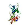
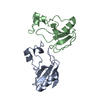
| Entry | Database: PDB / ID: 2pej | ||||||
|---|---|---|---|---|---|---|---|
| Title | Crystal structure of RbcX point mutant Y17A/Y20L | ||||||
 Components Components | ORF134 | ||||||
 Keywords Keywords | CHAPERONE / helix bundle / protein complex assembly | ||||||
| Function / homology |  Function and homology information Function and homology informationribulose bisphosphate carboxylase complex assembly / carboxysome / carbon fixation / photosynthesis / protein folding chaperone / protein homodimerization activity / cytoplasm Similarity search - Function | ||||||
| Biological species |  Synechococcus sp. (bacteria) Synechococcus sp. (bacteria) | ||||||
| Method |  X-RAY DIFFRACTION / SYNCHROTRON / MOLECULAR REPLACEMENT / Resolution: 3.4 Å X-RAY DIFFRACTION / SYNCHROTRON / MOLECULAR REPLACEMENT / Resolution: 3.4 Å | ||||||
 Authors Authors | Saschenbrecker, S. / Bracher, A. / Vasudeva Rao, K. / Vasudeva Rao, B. / Hartl, F.U. / Hayer-Hartl, M. | ||||||
 Citation Citation | Journal: Cell(Cambridge,Mass.) / Year: 2007 Title: Structure and Function of RbcX, an Assembly Chaperone for Hexadecameric Rubisco. Authors: Saschenbrecker, S. / Bracher, A. / Rao, K.V. / Rao, B.V. / Hartl, F.U. / Hayer-Hartl, M. | ||||||
| History |
|
- Structure visualization
| Structure viewer | Molecule: MolmilJmol/JSmol |
|---|
- Downloads & links
-Download
| PDBx/mmCIF format | 2pej.cif.gz | 129.2 KB | Display | PDBx/mmCIF format |
|---|---|---|---|---|
| PDB format | pdb2pej.ent.gz | 100.1 KB | Display | PDB format |
| PDBx/mmJSON format | 2pej.json.gz | Tree view | PDBx/mmJSON format | |
| Others |  Other downloads Other downloads |
-Validation report
| Arichive directory | https://data.pdbj.org/pub/pdb/validation_reports/pe/2pejftp://data.pdbj.org/pub/pdb/validation_reports/pe/2pej | HTTPS FTP |
|---|
-Related structure data
| Related structure data | 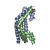 2peiC  2pekC 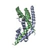 2pemC 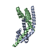 2penSC  2peoC 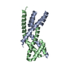 2peqC C: citing same article ( S: Starting model for refinement |
|---|---|
| Similar structure data |


-Links
 PDBj
PDBj
- Assembly
| Deposited unit | 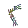
| ||||||||
|---|---|---|---|---|---|---|---|---|---|
| 1 | 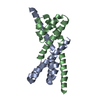
| ||||||||
| 2 | 
| ||||||||
| 3 | 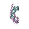
| ||||||||
| Unit cell |
| ||||||||
| Details | The biological unit of RbcX is a dimer. There are 3 biological units in the asymmetric unit (chains A & B, chains C & D and chains E & F). |
-Components
| #1: Protein | Mass: 15141.973 Da / Num. of mol.: 6 / Mutation: Y17A, Y20L Source method: isolated from a genetically manipulated source Source: (gene. exp.) Synechococcus sp. (bacteria) / Strain: PCC 7002 / Gene: RbcX / Plasmid: pET11a / Species (production host): Escherichia coli / Production host: |
|---|
-Experimental details
-Experiment
| Experiment | Method: X-RAY DIFFRACTION / Number of used crystals: 1 |
|---|
- Sample preparation
| Crystal | Density Matthews: 4.94 Å3/Da / Density % sol: 75.11 % |
|---|---|
| Crystal grow | Temperature: 293 K / Method: vapor diffusion, hanging drop / pH: 7.5 Details: 1.5-2.5 M Sodium acetate, 0.1 M HEPES-NaOH pH 7.5, VAPOR DIFFUSION, HANGING DROP, temperature 293K |
-Data collection
| Diffraction | Mean temperature: 100 K | ||||||||||||||||||||||||||||||||||||||||||||||||||||||||||||||||||||||||||||||||||||||||
|---|---|---|---|---|---|---|---|---|---|---|---|---|---|---|---|---|---|---|---|---|---|---|---|---|---|---|---|---|---|---|---|---|---|---|---|---|---|---|---|---|---|---|---|---|---|---|---|---|---|---|---|---|---|---|---|---|---|---|---|---|---|---|---|---|---|---|---|---|---|---|---|---|---|---|---|---|---|---|---|---|---|---|---|---|---|---|---|---|---|
| Diffraction source | Source: SYNCHROTRON / Site: ESRF  / Beamline: ID29 / Wavelength: 1 Å / Beamline: ID29 / Wavelength: 1 Å | ||||||||||||||||||||||||||||||||||||||||||||||||||||||||||||||||||||||||||||||||||||||||
| Detector | Type: ADSC QUANTUM 315 / Detector: CCD / Date: Aug 26, 2006 | ||||||||||||||||||||||||||||||||||||||||||||||||||||||||||||||||||||||||||||||||||||||||
| Radiation | Protocol: SINGLE WAVELENGTH / Monochromatic (M) / Laue (L): M / Scattering type: x-ray | ||||||||||||||||||||||||||||||||||||||||||||||||||||||||||||||||||||||||||||||||||||||||
| Radiation wavelength | Wavelength: 1 Å / Relative weight: 1 | ||||||||||||||||||||||||||||||||||||||||||||||||||||||||||||||||||||||||||||||||||||||||
| Reflection | Resolution: 3.4→103.142 Å / Num. obs: 26126 / % possible obs: 99.6 % / Redundancy: 3.4 % / Rmerge(I) obs: 0.089 / Rsym value: 0.089 / Net I/σ(I): 3.4 | ||||||||||||||||||||||||||||||||||||||||||||||||||||||||||||||||||||||||||||||||||||||||
| Reflection shell | Diffraction-ID: 1
|
- Processing
| Software |
| ||||||||||||||||||||||||||||||||||||||||||||||||||||||||||||||||||||||||||||||||||||||||||
|---|---|---|---|---|---|---|---|---|---|---|---|---|---|---|---|---|---|---|---|---|---|---|---|---|---|---|---|---|---|---|---|---|---|---|---|---|---|---|---|---|---|---|---|---|---|---|---|---|---|---|---|---|---|---|---|---|---|---|---|---|---|---|---|---|---|---|---|---|---|---|---|---|---|---|---|---|---|---|---|---|---|---|---|---|---|---|---|---|---|---|---|
| Refinement | Method to determine structure: MOLECULAR REPLACEMENT Starting model: PDB entry 2PEN Resolution: 3.4→20 Å / Cor.coef. Fo:Fc: 0.932 / Cor.coef. Fo:Fc free: 0.916 / SU B: 20.589 / SU ML: 0.342 / Cross valid method: THROUGHOUT / σ(F): 0 / ESU R: 0.799 / ESU R Free: 0.447 / Stereochemistry target values: MAXIMUM LIKELIHOOD / Details: HYDROGENS HAVE BEEN ADDED IN THE RIDING POSITIONS
| ||||||||||||||||||||||||||||||||||||||||||||||||||||||||||||||||||||||||||||||||||||||||||
| Solvent computation | Ion probe radii: 0.8 Å / Shrinkage radii: 0.8 Å / VDW probe radii: 1.2 Å / Solvent model: MASK | ||||||||||||||||||||||||||||||||||||||||||||||||||||||||||||||||||||||||||||||||||||||||||
| Displacement parameters | Biso mean: 106.428 Å2
| ||||||||||||||||||||||||||||||||||||||||||||||||||||||||||||||||||||||||||||||||||||||||||
| Refinement step | Cycle: LAST / Resolution: 3.4→20 Å
| ||||||||||||||||||||||||||||||||||||||||||||||||||||||||||||||||||||||||||||||||||||||||||
| Refine LS restraints |
| ||||||||||||||||||||||||||||||||||||||||||||||||||||||||||||||||||||||||||||||||||||||||||
| LS refinement shell | Resolution: 3.4→3.486 Å / Total num. of bins used: 20
|
