 Movie
Movie Controller


+ Open data

- Basic information
| Entry | Database: PDB / ID: 2z46 | ||||||
|---|---|---|---|---|---|---|---|
| Title | Crystal Structure of Native-ORF134 | ||||||
 Components Components | ORF134 | ||||||
 Keywords Keywords | CHAPERONE / helix bundle | ||||||
| Function / homology |  Function and homology information Function and homology informationribulose bisphosphate carboxylase complex assembly / carboxysome / carbon fixation / photosynthesis / protein folding chaperone / protein homodimerization activity / cytoplasm Similarity search - Function | ||||||
| Biological species |  Synechococcus sp. (bacteria) Synechococcus sp. (bacteria) | ||||||
| Method |  X-RAY DIFFRACTION / SYNCHROTRON / MOLECULAR REPLACEMENT / Resolution: 2.97 Å X-RAY DIFFRACTION / SYNCHROTRON / MOLECULAR REPLACEMENT / Resolution: 2.97 Å | ||||||
 Authors Authors | Tomimoto, Y. / Ihara, K. / Onizuka, T. / Kanai, S. / Ashida, H. / Yokota, A. / Tanaka, S. / Miyasaka, H. / Yamada, Y. / Kato, R. / Wakatsuki, S. | ||||||
 Citation Citation | Journal: To be Published Title: Crystal Structure of ORF134 Authors: Tomimoto, Y. / Ihara, K. / Onizuka, T. / Kanai, S. / Ashida, H. / Yokota, A. / Tanaka, S. / Miyasaka, H. / Yamada, Y. / Kato, R. / Wakatsuki, S. | ||||||
| History |
|
- Structure visualization
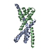
| Structure viewer | Molecule: MolmilJmol/JSmol |
|---|
- Downloads & links
-Download
| PDBx/mmCIF format | 2z46.cif.gz | 137.6 KB | Display | PDBx/mmCIF format |
|---|---|---|---|---|
| PDB format | pdb2z46.ent.gz | 111.2 KB | Display | PDB format |
| PDBx/mmJSON format | 2z46.json.gz | Tree view | PDBx/mmJSON format | |
| Others |  Other downloads Other downloads |
-Validation report
| Arichive directory | https://data.pdbj.org/pub/pdb/validation_reports/z4/2z46ftp://data.pdbj.org/pub/pdb/validation_reports/z4/2z46 | HTTPS FTP |
|---|
-Related structure data
| Related structure data | 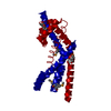 2z44SC  2z45C S: Starting model for refinement C: citing same article ( |
|---|---|
| Similar structure data |


-Links
 PDBj
PDBj
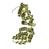
- Assembly
| Deposited unit | 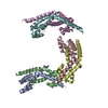
| ||||||||
|---|---|---|---|---|---|---|---|---|---|
| 1 | 
| ||||||||
| 2 | 
| ||||||||
| 3 | 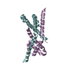
| ||||||||
| Unit cell |
|
-Components
| #1: Protein | Mass: 15284.084 Da / Num. of mol.: 6 Source method: isolated from a genetically manipulated source Source: (gene. exp.) Synechococcus sp. (bacteria) / Strain: PCC7002 / Description: In vitro translation / Plasmid: pIVEX3.2 / References: UniProt: Q44177 |
|---|
-Experimental details
-Experiment
| Experiment | Method: X-RAY DIFFRACTION / Number of used crystals: 1 |
|---|
- Sample preparation
| Crystal | Density Matthews: 4.92 Å3/Da / Density % sol: 74.99 % |
|---|---|
| Crystal grow | Temperature: 293 K / Method: vapor diffusion, hanging drop / pH: 6.5 Details: 1.4M Na acetate, 0.1M Na cacodylate , pH 6.5, VAPOR DIFFUSION, HANGING DROP, temperature 293K |
-Data collection
| Diffraction | Mean temperature: 95 K |
|---|---|
| Diffraction source | Source: SYNCHROTRON / Site: Photon Factory  / Beamline: AR-NW12A / Wavelength: 0.92 Å / Beamline: AR-NW12A / Wavelength: 0.92 Å |
| Detector | Type: ADSC QUANTUM 210 / Detector: CCD / Date: Jan 1, 2005 |
| Radiation | Monochromator: Si(111) / Protocol: SINGLE WAVELENGTH / Monochromatic (M) / Laue (L): M / Scattering type: x-ray |
| Radiation wavelength | Wavelength: 0.92 Å / Relative weight: 1 |
| Reflection | Resolution: 2.97→50 Å / Num. obs: 37069 / % possible obs: 99.3 % / Biso Wilson estimate: 90.4 Å2 / Rmerge(I) obs: 0.088 |
| Reflection shell | Resolution: 2.97→3.08 Å / Rmerge(I) obs: 0.726 / % possible all: 95.1 |
- Processing
| Software |
| ||||||||||||||||||||||||||||||||||||||||||||||||||||||||||||||||||||||||||||||||||||||||||||||||||||||||||||||||||||||||||||||||||||||||||||||||||||||||||||||||||||||||||
|---|---|---|---|---|---|---|---|---|---|---|---|---|---|---|---|---|---|---|---|---|---|---|---|---|---|---|---|---|---|---|---|---|---|---|---|---|---|---|---|---|---|---|---|---|---|---|---|---|---|---|---|---|---|---|---|---|---|---|---|---|---|---|---|---|---|---|---|---|---|---|---|---|---|---|---|---|---|---|---|---|---|---|---|---|---|---|---|---|---|---|---|---|---|---|---|---|---|---|---|---|---|---|---|---|---|---|---|---|---|---|---|---|---|---|---|---|---|---|---|---|---|---|---|---|---|---|---|---|---|---|---|---|---|---|---|---|---|---|---|---|---|---|---|---|---|---|---|---|---|---|---|---|---|---|---|---|---|---|---|---|---|---|---|---|---|---|---|---|---|---|---|
| Refinement | Method to determine structure: MOLECULAR REPLACEMENT Starting model: PDB ENTRY 2Z44 Resolution: 2.97→50 Å / Cor.coef. Fo:Fc: 0.924 / Cor.coef. Fo:Fc free: 0.899 / SU B: 13.646 / SU ML: 0.252 / Cross valid method: THROUGHOUT / ESU R: 0.438 / ESU R Free: 0.318 / Stereochemistry target values: MAXIMUM LIKELIHOOD / Details: HYDROGENS HAVE BEEN ADDED IN THE RIDING POSITIONS
| ||||||||||||||||||||||||||||||||||||||||||||||||||||||||||||||||||||||||||||||||||||||||||||||||||||||||||||||||||||||||||||||||||||||||||||||||||||||||||||||||||||||||||
| Solvent computation | Ion probe radii: 0.8 Å / Shrinkage radii: 0.8 Å / VDW probe radii: 1.2 Å / Solvent model: BABINET MODEL WITH MASK | ||||||||||||||||||||||||||||||||||||||||||||||||||||||||||||||||||||||||||||||||||||||||||||||||||||||||||||||||||||||||||||||||||||||||||||||||||||||||||||||||||||||||||
| Displacement parameters | Biso mean: 71.089 Å2
| ||||||||||||||||||||||||||||||||||||||||||||||||||||||||||||||||||||||||||||||||||||||||||||||||||||||||||||||||||||||||||||||||||||||||||||||||||||||||||||||||||||||||||
| Refinement step | Cycle: LAST / Resolution: 2.97→50 Å
| ||||||||||||||||||||||||||||||||||||||||||||||||||||||||||||||||||||||||||||||||||||||||||||||||||||||||||||||||||||||||||||||||||||||||||||||||||||||||||||||||||||||||||
| Refine LS restraints |
| ||||||||||||||||||||||||||||||||||||||||||||||||||||||||||||||||||||||||||||||||||||||||||||||||||||||||||||||||||||||||||||||||||||||||||||||||||||||||||||||||||||||||||
| LS refinement shell | Resolution: 2.97→3.047 Å / Total num. of bins used: 20
|
