 Movie
Movie Controller

[English] 日本語
 Yorodumi
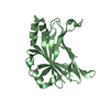
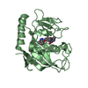
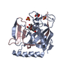
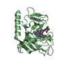
- PDB-6g6f: Crystal structure of a parallel eight-helix coiled coil CC-Type2-LF -
+ Open data

- Basic information
| Entry | Database: PDB / ID: 6g6f | |||||||||
|---|---|---|---|---|---|---|---|---|---|---|
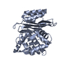
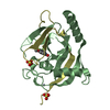
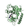
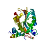
| Title | Crystal structure of a parallel eight-helix coiled coil CC-Type2-LF | |||||||||
 Components Components | CC-Type2-LF | |||||||||
 Keywords Keywords | DE NOVO PROTEIN / de novo / coiled coil / alpha-helical bundle / synthetic construct | |||||||||
| Biological species | synthetic construct (others) | |||||||||
| Method |  X-RAY DIFFRACTION / SYNCHROTRON / MOLECULAR REPLACEMENT / molecular replacement / Resolution: 1.7 Å X-RAY DIFFRACTION / SYNCHROTRON / MOLECULAR REPLACEMENT / molecular replacement / Resolution: 1.7 Å | |||||||||
 Authors Authors | Rhys, G.G. / Brady, R.L. / Woolfson, D.N. | |||||||||
| Funding support |  United Kingdom, 2items United Kingdom, 2items
| |||||||||
 Citation Citation | Journal: Nat Commun / Year: 2018 Title: Maintaining and breaking symmetry in homomeric coiled-coil assemblies. Authors: Rhys, G.G. / Wood, C.W. / Lang, E.J.M. / Mulholland, A.J. / Brady, R.L. / Thomson, A.R. / Woolfson, D.N. | |||||||||
| History |
|
- Structure visualization
| Structure viewer | Molecule:  MolmilJmol/JSmol MolmilJmol/JSmol |
|---|
- Downloads & links
-Download
| PDBx/mmCIF format | 6g6f.cif.gz | 64.9 KB | Display | PDBx/mmCIF format |
|---|---|---|---|---|
| PDB format | pdb6g6f.ent.gz | 49.8 KB | Display | PDB format |
| PDBx/mmJSON format | 6g6f.json.gz | Tree view | PDBx/mmJSON format | |
| Others |  Other downloads Other downloads |
-Validation report
| Summary document | 6g6f_validation.pdf.gz | 485.2 KB | Display | wwPDB validaton report |
|---|---|---|---|---|
| Full document | 6g6f_full_validation.pdf.gz | 485.9 KB | Display | |
| Data in XML | 6g6f_validation.xml.gz | 14.9 KB | Display | |
| Data in CIF | 6g6f_validation.cif.gz | 20.9 KB | Display | |
| Arichive directory | https://data.pdbj.org/pub/pdb/validation_reports/g6/6g6fftp://data.pdbj.org/pub/pdb/validation_reports/g6/6g6f | HTTPS FTP |
-Related structure data
| Related structure data | 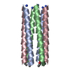 6g65C 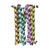 6g66C 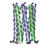 6g67C 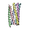 6g68C 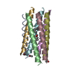 6g69C 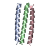 6g6aC 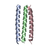 6g6bC 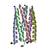 6g6cC 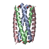 6g6dC 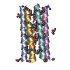 6g6eC 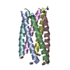 6g6gC 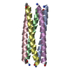 6g6hC C: citing same article ( |
|---|---|
| Similar structure data |


-Links
 PDBj
PDBj
- Assembly
| Deposited unit | 
| ||||||||
|---|---|---|---|---|---|---|---|---|---|
| 1 |
| ||||||||
| Unit cell |
|
-Components
| #1: Protein/peptide | Mass: 3370.935 Da / Num. of mol.: 8 / Source method: obtained synthetically Details: solid-phase peptide synthesis using the fmoc-based strategy Source: (synth.) synthetic construct (others) #2: Chemical | ChemComp-TRS / |   Mass: 122.143 Da / Num. of mol.: 1 / Source method: obtained synthetically / Formula: C4H12NO3 / Comment: pH buffer*YM Mass: 122.143 Da / Num. of mol.: 1 / Source method: obtained synthetically / Formula: C4H12NO3 / Comment: pH buffer*YM#3: Water | ChemComp-HOH / |  Mass: 18.015 Da / Num. of mol.: 237 / Source method: isolated from a natural source / Formula: H2O Mass: 18.015 Da / Num. of mol.: 237 / Source method: isolated from a natural source / Formula: H2OHas protein modification | Y | |
|---|
-Experimental details
-Experiment
| Experiment | Method: X-RAY DIFFRACTION / Number of used crystals: 1 |
|---|
- Sample preparation
| Crystal | Density Matthews: 2.04 Å3/Da / Density % sol: 39.76 % / Mosaicity: 0.26 ° |
|---|---|
| Crystal grow | Temperature: 293 K / Method: vapor diffusion, sitting drop / pH: 8.5 / Details: 50 mM Tris and 10 % w/v PEG 6000 |
-Data collection
| Diffraction | Mean temperature: 80 K | ||||||||||||||||||||||||
|---|---|---|---|---|---|---|---|---|---|---|---|---|---|---|---|---|---|---|---|---|---|---|---|---|---|
| Diffraction source | Source: SYNCHROTRON / Site: Diamond / Beamline: I04 / Wavelength: 0.97949 Å | ||||||||||||||||||||||||
| Detector | Type: DECTRIS PILATUS 6M-F / Detector: PIXEL / Date: Jul 31, 2015 | ||||||||||||||||||||||||
| Radiation | Protocol: SINGLE WAVELENGTH / Monochromatic (M) / Laue (L): M / Scattering type: x-ray | ||||||||||||||||||||||||
| Radiation wavelength | Wavelength: 0.97949 Å / Relative weight: 1 | ||||||||||||||||||||||||
| Reflection | Resolution: 1.7→58.99 Å / Num. obs: 22185 / % possible obs: 92.6 % / Redundancy: 5.7 % / CC1/2: 1 / Rmerge(I) obs: 0.028 / Rpim(I) all: 0.012 / Rrim(I) all: 0.031 / Net I/σ(I): 36.7 / Num. measured all: 125674 / Scaling rejects: 215 | ||||||||||||||||||||||||
| Reflection shell | Diffraction-ID: 1
|
-Phasing
| Phasing | Method: molecular replacement | |||||||||
|---|---|---|---|---|---|---|---|---|---|---|
| Phasing MR | Model details: Phaser MODE: MR_AUTO
|
- Processing
| Software |
| |||||||||||||||||||||||||||||||||||||||||||||||||||||||||||||||
|---|---|---|---|---|---|---|---|---|---|---|---|---|---|---|---|---|---|---|---|---|---|---|---|---|---|---|---|---|---|---|---|---|---|---|---|---|---|---|---|---|---|---|---|---|---|---|---|---|---|---|---|---|---|---|---|---|---|---|---|---|---|---|---|---|
| Refinement | Method to determine structure: MOLECULAR REPLACEMENT / Resolution: 1.7→44.452 Å / SU ML: 0.15 / Cross valid method: THROUGHOUT / σ(F): 1.48 / Phase error: 19.34
| |||||||||||||||||||||||||||||||||||||||||||||||||||||||||||||||
| Solvent computation | Shrinkage radii: 0.9 Å / VDW probe radii: 1.11 Å | |||||||||||||||||||||||||||||||||||||||||||||||||||||||||||||||
| Displacement parameters | Biso max: 60.78 Å2 / Biso mean: 18.19 Å2 / Biso min: 4.37 Å2 | |||||||||||||||||||||||||||||||||||||||||||||||||||||||||||||||
| Refinement step | Cycle: final / Resolution: 1.7→44.452 Å
| |||||||||||||||||||||||||||||||||||||||||||||||||||||||||||||||
| Refine LS restraints |
| |||||||||||||||||||||||||||||||||||||||||||||||||||||||||||||||
| LS refinement shell | Refine-ID: X-RAY DIFFRACTION / Rfactor Rfree error: 0 / Total num. of bins used: 8
|
