 Movie
Movie Controller

[English] 日本語
 Yorodumi
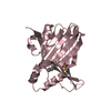
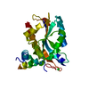
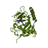
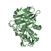
- PDB-6e5g: Crystal structure of human TEAD2-Yap binding domain covalently bo... -
+ Open data

- Basic information
| Entry | Database: PDB / ID: 6e5g | ||||||
|---|---|---|---|---|---|---|---|
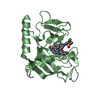
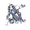
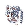
| Title | Crystal structure of human TEAD2-Yap binding domain covalently bound to an allosteric regulator | ||||||
 Components Components | Transcriptional enhancer factor TEF-4 | ||||||
 Keywords Keywords | TRANSCRIPTION/INHIBITOR / inhibitor / TRANSCRIPTION-INHIBITOR complex | ||||||
| Function / homology |  Function and homology information Function and homology informationTEAD-YAP complex / mesenchyme development / RUNX3 regulates YAP1-mediated transcription / YAP1- and WWTR1 (TAZ)-stimulated gene expression / hippo signaling / Formation of axial mesoderm / embryonic organ development / Regulation of PD-L1(CD274) transcription / transcription coactivator binding / disordered domain specific binding ...TEAD-YAP complex / mesenchyme development / RUNX3 regulates YAP1-mediated transcription / YAP1- and WWTR1 (TAZ)-stimulated gene expression / hippo signaling / Formation of axial mesoderm / embryonic organ development / Regulation of PD-L1(CD274) transcription / transcription coactivator binding / disordered domain specific binding / sequence-specific double-stranded DNA binding / nervous system development / protein-containing complex assembly / transcription regulator complex / DNA-binding transcription factor activity, RNA polymerase II-specific / RNA polymerase II cis-regulatory region sequence-specific DNA binding / DNA-binding transcription factor activity / regulation of transcription by RNA polymerase II / regulation of DNA-templated transcription / positive regulation of DNA-templated transcription / chromatin / DNA-templated transcription / nucleoplasm / nucleus Similarity search - Function | ||||||
| Biological species |  Homo sapiens (human) Homo sapiens (human) | ||||||
| Method |  X-RAY DIFFRACTION / SYNCHROTRON / MOLECULAR REPLACEMENT / Resolution: 2.43 Å X-RAY DIFFRACTION / SYNCHROTRON / MOLECULAR REPLACEMENT / Resolution: 2.43 Å | ||||||
 Authors Authors | Bum-Erdene, K. / Gonzalez-Gutierrez, G. / Meroueh, S.O. | ||||||
| Funding support |  United States, 1items United States, 1items
| ||||||
 Citation Citation | Journal: Cell Chem Biol / Year: 2019 Title: Small-Molecule Covalent Modification of Conserved Cysteine Leads to Allosteric Inhibition of the TEAD⋅Yap Protein-Protein Interaction. Authors: Bum-Erdene, K. / Zhou, D. / Gonzalez-Gutierrez, G. / Ghozayel, M.K. / Si, Y. / Xu, D. / Shannon, H.E. / Bailey, B.J. / Corson, T.W. / Pollok, K.E. / Wells, C.D. / Meroueh, S.O. #1: Journal: Acta Crystallogr. D Biol. Crystallogr. / Year: 2012 Title: Towards automated crystallographic structure refinement with phenix.refine. Authors: Afonine, P.V. / Grosse-Kunstleve, R.W. / Echols, N. / Headd, J.J. / Moriarty, N.W. / Mustyakimov, M. / Terwilliger, T.C. / Urzhumtsev, A. / Zwart, P.H. / Adams, P.D. #2: Journal: Acta Crystallogr D Biol Crystallogr / Year: 2010 Title: PHENIX: a comprehensive Python-based system for macromolecular structure solution. Authors: Paul D Adams / Pavel V Afonine / Gábor Bunkóczi / Vincent B Chen / Ian W Davis / Nathaniel Echols / Jeffrey J Headd / Li-Wei Hung / Gary J Kapral / Ralf W Grosse-Kunstleve / Airlie J McCoy ...Authors: Paul D Adams / Pavel V Afonine / Gábor Bunkóczi / Vincent B Chen / Ian W Davis / Nathaniel Echols / Jeffrey J Headd / Li-Wei Hung / Gary J Kapral / Ralf W Grosse-Kunstleve / Airlie J McCoy / Nigel W Moriarty / Robert Oeffner / Randy J Read / David C Richardson / Jane S Richardson / Thomas C Terwilliger / Peter H Zwart / Abstract: Macromolecular X-ray crystallography is routinely applied to understand biological processes at a molecular level. However, significant time and effort are still required to solve and complete many ...Macromolecular X-ray crystallography is routinely applied to understand biological processes at a molecular level. However, significant time and effort are still required to solve and complete many of these structures because of the need for manual interpretation of complex numerical data using many software packages and the repeated use of interactive three-dimensional graphics. PHENIX has been developed to provide a comprehensive system for macromolecular crystallographic structure solution with an emphasis on the automation of all procedures. This has relied on the development of algorithms that minimize or eliminate subjective input, the development of algorithms that automate procedures that are traditionally performed by hand and, finally, the development of a framework that allows a tight integration between the algorithms. | ||||||
| History |
|
- Structure visualization
| Structure viewer | Molecule: MolmilJmol/JSmol |
|---|
- Downloads & links
-Download
| PDBx/mmCIF format | 6e5g.cif.gz | 172.2 KB | Display | PDBx/mmCIF format |
|---|---|---|---|---|
| PDB format | pdb6e5g.ent.gz | 138.1 KB | Display | PDB format |
| PDBx/mmJSON format | 6e5g.json.gz | Tree view | PDBx/mmJSON format | |
| Others |  Other downloads Other downloads |
-Validation report
| Arichive directory | https://data.pdbj.org/pub/pdb/validation_reports/e5/6e5gftp://data.pdbj.org/pub/pdb/validation_reports/e5/6e5g | HTTPS FTP |
|---|
-Related structure data
| Related structure data |  5emvS S: Starting model for refinement |
|---|---|
| Similar structure data |


-Links
 PDBj
PDBj- Assembly
| Deposited unit | 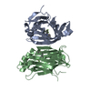
| ||||||||
|---|---|---|---|---|---|---|---|---|---|
| 1 | 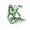
| ||||||||
| 2 | 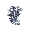
| ||||||||
| Unit cell |
|
-Components
| #1: Protein | Mass: 26714.248 Da / Num. of mol.: 2 Source method: isolated from a genetically manipulated source Source: (gene. exp.) Homo sapiens (human) / Gene: TEAD2, TEF4 / Production host:  #2: Chemical | 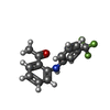  Mass: 279.257 Da / Num. of mol.: 2 / Source method: obtained synthetically / Formula: C15H12F3NO Mass: 279.257 Da / Num. of mol.: 2 / Source method: obtained synthetically / Formula: C15H12F3NO#3: Water | ChemComp-HOH / |  Mass: 18.015 Da / Num. of mol.: 63 / Source method: isolated from a natural source / Formula: H2O Mass: 18.015 Da / Num. of mol.: 63 / Source method: isolated from a natural source / Formula: H2OHas protein modification | Y | |
|---|
-Experimental details
-Experiment
| Experiment | Method: X-RAY DIFFRACTION / Number of used crystals: 1 |
|---|
- Sample preparation
| Crystal | Density Matthews: 2.53 Å3/Da / Density % sol: 51.31 % |
|---|---|
| Crystal grow | Temperature: 294 K / Method: vapor diffusion, hanging drop / Details: 2.4-2.8 M sodium formate, HEPES, pH 7.2- 7.4 / PH range: 7.2 - 7.4 |
-Data collection
| Diffraction | Mean temperature: 100 K |
|---|---|
| Diffraction source | Source: SYNCHROTRON / Site: ALS / Beamline: 4.2.2 / Wavelength: 0.97625 Å |
| Detector | Type: RDI CMOS_8M / Detector: CMOS / Date: May 17, 2018 |
| Radiation | Monochromator: double crystal Si(111) / Protocol: SINGLE WAVELENGTH / Monochromatic (M) / Laue (L): M / Scattering type: x-ray |
| Radiation wavelength | Wavelength: 0.97625 Å / Relative weight: 1 |
| Reflection | Resolution: 2.43→48.17 Å / Num. obs: 20219 / % possible obs: 99.7 % / Redundancy: 3.6 % / CC1/2: 0.996 / Rmerge(I) obs: 0.098 / Rpim(I) all: 0.061 / Rrim(I) all: 0.116 / Χ2: 1.08 / Net I/σ(I): 9.8 |
| Reflection shell | Resolution: 2.43→2.52 Å / Redundancy: 3.5 % / Rmerge(I) obs: 1.198 / Mean I/σ(I) obs: 1 / Num. unique obs: 2112 / CC1/2: 0.601 / Rpim(I) all: 0.76 / Rrim(I) all: 1.423 / Χ2: 0.96 / % possible all: 100 |
- Processing
| Software |
| ||||||||||||||||||||||||||||||||||||||||||||||||||||||||
|---|---|---|---|---|---|---|---|---|---|---|---|---|---|---|---|---|---|---|---|---|---|---|---|---|---|---|---|---|---|---|---|---|---|---|---|---|---|---|---|---|---|---|---|---|---|---|---|---|---|---|---|---|---|---|---|---|---|
| Refinement | Method to determine structure: MOLECULAR REPLACEMENT Starting model: PDB entry 5EMV Resolution: 2.43→38.89 Å / SU ML: 0.38 / Cross valid method: FREE R-VALUE / σ(F): 1.34 / Phase error: 30.59
| ||||||||||||||||||||||||||||||||||||||||||||||||||||||||
| Solvent computation | Shrinkage radii: 0.9 Å / VDW probe radii: 1.11 Å | ||||||||||||||||||||||||||||||||||||||||||||||||||||||||
| Refinement step | Cycle: LAST / Resolution: 2.43→38.89 Å
| ||||||||||||||||||||||||||||||||||||||||||||||||||||||||
| Refine LS restraints |
| ||||||||||||||||||||||||||||||||||||||||||||||||||||||||
| LS refinement shell |
|
