 Movie
Movie Controller

[English] 日本語
 Yorodumi
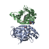
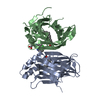
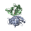
- PDB-5b60: Crystal structure of PtLCIB4 S47R mutant, a homolog of the limiti... -
+ Open data

- Basic information
| Entry | Database: PDB / ID: 5b60 | ||||||
|---|---|---|---|---|---|---|---|
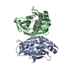
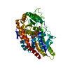
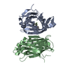
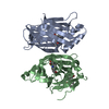
| Title | Crystal structure of PtLCIB4 S47R mutant, a homolog of the limiting CO2-inducible protein LCIB | ||||||
 Components Components | PtLCIB4 S47R mutant | ||||||
 Keywords Keywords | METAL BINDING PROTEIN / metalloenzyme | ||||||
| Function / homology | Limiting CO2-inducible protein B/C, beta carbonyic anhydrase domain / Limiting CO2-inducible proteins B/C beta carbonyic anhydrases / metal ion binding / Limiting CO2-inducible protein B/C beta carbonyic anhydrase domain-containing protein Function and homology information Function and homology information | ||||||
| Biological species |  | ||||||
| Method |  X-RAY DIFFRACTION / SYNCHROTRON / MOLECULAR REPLACEMENT / Resolution: 2.2 Å X-RAY DIFFRACTION / SYNCHROTRON / MOLECULAR REPLACEMENT / Resolution: 2.2 Å | ||||||
 Authors Authors | Jin, S. / Sun, J. / Wunder, T. / Tang, D. / Mueller-Caja, O.M. / Gao, Y. | ||||||
| Funding support |  Singapore, 1items Singapore, 1items
| ||||||
 Citation Citation | Journal: Proc. Natl. Acad. Sci. U.S.A. / Year: 2016 Title: Structural insights into the LCIB protein family reveals a new group of beta-carbonic anhydrases Authors: Jin, S. / Sun, J. / Wunder, T. / Tang, D. / Cousins, A.B. / Sze, S.K. / Mueller-Cajar, O. / Gao, Y.G. | ||||||
| History |
|
- Structure visualization
| Structure viewer | Molecule: MolmilJmol/JSmol |
|---|
- Downloads & links
-Download
| PDBx/mmCIF format | 5b60.cif.gz | 58 KB | Display | PDBx/mmCIF format |
|---|---|---|---|---|
| PDB format | pdb5b60.ent.gz | 38.7 KB | Display | PDB format |
| PDBx/mmJSON format | 5b60.json.gz | Tree view | PDBx/mmJSON format | |
| Others |  Other downloads Other downloads |
-Validation report
| Arichive directory | https://data.pdbj.org/pub/pdb/validation_reports/b6/5b60ftp://data.pdbj.org/pub/pdb/validation_reports/b6/5b60 | HTTPS FTP |
|---|
-Related structure data
| Related structure data | 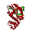 5b5xC 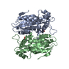 5b5ySC 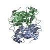 5b5zC 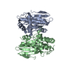 5k5wC C: citing same article ( S: Starting model for refinement |
|---|---|
| Similar structure data |


-Links
 PDBj
PDBj- Assembly
| Deposited unit | 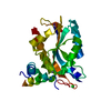
| ||||||||
|---|---|---|---|---|---|---|---|---|---|
| 1 | 
| ||||||||
| Unit cell |
|
-Components
| #1: Protein | Mass: 30377.990 Da / Num. of mol.: 1 / Mutation: S47R Source method: isolated from a genetically manipulated source Source: (gene. exp.)  |
|---|---|
| #2: Chemical | ChemComp-ZN /   Mass: 65.409 Da / Num. of mol.: 1 Mass: 65.409 Da / Num. of mol.: 1Source method: isolated from a genetically manipulated source Formula: Zn |
| #3: Chemical | ChemComp-CL /   Mass: 35.453 Da / Num. of mol.: 1 Mass: 35.453 Da / Num. of mol.: 1Source method: isolated from a genetically manipulated source Formula: Cl |
| #4: Water | ChemComp-HOH /  Mass: 18.015 Da / Num. of mol.: 24 / Source method: isolated from a natural source / Formula: H2O Mass: 18.015 Da / Num. of mol.: 24 / Source method: isolated from a natural source / Formula: H2O |
| Sequence details | The sequence database of this protein is BAV00141 in GenBank. The residues (-12)-1 are N-terminal ...The sequence database of this protein is BAV00141 in GenBank. The residues (-12)-1 are N-terminal expression tags. This structure is S47R mutant. |
-Experimental details
-Experiment
| Experiment | Method: X-RAY DIFFRACTION / Number of used crystals: 1 |
|---|
- Sample preparation
| Crystal | Density Matthews: 2.53 Å3/Da / Density % sol: 51.35 % |
|---|---|
| Crystal grow | Temperature: 293.15 K / Method: vapor diffusion, hanging drop / pH: 6.5 Details: 200 mM sodium chloride, 100 mM Bis-Tris, 25% w/v PEG3350 |
-Data collection
| Diffraction | Mean temperature: 100 K |
|---|---|
| Diffraction source | Source: SYNCHROTRON / Site: Australian Synchrotron  / Beamline: MX2 / Wavelength: 1 Å / Beamline: MX2 / Wavelength: 1 Å |
| Detector | Type: ADSC QUANTUM 210r / Detector: CCD / Date: Feb 25, 2016 |
| Radiation | Protocol: SINGLE WAVELENGTH / Monochromatic (M) / Laue (L): M / Scattering type: x-ray |
| Radiation wavelength | Wavelength: 1 Å / Relative weight: 1 |
| Reflection | Resolution: 2.2→67.8 Å / Num. obs: 15494 / % possible obs: 99.5 % / Redundancy: 8.8 % / Net I/σ(I): 11 |
| Reflection shell | Resolution: 2.2→2.32 Å |
- Processing
| Software |
| |||||||||||||||||||||||||||||||||||||||||||||||||
|---|---|---|---|---|---|---|---|---|---|---|---|---|---|---|---|---|---|---|---|---|---|---|---|---|---|---|---|---|---|---|---|---|---|---|---|---|---|---|---|---|---|---|---|---|---|---|---|---|---|---|
| Refinement | Method to determine structure: MOLECULAR REPLACEMENT Starting model: 5B5Y Resolution: 2.2→47.159 Å / SU ML: 0.29 / Cross valid method: FREE R-VALUE / σ(F): 1.64 / Phase error: 27.48
| |||||||||||||||||||||||||||||||||||||||||||||||||
| Solvent computation | Shrinkage radii: 0.9 Å / VDW probe radii: 1.11 Å | |||||||||||||||||||||||||||||||||||||||||||||||||
| Refinement step | Cycle: LAST / Resolution: 2.2→47.159 Å
| |||||||||||||||||||||||||||||||||||||||||||||||||
| Refine LS restraints |
| |||||||||||||||||||||||||||||||||||||||||||||||||
| LS refinement shell |
|
