 Movie
Movie Controller


+ Open data

- Basic information
| Entry | Database: PDB / ID: 2okh | ||||||
|---|---|---|---|---|---|---|---|
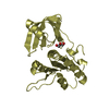
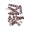
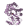
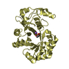
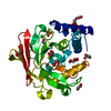
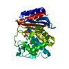
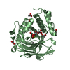
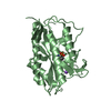
| Title | Crystal structure of dimeric form of PfFabZ in crystal form3 | ||||||
 Components Components | Beta-hydroxyacyl-ACP dehydratase | ||||||
 Keywords Keywords | LYASE / fabz / hotdog fold / non-isomorphism / plasmodium | ||||||
| Function / homology |  Function and homology information Function and homology information3-hydroxyacyl-[acyl-carrier-protein] dehydratase / (3R)-hydroxyacyl-[acyl-carrier-protein] dehydratase activity / lipid A biosynthetic process / fatty acid biosynthetic process / membrane / cytoplasm Similarity search - Function | ||||||
| Biological species |  | ||||||
| Method |  X-RAY DIFFRACTION / MOLECULAR REPLACEMENT / Resolution: 3 Å X-RAY DIFFRACTION / MOLECULAR REPLACEMENT / Resolution: 3 Å | ||||||
 Authors Authors | Swarnamukhi, P.L. / Sharma, S.K. / Padala, P. / Surolia, N. / Surolia, A. / Suguna, K. | ||||||
 Citation Citation | Journal: ACTA CRYSTALLOGR.,SECT.D / Year: 2007 Title: Packing and loop-structure variations in non-isomorphous crystals of FabZ from Plasmodium falciparum Authors: Swarnamukhi, P.L. / Sharma, S.K. / Padala, P. / Surolia, N. / Surolia, A. / Suguna, K. | ||||||
| History |
|
- Structure visualization
| Structure viewer | Molecule: MolmilJmol/JSmol |
|---|
- Downloads & links
-Download
| PDBx/mmCIF format | 2okh.cif.gz | 58.9 KB | Display | PDBx/mmCIF format |
|---|---|---|---|---|
| PDB format | pdb2okh.ent.gz | 42.5 KB | Display | PDB format |
| PDBx/mmJSON format | 2okh.json.gz | Tree view | PDBx/mmJSON format | |
| Others |  Other downloads Other downloads |
-Validation report
| Arichive directory | https://data.pdbj.org/pub/pdb/validation_reports/ok/2okhftp://data.pdbj.org/pub/pdb/validation_reports/ok/2okh | HTTPS FTP |
|---|
-Related structure data
| Related structure data |  2okiC  1zhgS C: citing same article ( S: Starting model for refinement |
|---|---|
| Similar structure data |


-Links
 PDBj
PDBj
- Assembly
| Deposited unit | 
| ||||||||
|---|---|---|---|---|---|---|---|---|---|
| 1 |
| ||||||||
| Unit cell |
|
-Components
| #1: Protein | Mass: 14990.649 Da / Num. of mol.: 2 / Fragment: residues 94-229 Source method: isolated from a genetically manipulated source Source: (gene. exp.) Gene: fabZ / Plasmid: PET-28A(+) / Species (production host): Escherichia coli / Production host:  References: UniProt: Q965D7, Lyases; Carbon-oxygen lyases; Hydro-lyases #2: Water | ChemComp-HOH / |  Mass: 18.015 Da / Num. of mol.: 20 / Source method: isolated from a natural source / Formula: H2O Mass: 18.015 Da / Num. of mol.: 20 / Source method: isolated from a natural source / Formula: H2O |
|---|
-Experimental details
-Experiment
| Experiment | Method: X-RAY DIFFRACTION / Number of used crystals: 1 |
|---|
- Sample preparation
| Crystal | Density Matthews: 1.7 Å3/Da / Density % sol: 27 % |
|---|---|
| Crystal grow | Temperature: 300 K / Method: vapor diffusion, hanging drop / pH: 4.5 Details: 30% PEG 4000, 0.1M acetate buffer pH4.5, 0.2M sodium acetate, VAPOR DIFFUSION, HANGING DROP, temperature 300K |
-Data collection
| Diffraction | Mean temperature: 100 K |
|---|---|
| Diffraction source | Source: ROTATING ANODE / Type: RIGAKU / Wavelength: 1.5418 Å |
| Detector | Type: MAR scanner 345 mm plate / Detector: IMAGE PLATE / Date: Apr 5, 2003 / Details: osmic mirror |
| Radiation | Monochromator: osmic mirror / Protocol: SINGLE WAVELENGTH / Monochromatic (M) / Laue (L): M / Scattering type: x-ray |
| Radiation wavelength | Wavelength: 1.5418 Å / Relative weight: 1 |
| Reflection | Resolution: 3→30 Å / Num. all: 4939 / Num. obs: 4840 / % possible obs: 98 % / Observed criterion σ(F): 0 / Observed criterion σ(I): 0 / Biso Wilson estimate: 51.4 Å2 / Rmerge(I) obs: 0.085 / Net I/σ(I): 14.5 |
| Reflection shell | Resolution: 3→3.11 Å / Rmerge(I) obs: 0.3 / Mean I/σ(I) obs: 3.31 / Num. unique all: 476 / % possible all: 92.4 |
- Processing
| Software |
| ||||||||||||||||||||||||||||||||||||
|---|---|---|---|---|---|---|---|---|---|---|---|---|---|---|---|---|---|---|---|---|---|---|---|---|---|---|---|---|---|---|---|---|---|---|---|---|---|
| Refinement | Method to determine structure: MOLECULAR REPLACEMENT Starting model: 1ZHG Resolution: 3→19.64 Å / Rfactor Rfree error: 0.02 / Data cutoff high absF: 1429838.91 / Data cutoff low absF: 0 / Isotropic thermal model: RESTRAINED / Cross valid method: THROUGHOUT / σ(F): 0 / σ(I): 0
| ||||||||||||||||||||||||||||||||||||
| Solvent computation | Solvent model: FLAT MODEL / Bsol: 38.4742 Å2 / ksol: 0.344079 e/Å3 | ||||||||||||||||||||||||||||||||||||
| Displacement parameters | Biso mean: 51.4 Å2
| ||||||||||||||||||||||||||||||||||||
| Refine analyze |
| ||||||||||||||||||||||||||||||||||||
| Refinement step | Cycle: LAST / Resolution: 3→19.64 Å
| ||||||||||||||||||||||||||||||||||||
| Refine LS restraints |
| ||||||||||||||||||||||||||||||||||||
| LS refinement shell | Resolution: 3→3.19 Å / Rfactor Rfree error: 0.07 / Total num. of bins used: 6
| ||||||||||||||||||||||||||||||||||||
| Xplor file |
|
